Review
. 2022 Mar 29;23(7):3732.
doi: 10.3390/ijms23073732.

Enteroendocrine System and Gut Barrier in Metabolic Disorders

Affiliations
Review

Enteroendocrine System and Gut Barrier in Metabolic Disorders

Céline Osinski et al. Int J Mol Sci. .

Abstract

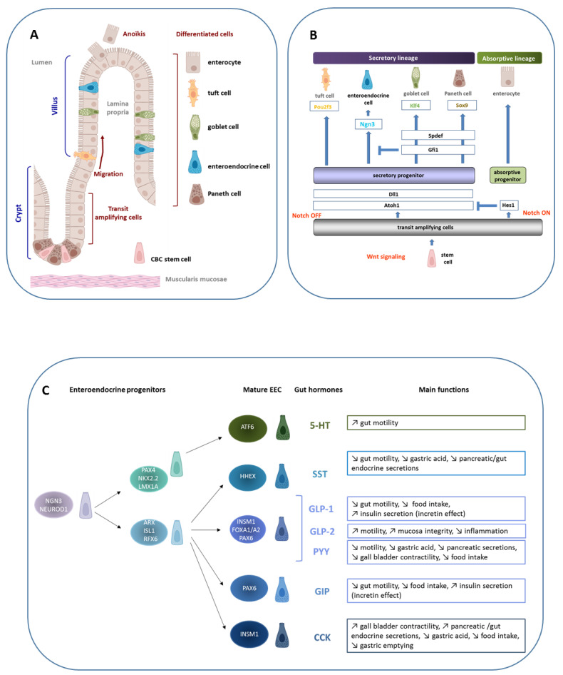
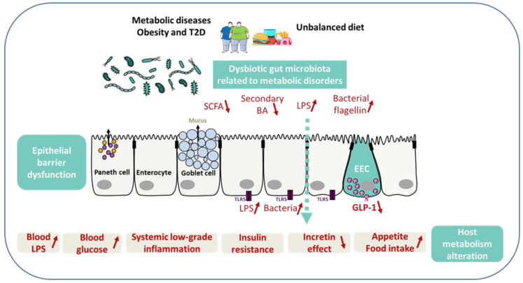
With the continuous rise in the worldwide prevalence of obesity and type 2 diabetes, developing therapies regulating body weight and glycemia has become a matter of great concern. Among the current treatments, evidence now shows that the use of intestinal hormone analogs (e.g., GLP1 analogs and others) helps to control glycemia and reduces body weight. Indeed, intestinal endocrine cells produce a large variety of hormones regulating metabolism, including appetite, digestion, and glucose homeostasis. Herein, we discuss how the enteroendocrine system is affected by local environmental and metabolic signals. These signals include those arising from unbalanced diet, gut microbiota, and the host metabolic organs and their complex cross-talk with the intestinal barrier integrity.

Keywords: cell lineage; diets; enteroendocrine cells; gut barrier integrity; intestinal hormones; microbiota; obesity; type 2 diabetes.


Conflict of interest statement

The authors declare no potential conflict of interest with respect to the research, authorship, and/or publication of this article.

Figures

Figure 1
Figure 1
Intestinal epithelium homeostasis, enteroendocrine cell lineage, and functions. (A) The intestinal epithelium is composed of a single layer of cells lying on a mesenchymal tissue, the lamina propria, which contains connective tissue and vascular structure. In the small intestine, the epithelium is organized into villi that project in the intestinal lumen, with crypts which invaginate in the mucosa. This structural unit rests on a thin layer of smooth muscle, the muscularis mucosa. The intestinal epithelium is completely renewed in 3 days in mice and 5 days in humans from crypt-based columnar (CBC) stems cells located in the crypt bottom. After division, proliferating cells start to differentiate and migrate toward the villus tip where they are exfoliated after apoptosis, a process called anoïkis. Except for the Paneth cells that are located in the crypt bottom and intercalated between CBC stem cells, participating in the formation of the «niche», the other differentiated cells are located along the villus, enterocytes, tuft cells, goblet cells, and enteroendocrine cells (EEC). (B) Schematic demonstrating that under the Wnt signal, CBC stem cells proliferate and give rise to transit amplifying cells. Under the Notch signal, the transcription factor Hes1 is expressed and allows the progenitors to differentiate into enterocytes, the absorptive cells. When the Notch signal is OFF, Atoh1 is expressed and gives rise to the secretory lineage. The differentiation of secretory progenitors is under the control of specific transcription factors. The expression of Ngn3 leads to enteroendocrine progenitors. (C) A focus on the EEC linage shows its complexity. EEC lineage is under the control of a transcription factor network that acts sequentially leading to a collection of mature EEC characterized by the hormone they produce. Although EEC represent only 1% of the total intestinal epithelial cells, the large variety of hormones they secrete makes the intestine a major endocrine organ. Examples of gut hormones and their main biological functions are listed: serotonin (5-HT), somatostatin (SST), GLP-1 and GLP-2, PYY, GIP, and CCK.
Figure 2
Figure 2
Gut barrier, enteroendocrine cells, and metabolic signals related to metabolic diseases. Unbalanced diet and dysbiotic gut microbiota related to metabolic disorders may result in dysfunction of intestinal barrier integrity, local and systemic inflammation, and reduced production of both SCFA and secondary bile acids, leading to less GLP-1 secretion by EEC. The reduced GLP-1 secretion leads in turn to impaired incretin effect, and an increase of blood glucose, appetite, and food intake. When in excess, LPS prompt intestinal and systemic inflammation and insulin resistance. TLR5, expressed mainly on the basolateral membrane of intestinal epithelial cells, identifies the bacterial locomotion component flagellin for uncovering whether bacteria have crossed the gut epithelia. Aberrantly elevated TLR5 activation might result in damage of the epithelial barrier integrity and inflammation.

References

    1. Darwich A.S., Aslam U., Ashcroft D.M., Rostami-Hodjegan A. Meta-Analysis of the Turnover of Intestinal Epithelia in Preclinical Animal Species and Humans. Drug Metab. Dispos. 2014;42:2016–2022. doi: 10.1124/dmd.114.058404. - DOI - PubMed
    1. Clevers H. The Intestinal Crypt, a Prototype Stem Cell Compartment. Cell. 2013;154:274–284. doi: 10.1016/j.cell.2013.07.004. - DOI - PubMed
    1. Beumer J., Clevers H. Regulation and Plasticity of Intestinal Stem Cells during Homeostasis and Regeneration. Development. 2016;143:3639–3649. doi: 10.1242/dev.133132. - DOI - PubMed
    1. Gerbe F., Legraverend C., Jay P. The Intestinal Epithelium Tuft Cells: Specification and Function. Cell Mol. Life Sci. 2012;69:2907–2917. doi: 10.1007/s00018-012-0984-7. - DOI - PMC - PubMed
    1. Farin H.F., Van Es J.H., Clevers H. Redundant Sources of Wnt Regulate Intestinal Stem Cells and Promote Formation of Paneth Cells. Gastroenterology. 2012;143:1518–1529.e7. doi: 10.1053/j.gastro.2012.08.031. - DOI - PubMed