. 2022 Mar 29;23(7):3756.
doi: 10.3390/ijms23073756.

Identification, Isolation, and Characterization of Melanocyte Precursor Cells in the Human Limbal Stroma

Affiliations

Identification, Isolation, and Characterization of Melanocyte Precursor Cells in the Human Limbal Stroma

Shen Li et al. Int J Mol Sci. .

Abstract

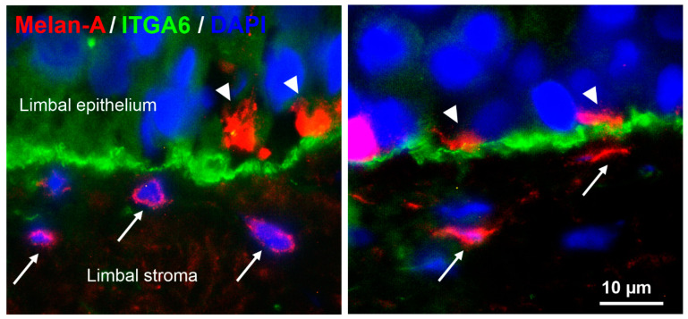
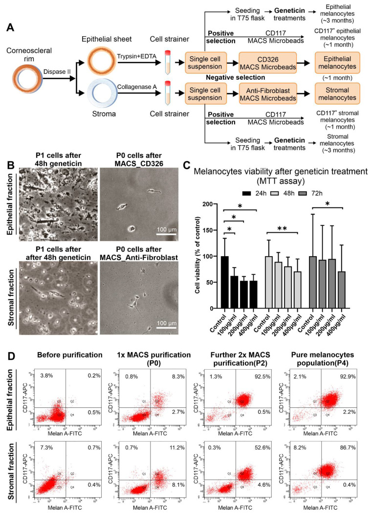
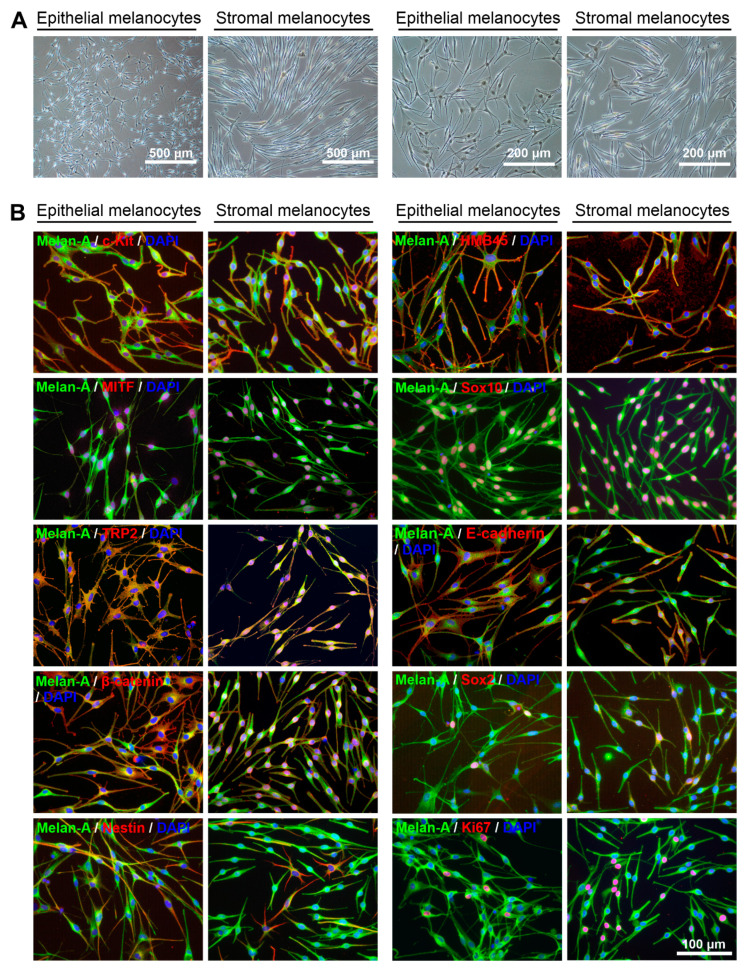
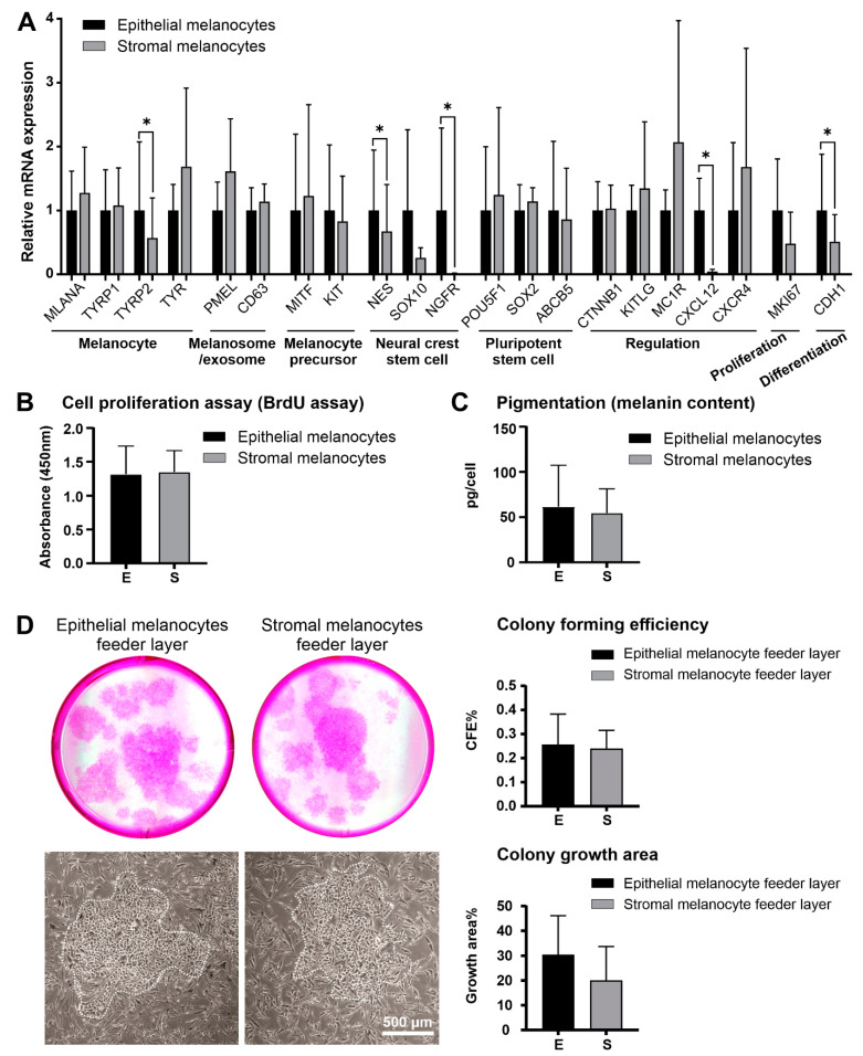
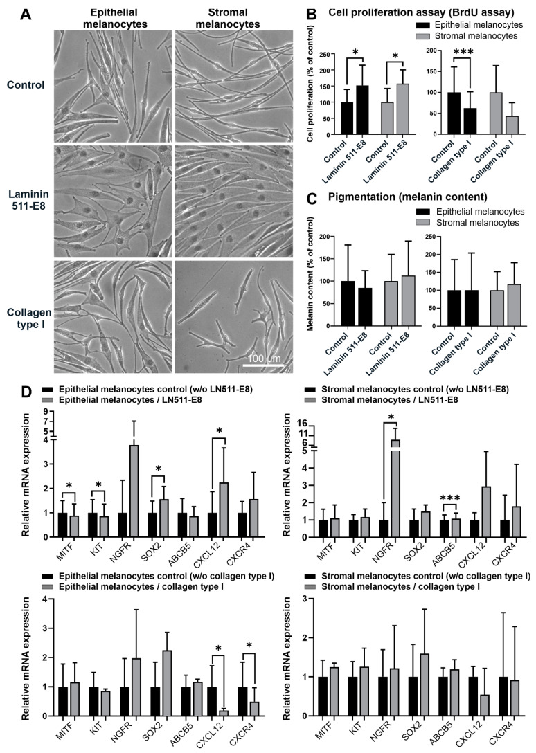
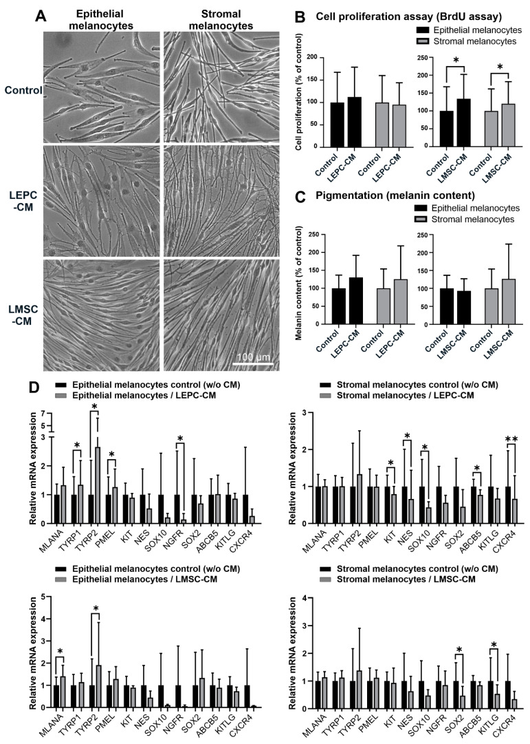
Given their vital role in the homeostasis of the limbal stem cell niche, limbal melanocytes have emerged as promising candidates for tissue engineering applications. This study aimed to isolate and characterize a population of melanocyte precursors in the limbal stroma, compared with melanocytes originating from the limbal epithelium, using magnetic-activated cell sorting (MACS) with positive (CD117/c-Kit microbeads) or negative (CD326/EpCAM or anti-fibroblast microbeads) selection approaches. Both approaches enabled fast and easy isolation and cultivation of pure limbal epithelial and stromal melanocyte populations, which differed in phenotype and gene expression, but exhibited similar functional properties regarding proliferative potential, pigmentation, and support of clonal growth of limbal epithelial stem/progenitor cells (LEPCs). In both melanocyte populations, limbus-specific matrix (laminin 511-E8) and soluble factors (LEPC-derived conditioned medium) stimulated melanocyte adhesion, dendrite formation, melanogenesis, and expression of genes involved in UV protection and immune regulation. The findings provided not only a novel protocol for the enrichment of pure melanocyte populations from limbal tissue applying easy-to-use MACS technology, but also identified a population of stromal melanocyte precursors, which may serve as a reservoir for the replacement of damaged epithelial melanocytes and an alternative resource for tissue engineering applications.

Keywords: cultivation; laminin 511-E8; limbal stem cell niche; limbal stem cells; limbal stroma; magnetic-activated cell sorting; melanocytes.


Conflict of interest statement

The authors declare no conflict of interest.

Figures

Figure 1
Figure 1
Localization of epithelial and stromal melanocytes at the human limbus in situ. Immunofluorescence double labeling of corneoscleral tissue sections prepared from two donor corneas (age 65 and 72 years) showing Melan-A+ melanocyte processes (arrowheads) in the basal limbal epithelium expressing integrin α6 (ITGA6) and additional Melan-A+ melanocytes (arrows) in the subepithelial limbal stroma; nuclear counterstaining with 4′,6-diamidino-2-phenylindole (DAPI).
Figure 2
Figure 2
Isolation of limbal epithelial and stromal melanocyte populations using conventional geneticin treatment and magnetic-activated cell sorting. (A). Graphical representation of limbal melanocyte isolation: Differential enzymatic digestion of limbal tissue generated single-cell suspensions of epithelial and stromal cells; melanocytes were enriched from both suspensions either by conventional geneticin treatments or by magnetic-activated cell sorting (MACS) technology using either a positive selection approach with CD117 (c-Kit) Microbeads or a negative selection approach with CD326 (EpCAM) Microbeads and Anti-Fibroblast Microbeads, respectively. (B). Phase-contrast images showing epithelial (B) and stromal (C) cell fractions after 48 h of geneticin treatment at passage 1 (P1) and after MACS negative selection at passage 0 (P0). (C). Cell viability (MTT) assay after 24, 48, and 72 h of treatment with different concentrations of geneticin (100-400 µg/mL). Data (% of control) are expressed as means ± SD (n = 3) relative to control (* p < 0.05; ** p < 0.01; paired t-test). (D). Flow cytometry analysis showing an increase of Melan-A+/CD117+ cells in epithelial and stromal fractions after MACS purification steps.
Figure 3
Figure 3
Phenotypic and immunocytochemical characterization of limbal epithelial and stromal melanocytes. (A). Phase-contrast images showing pure populations of epithelial and stromal melanocytes at passage 4 at two different magnifications. (B). Immunofluorescence double labeling of epithelial and stromal melanocytes for Melan-A and additional melanocyte-associated markers c-Kit (CD117), HMB45 (PMEL, premelanosome protein), MITF (microphthalmia-associated transcription factor), Sox10 (SRY-box transcription factor 10), TRP2 (tyrosine-related protein 2), E-cadherin, ß-catenin, Sox2, nestin, and Ki-67; nuclear counterstaining with 4′,6-diamidino-2-phenylindole (DAPI). The magnification bar at the bottom-right corner applies to all images.
Figure 4
Figure 4
Comparative analysis of limbal epithelial and stromal melanocytes. (A). Quantitative real-time polymerase chain reaction (qRT-PCR) primer assays showing relative expression levels of MLANA (Melan-A), TYRP1 and TYRP2 (tyrosinase-related protein 1 and 2), TYR (tyrosinase), PMEL (premelanosome protein), CD63 (CD63 antigen), MITF (microphthalmia-associated transcription factor), KIT (c-Kit/CD117), NES (nestin), SOX10 (SRY-Box transcription factor 10), NGFR (nerve growth factor receptor), POU5F1 (POU class 5 homeobox 1), SOX2 (SRY-Box transcription factor 2), ABCB5 (ATP binding cassette subfamily B member 5), CTNNB1 (catenin beta 1), KITLG (Kit ligand), MC1R (melanocortin 1 receptor), CXCL12 (C-X-C Motif Chemokine Ligand 12), CXCR4 (stromal cell-derived factor 1 receptor), MKI67 (Ki-67), and CDH1 (E-cadherin) in limbal epithelial and stromal melanocytes. Data are normalized to GAPDH and ACTB and expressed as means (2−ΔCT × 1000) ± SD (n = 5) relative to epithelial melanocytes (* p < 0.05; paired t-test). (B). Proliferative capacity of limbal epithelial and stromal melanocytes as determined by BrdU incorporation assay. Data are expressed as means ± SD (n = 5). (C). Quantification of cellular melanin content in limbal epithelial and stromal melanocytes. Data are expressed as means ± SD (n = 5). (D). Formation of limbal epithelial stem cell clones (dotted lines) expanded on feeder layers of limbal epithelial and stromal melanocytes, followed by quantitative comparative analysis of colony-forming efficiency (CFE) and colony growth area. Data represent means ± SD (n = 3).
Figure 5
Figure 5
Effects of extracellular matrix components on limbal epithelial and stromal melanocytes. (A). Phase-contrast images of limbal epithelial and stromal melanocytes cultured on laminin 511-E8 and collagen type I (the magnification bar at the bottom-right corner applies to all images). (B). The effect of laminin 511-E8 and collagen type I on the proliferation of limbal epithelial and stromal melanocytes was analyzed by BrdU incorporation assay after 72 h. Data are expressed as means ± SD (n = 4) relative to control (* p < 0.05; *** p < 0.001; paired t-test). (C). The effect of laminin 511-E8 and collagen type I on the melanin content of limbal epithelial and stromal melanocytes was analyzed by pigmentation assay after 72 h. Data are expressed as means ± SD (n = 4) relative to control. (D). Quantitative real-time polymerase chain reaction (qRT-PCR) primer assays showing relative expression levels of MITF (microphthalmia-associated transcription factor), KIT (c-Kit/CD117), NGFR (nerve growth factor receptor), SOX2 (SRY-Box transcription factor 2), ABCB5 (ATP binding cassette subfamily B member 5), CXCL12 (C-X-C Motif Chemokine Ligand 12), and CXCR4 (stromal cell-derived factor 1 receptor) in limbal epithelial and stromal melanocytes cultured on laminin 511-E8 and collagen type I. Data are normalized to GAPDH and ACTB and expressed as means (2−ΔCT × 1000) ± SD (n = 3) relative to control (* p < 0.05; *** p < 0.001; paired t-test).
Figure 6
Figure 6
Effects of soluble factors on limbal epithelial and stromal melanocytes. (A). Phase-contrast images of limbal epithelial and stromal melanocytes cultured in conditioned media derived from limbal epithelial stem/progenitor cells (LEPC-CM) and limbal mesenchymal stem cells (LMSC-CM) (the magnification bar at the bottom-right corner applies to all images). (B). The effect of LEPC-CM and LMSC-CM on the proliferation of limbal epithelial and stromal melanocytes was analyzed by BrdU incorporation assay after 72 h. Data are expressed as means ± SD (n = 4) relative to control (* p < 0.05; paired t-test). (C). The effect of LEPC-CM and LMSC-CM on the melanin content of limbal epithelial and stromal melanocytes was analyzed by pigmentation assay after 72 h. Data are expressed as means ± SD (n = 4) relative to control. (D). Quantitative real-time polymerase chain reaction (qRT-PCR) primer assays showing relative expression levels of MLANA (Melan-A), TYRP1 and TYRP2 (tyrosinase-related protein 1 and 2), PMEL (premelanosome protein), KIT (c-Kit/CD117), NES (nestin), SOX10 (SRY-Box transcription factor 10), NGFR (nerve growth factor receptor), SOX2 (SRY-Box transcription factor 2), ABCB5 (ATP binding cassette subfamily B member 5), KITLG (Kit ligand), and CXCR4 (stromal cell-derived factor 1 receptor) in limbal epithelial and stromal melanocytes cultured in LEPC-CM and LMSC-CM. Data are normalized to GAPDH and ACTB and expressed as means (2−ΔCT × 1000) ± SD (n = 4) relative to control (* p < 0.05; ** p < 0.01; paired t-test).

References

    1. Bonnet C., González S., Roberts J.S., Robertson S.Y., Ruiz M., Zheng J., Deng S.X. Human limbal epithelial stem cell regulation, bioengineering and function. Prog. Retin. Eye Res. 2021;85:100956. doi: 10.1016/j.preteyeres.2021.100956. - DOI - PMC - PubMed
    1. Gonzalez G., Sasamoto Y., Ksander B.R., Frank M., Frank N.Y. Limbal stem cells: Identity, developmental origin, and therapeutic potential. Wiley Interdiscip. Rev. Dev. Biol. 2017;7:e303. doi: 10.1002/wdev.303. - DOI - PMC - PubMed
    1. Shortt A.J., Secker G.A., Munro P.M., Khaw P.T., Tuft S.J., Daniels J.T. Characterization of the Limbal Epithelial Stem Cell Niche: Novel Imaging Techniques Permit In Vivo Observation and Targeted Biopsy of Limbal Epithelial Stem Cells. Stem Cells. 2007;25:1402–1409. doi: 10.1634/stemcells.2006-0580. - DOI - PubMed
    1. Li W., Hayashida Y., Chen Y.-T., Tseng S.C.G. Niche regulation of corneal epithelial stem cells at the limbus. Cell Res. 2007;17:26–36. doi: 10.1038/sj.cr.7310137. - DOI - PMC - PubMed
    1. Ordonez P., Di Girolamo N. Limbal Epithelial Stem Cells: Role of the Niche Microenvironment. Stem Cells. 2012;30:100–107. doi: 10.1002/stem.794. - DOI - PubMed