Review
. 2022 Mar 29;23(7):3785.
doi: 10.3390/ijms23073785.

Brain Metabolic Alterations in Alzheimer's Disease

Affiliations
Review

Brain Metabolic Alterations in Alzheimer's Disease

Carlos G Ardanaz et al. Int J Mol Sci. .

Abstract

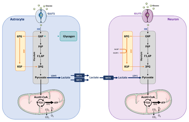
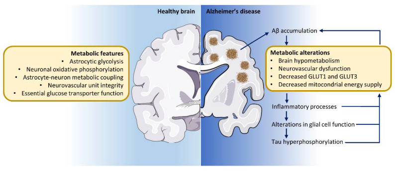
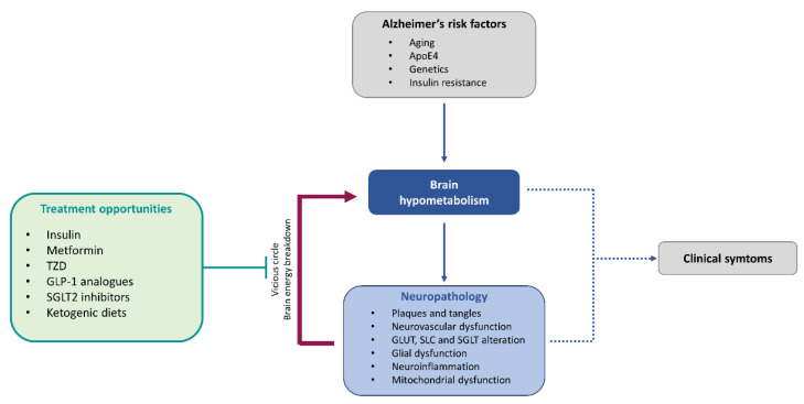
The brain is one of the most energy-consuming organs in the body. Satisfying such energy demand requires compartmentalized, cell-specific metabolic processes, known to be complementary and intimately coupled. Thus, the brain relies on thoroughly orchestrated energy-obtaining agents, processes and molecular features, such as the neurovascular unit, the astrocyte-neuron metabolic coupling, and the cellular distribution of energy substrate transporters. Importantly, early features of the aging process are determined by the progressive perturbation of certain processes responsible for adequate brain energy supply, resulting in brain hypometabolism. These age-related brain energy alterations are further worsened during the prodromal stages of neurodegenerative diseases, namely Alzheimer's disease (AD), preceding the onset of clinical symptoms, and are anatomically and functionally associated with the loss of cognitive abilities. Here, we focus on concrete neuroenergetic features such as the brain's fueling by glucose and lactate, the transporters and vascular system guaranteeing its supply, and the metabolic interactions between astrocytes and neurons, and on its neurodegenerative-related disruption. We sought to review the principles underlying the metabolic dimension of healthy and AD brains, and suggest that the integration of these concepts in the preventive, diagnostic and treatment strategies for AD is key to improving the precision of these interventions.

Keywords: GLUTs; astrocyte; astrocyte–neuron lactate shuttle (ANLS); glucose; hypometabolism; lactate; neurodegeneration.


Conflict of interest statement

The authors declare no conflict of interest.

Figures

Figure 1
Figure 1
Different metabolic strategies used by neurons and astrocytes. In astrocytes, glucose is imported through glucose transporter 1 (GLUT1) and preferentially stored as glycogen, or metabolized via glycolysis. The generated pyruvate is converted to lactate thanks to the expression of lactate dehydrogenase 5 (LDH5). Glucose enters neurons via GLUT3, and once inside the cell is phosphorylated by hexokinase (HK), resulting in glucose-6-phosphate (G6P) subsequently directed to the pentose phosphate pathway (PPP) and the glycolytic pathway. The final product of glycolysis is pyruvate, which after entering the mitochondria will be subjected to the tricarboxylic acid (TCA) cycle, and subsequently to oxidative phosphorylation consisting of the electron transport chain (ETC). This process consumes O2 and leads to the production of ATP and CO2. G6P undergoing PPP is transformed into 6-phosphogluconate (6PG), and is thereafter converted to ribulose-5-phosphate (R5P). In this process, nicotinamide adenine dinucleotide phosphate (NADPH) is produced, an essential molecule to regenerate oxidized antioxidants. The weak glycolytic activity of neurons may reduce pyruvate formation and, therefore, obtain limited energy production in mitochondria from glucose metabolism. However, this may be compensated by the uptake of lactate from astrocytes, given that glutamate stimulates lactate release from astrocytes (astrocyte–neuron lactate shuttle; ANLS). Abbreviations are as follows: 3PG, 3-phosphoglycerate; 6PG, 6-phosphogluconate; ETC, electron transport chain; F6P, fructose-6-phosphate; F1,6P, fructose-1,6-diphosphate; G6P, glucose-6-phosphate; LDH, lactate dehydrogenase; MCT, monocarboxylic acid transporter; NADPH, nicotinamide adenine dinucleotide phosphate; PPP, pentose phosphate pathway; R5P, ribulose-5-phosphate; TCA, tricarboxylic acid.
Figure 2
Figure 2
Metabolic alterations contribute to Alzheimer’s disease pathology. The metabolic coupling between astrocytes and neurons sustains proper brain function in the healthy brain. In the course of Alzheimer’s disease, at least 15 years before the onset of symptoms, a marked glucose hypometabolism is detected in specific brain regions. The accumulation of Aβ exacerbates brain glucose hypometabolism, both directly within the area of Aβ accumulation as well as in remote regions. In turn, this hypometabolic state triggers inflammatory processes and cellular damage. Moreover, alterations in glial cell function together with tau phosphorylation and subsequent accumulation exacerbates brain metabolic breakdown. Together, these features perpetuate a vicious cycle of neurodegeneration and declining brain glucose metabolism that contributes not only to the deterioration of memory and cognition but also to abnormal behavior in affected patients.
Figure 3
Figure 3
Central contribution of brain energy hypometabolism in Alzheimer’s disease pathology and treatment opportunities. Glucose hypometabolism occurring at early stages of the disease, together with the neuropathological features, induces a vicious cycle leading to brain energy breakdown and dysfunction. Treatment opportunities based on energy rescue strategies attempt to break this vicious circle and improve brain energy metabolism to, in last instance, prevent or revert clinical symptoms.

References

    1. Kety S.S. The general metabolism of the brain in vivo. In: Richter D., editor. Metabolism of the Nervous System. Peramon; London, UK: 1957. pp. 221–237.
    1. Sokoloff L., Reivich M., Kennedy C., Des Rosiers M.H., Patlak C.S., Pettigrew K.D., Sakurada O., Shinohara M. The [14C]deoxyglucose method for the measurement of local cerebral glucose utilization: Theory, procedure, and normal values in the conscious and anesthetized albino rat. J. Neurochem. 1977;28:897–916. doi: 10.1111/j.1471-4159.1977.tb10649.x. - DOI - PubMed
    1. Attwell D., Laughlin S.B. An energy budget for signaling in the grey matter of the brain. J. Cereb. Blood Flow Metab. 2001;21:1133–1145. doi: 10.1097/00004647-200110000-00001. - DOI - PubMed
    1. Alle H., Roth A., Geiger J.R. Energy-efficient action potentials in hippocampal mossy fibers. Science. 2009;325:1405–1408. doi: 10.1126/science.1174331. - DOI - PubMed
    1. Rangaraju V., Lauterbach M., Schuman E.M. Spatially stable mitochondrial compartments fuel local translation during plasticity. Cell. 2019;176:73–84. doi: 10.1016/j.cell.2018.12.013. - DOI - PubMed