Review
. 2022 Mar 31;23(7):3847.
doi: 10.3390/ijms23073847.

Metastasis and MAPK Pathways

Affiliations
Review

Metastasis and MAPK Pathways

Mateusz Kciuk et al. Int J Mol Sci. .

Abstract

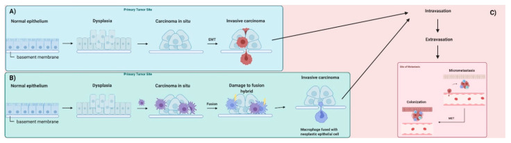
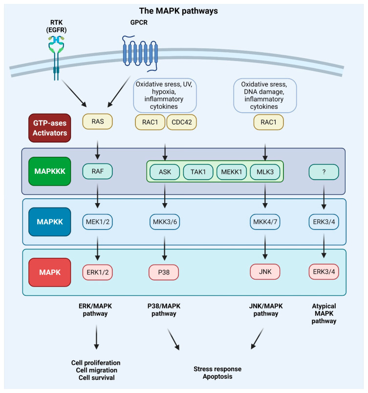
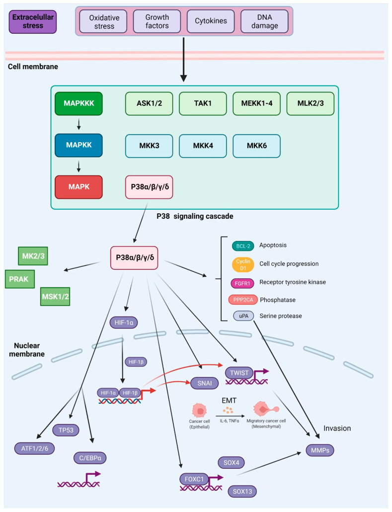
Cancer is a leading cause of death worldwide. In many cases, the treatment of the disease is limited due to the metastasis of cells to distant locations of the body through the blood and lymphatic drainage. Most of the anticancer therapeutic options focus mainly on the inhibition of tumor cell growth or the induction of cell death, and do not consider the molecular basis of metastasis. The aim of this work is to provide a comprehensive review focusing on cancer metastasis and the mitogen-activated protein kinase (MAPK) pathway (ERK/JNK/P38 signaling) as a crucial modulator of this process.

Keywords: c-JUN N-terminal kinase (JNK); cancer; extracellular signal-regulated kinase (ERK); metastasis; mitogen-activated kinases (MAPKs).


Conflict of interest statement

The authors declare no conflict of interest.

Figures

Figure 1
Figure 1
Overview of tumor metastasis. (A) Conventional model of metastasis. Normal epithelia are lined with a basement membrane that separates epithelia or endothelia from the connective tissues located beneath. Epithelial cells proliferate and become dysplastic. Accumulation of genetic and epigenetic changes leads to the formation of carcinoma in situ also lined with the basement membrane. During epithelial–mesenchymal transition (EMT), cells acquire abilities that allow them to move through the basement membrane. (B) The metastatic process according to the fusion hybrid hypothesis. Metastatic cancer cells are formed as a result of direct transformation or as a result of fusion of neoplastic epithelial cells and myeloid cells such as macrophages. (C) Metastatic cancer cells intravasate into small blood (shown here) and lymph vessels, where they travel with circulation to distant sites. At secondary sites, carcinoma cells extravasate, form micrometastases, and in the process of mesenchymal–epithelial transition (MET) colonize the foreign tissue (macrometastasis). Based on [33].
Figure 2
Figure 2
Major metabolic adaptations of cancer cells during metastasis with special focus on ways by which cancer cells avoid repercussions resulting from hypoxia and lack of nutrients. Hypoxia-inducible transcription factor α (HIF1α) is a transcription factor for many genes involved in anaerobic metabolisms, angiogenesis, and metastasis. HIFα subunit is targeted for degradation by the von Hippel–Lindau tumor suppressor (VHL) under normoxic conditions; however, the occurrence of hypoxia allows the translocation of the HIFα subunit to the nucleus and formation of transcription factor complexes with constitutively expressed HIFβ subunit. The transcription factor regulates the expression of various genes containing HIF-responsive elements (HRE) such as carbonic anhydrases, glucose transporters, and vascular endothelial growth factor (VEGF). MCT1–MCT4 are proton-coupled monocarboxylate (lactate, pyruvate, and ketone) transporters. Sodium hydrogen ion exchangers (NHE) regulate the intracellular pH regulation via electroneutral, 1:1, exchange of Na+ and H+ along their gradients. Vacuolar-type H+ adenosine triphosphatases (V-ATPases) transport H+ across membranes in an active process. Carbonic anhydrases (CA) are zinc metalloproteins that catalyze the reversible hydration of CO2 to form HCO3 and H+. CO2 is also the main byproduct of the Krebs cycle and may be generated from HCO3 in reactions catalyzed by CAII. HIF transcription factor governs the expression of MCT1/4, glucose transporters (GLUT1/3), amino acid transporters—sodium-coupled neutral amino acid transporter 2 (SNAT2), large neutral amino acid transporter 1 (LAT1), and CAIX/CAXII. Cl/HCO3—chloride bicarbonate anion exchangers; Na+/HCO3—sodium-dependent bicarbonate cotransporters.
Figure 3
Figure 3
Integration network of epidermal growth factor receptor (EGFR), mitogen-activated protein kinase (ERK/MAPK), and transforming growth factor-β (TGFβ) signaling pathways involved in metastasis processes. Several signaling pathways are involved in the progress of the epithelial–mesenchymal transition (EMT), and these pathways can work together to elicit complete EMT responses. Apart from its ability to promote EMT through the expression of mothers against decapentaplegic (SMAD) proteins, transforming growth factor β (TGF-β) can also activate the phosphotyrosine 3-kinase/RAC-alpha serine/threonine-protein kinase (PI3K–AKT), ERK–MAPK, P38–MAPK, and c-JUN N-terminal kinase (JNK) signaling pathways. After TβRI phosphorylates the adaptor protein SRC homology 2 domain-containing-transforming A (SHCA), it provides a docking site for the growth factor receptor-bound protein 2 (GRB2) and the son of sevenless (SOS), an event that signals the initiation of the MAPK cascade involving RAS, RAF, MEK, and ERK. Interaction of TNF receptor-associated factor 6 (TRAF6) with the TGF receptor complex stimulates TGF-activated kinase 1 (TAK1), which contributes to activation of P38 and JNK. The EMT is facilitated by the activation of ERK1 and ERK2 MAPK, which increase the expression of EMT transcription factors and proteins involved in cell motility or invasion, such as RHO GTPases that activate the RHO-associated kinase (ROCK) protein to confer cell contraction. Hypoxia-inducible factor 1 (HIF1) (not shown here) can promote EMT by activating the expression of the TWIST protein, which acts as a transcription factor. TGFβ signaling involves activation of SMAD proteins that stimulates expression SNAI/L and down-regulates the expression of VE-cadherin, CD31, and claudin 5 to facilitate the metastasis process. Other EMT-inducing transcription factors include forkhead box protein C2 (FOXC2), paired mesoderm homeobox protein 1 (PRRX1), SLUG, and zinc finger E-box-binding homeobox (ZEB). Furthermore, the PI3K–AKT signaling pathway may be activated both by EGFR and TGFβ signaling.
Figure 4
Figure 4
Mitogen-activated protein kinase (MAPK) signaling pathways. Extracellular signals such as growth factors and cytokines, as well as intracellular signals such as oxidative and DNA damage, activate MAPK pathways. GTPases (activators) such as RAS, RAS-related protein (RAC), and cell division control protein 42 homolog (CDC42) constitute the first layer of MAPK signaling cascade, which conveys the signal to downstream protein kinases. The MAPK signaling cascades consist of three kinases: mitogen-activated protein kinase kinase kinase (MAPKKK), a mitogen-activated protein kinase kinase (MAPKK), and mitogen-activated protein kinase (MAPK) and results in proliferation, migration, differentiation, survival, or apoptosis. Mammalian MAPK pathways include ERK MAPK, P38 MAPK, and JNK MAPK signaling events. ERK/MAPK pathway is activated by RAS, which is attracted to the plasma membrane through receptor tyrosine kinases (RTKs) and G protein-coupled receptor (GPCRs) activation. In this cascade, MAPK/ERK kinase 1/2 (MEK1/2) activates extracellular signal-regulated kinase 1/2 (ERK1/2). P38/MAPK and JNK/MAPK pathways are triggered by various insults that activate signaling through MKK3/6 or MKK4/7 (MAPKKs), respectively, that are activated upon MAPKKK s such apoptosis signal-regulating kinase 1 (ASK1), transforming growth factor-β-activated kinase 1 (TAK1), MEKK1 (MAPKKK), and MLK3 (MAPKKK). ERK3/4 are considered atypical MAPK kinases [156].
Figure 5
Figure 5
Activation and downstream targets of P38 MAPK. P38 MAPK is activated through several mechanisms. The canonical MAPK signaling module involves sequential phosphorylation and activation events that pass down from MAP3Ks to MAP2Ks, and from MAP2Ks to P38 MAPK. In response to various external stresses and signals (e.g., oxidative stress, UV irradiation, DNA-damage chemotherapeutic agents, and cytokines), several MAP3Ks can trigger activation of P38 signaling, such as TAK1, MEKK1-4, MLK2/3, and ASK1/2. Three MAP2Ks, namely MKK3, MKK6, and MKK4, are direct upstream activators of P38 MAPK. In addition to canonical activation, P38a, the best-characterized member of the P38 kinase family, can also be activated through autophosphorylation. P38 MAPK has been reported to phosphorylate more than 100 proteins, highlighting the versatility of this signaling pathway. Prominent downstream targets include transcription factors, protein kinases, and phosphatases, growth factor receptors, as well as key regulators of cell cycle and apoptosis (depicted in the main text of the article). Based on [181].

Similar articles

Cited by

References

    1. Welch D.R., Hurst D.R. Defining the Hallmarks of Metastasis. Cancer Res. 2019;79:3011–3027. doi: 10.1158/0008-5472.CAN-19-0458. - DOI - PMC - PubMed
    1. Patel S.A., Vanharanta S. Epigenetic determinants of metastasis. Mol. Oncol. 2017;11:79–96. doi: 10.1016/j.molonc.2016.09.008. - DOI - PMC - PubMed
    1. Eyles J., Puaux A.-L., Wang X., Toh B., Prakash C., Hong M., Tan T.G., Zheng L., Ong L.C., Jin Y., et al. Tumor cells disseminate early, but immunosurveillance limits metastatic outgrowth, in a mouse model of melanoma. J. Clin. Investig. 2010;120:2030–2039. doi: 10.1172/JCI42002. - DOI - PMC - PubMed
    1. Yurchenco P.D. Basement membranes: Cell scaffoldings and signaling platforms. Cold Spring Harb. Perspect. Biol. 2011;3:a004911. doi: 10.1101/cshperspect.a004911. - DOI - PMC - PubMed
    1. Hohenester E., Yurchenco P.D. Laminins in basement membrane assembly. Cell Adhes. Migr. 2013;7:56–63. doi: 10.4161/cam.21831. - DOI - PMC - PubMed

MeSH terms

Substances