. 2022 Mar 31;23(7):3889.
doi: 10.3390/ijms23073889.

Curcuminoids Inhibit Angiogenic Behaviors of Human Umbilical Vein Endothelial Cells via Endoglin/Smad1 Signaling

Affiliations

Curcuminoids Inhibit Angiogenic Behaviors of Human Umbilical Vein Endothelial Cells via Endoglin/Smad1 Signaling

Yi-Fan Chou et al. Int J Mol Sci. .

Abstract

Background: Angiogenesis is primarily attributed to the excessive proliferation and migration of endothelial cells. Targeting the vascular endothelial growth factor (VEGF) is therefore significant in anti-angiogenic therapy. Although these treatments have not reached clinical expectations, the upregulation of alternative angiogenic pathways (endoglin/Smad1) may play a critical role in drug (VEGF-neutralizing agents) resistance. Enhanced endoglin expression following a VEGF-neutralizing therapy (semaxanib®) was noted in patients. Treatment with an endoglin-targeting antibody augmented VEGF expression in human umbilical vein endothelial cells (HUVECs). Therefore, approaches that inhibit both the androgen and VEGF pathways enhance the HUVECs cytotoxicity and reverse semaxanib resistance. The purpose of this study was to find natural-occurring compounds that inhibited the endoglin-targeting pathway.

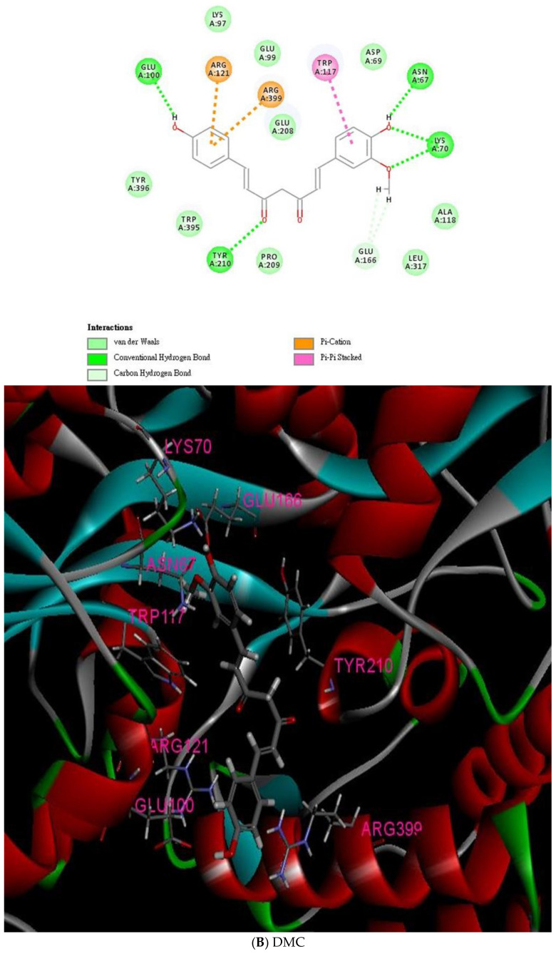
Methods: Curcuminoids targeting endoglin were recognized from two thousand compounds in the Traditional Chinese Medicine Database@Taiwan (TCM Database@Taiwan) using Discovery Studio 4.5.

Results: Our results, obtained using cytotoxicity, migration/invasion, and flow cytometry assays, showed that curcumin (Cur) and demethoxycurcumin (DMC) reduced angiogenesis. In addition, Cur and DMC downregulated endoglin/pSmad1 phosphorylation.

Conclusions: The study first showed that Cur and DMC demonstrated antiangiogenic activity via the inhibition of endoglin/Smad1 signaling. Synergistic effects of curcuminoids (i.e., curcumin and DMC) and semaxanib on HUVECs were found. This might be attributed to endoglin/pSmad1 downregulation in HUVECs. Combination treatment with curcuminoids and a semaxanib is therefore expected to reverse semaxanib resistance.

Keywords: Smad1; VEGF; angiogenesis; curcuminoids; endoglin.


Conflict of interest statement

We declare no conflict of interest.

Figures

Figure 1
Figure 1
Binding mode of the interactions between curcuminoids (i.e., curcumin, DMC, and bDMC) and the endoglin residues. (A) Left panel: interaction between curcumin and endoglin (2D visualization). Right panel: interaction between curcumin and endoglin adduct (3D visualization). (B) Left panel: interaction between DMC and endoglin (2D visualization). Right panel: interaction between DMC and endoglin adduct (3D visualization). (C) Left panel: interaction between bDMC and endoglin (2D visualization). Right panel: interaction between bDMC and endoglin adduct (3D visualization).
Figure 1
Figure 1
Binding mode of the interactions between curcuminoids (i.e., curcumin, DMC, and bDMC) and the endoglin residues. (A) Left panel: interaction between curcumin and endoglin (2D visualization). Right panel: interaction between curcumin and endoglin adduct (3D visualization). (B) Left panel: interaction between DMC and endoglin (2D visualization). Right panel: interaction between DMC and endoglin adduct (3D visualization). (C) Left panel: interaction between bDMC and endoglin (2D visualization). Right panel: interaction between bDMC and endoglin adduct (3D visualization).
Figure 2
Figure 2
Inhibitory effects of curcuminoids (i.e., curcumin, DMC, and bDMC) alone and combined with semaxanib on HUVECs. (AC) Cell-cytotoxic effects of curcumin, DMC, and bDMC on HUVECs according to SRB assay. (D) Cell-cytotoxic effects of semaxanib on HUVECs. (E) Combination treatment with curcumin and semaxanib demonstrated significant inhibitory effects on HUVECs compared with the semaxanib-treated group. (F) Combination treatment with DMC and semaxanib showed considerable inhibitory effects on HUVECs compared to the semaxanib-treated group. * p < 0.05, ** p < 0.01, and *** p < 0.001 compared to the control group (treated with 10% FBS alone). # p < 0.05, ## p < 0.01, and ### p < 0.001 compared to the semaxanib-treated group.
Figure 3
Figure 3
Effect of curcuminoids (i.e., curcumin and DMC) on the cell cycle distribution of HUVECs. (A) The cell-cycle distributions were analyzed in the HUVECs treated with curcumin. The cells were treated with several concentrations (0, 0.625, 1.25, 2.5, 5, and 10 µM) of curcumin for 72 h, and flow cytometry was used to analyze cell cycle distributions. (B) The cell cycle distributions were assessed in the HUVECs administered with DMC. The cells were administered with several different concentrations (0, 0.625, 1.25, 2.5, 5, 10, and 20 µM) of DMC for 72 h, and cell cycle distributions were analyzed by flow cytometry.
Figure 3
Figure 3
Effect of curcuminoids (i.e., curcumin and DMC) on the cell cycle distribution of HUVECs. (A) The cell-cycle distributions were analyzed in the HUVECs treated with curcumin. The cells were treated with several concentrations (0, 0.625, 1.25, 2.5, 5, and 10 µM) of curcumin for 72 h, and flow cytometry was used to analyze cell cycle distributions. (B) The cell cycle distributions were assessed in the HUVECs administered with DMC. The cells were administered with several different concentrations (0, 0.625, 1.25, 2.5, 5, 10, and 20 µM) of DMC for 72 h, and cell cycle distributions were analyzed by flow cytometry.
Figure 4
Figure 4
Inhibitory effects of curcuminoids (i.e., curcumin and DMC) on migration and invasion of endothelial cells. Transwell migration and Matrigel invasion assays were applied for analyzing the behavior of migration and invasion, respectively, of HUVECs administered with curcuminoids (i.e., curcumin and DMC). (A) The migration assay indicates that curcumin significantly inhibited the HUVECs migration. The left panel displays the image captured by a microscope at 100× magnification. The right panel displays the statistical study. (B) Matrigel invasion assays showed that curcumin significantly inhibited the HUVECs invasion. The left panel shows the image acquired with a microscope at 100× magnification. The right panel shows the statistical analysis. (C) The migration assay indicates that DMC significantly inhibited the HUVECs migration. The left panel shows the image acquired with a microscope at 100× magnification. (D) The Matrigel invasion assay indicates that DMC significantly inhibited the HUVECs migration. The left panel displays the image taken with a microscope at 100× magnification. * p < 0.05, ** p < 0.01, and *** p < 0.001 compared to the control group (treated with 10% FBS alone). All the scale bars indicate a length of 200 μM.
Figure 4
Figure 4
Inhibitory effects of curcuminoids (i.e., curcumin and DMC) on migration and invasion of endothelial cells. Transwell migration and Matrigel invasion assays were applied for analyzing the behavior of migration and invasion, respectively, of HUVECs administered with curcuminoids (i.e., curcumin and DMC). (A) The migration assay indicates that curcumin significantly inhibited the HUVECs migration. The left panel displays the image captured by a microscope at 100× magnification. The right panel displays the statistical study. (B) Matrigel invasion assays showed that curcumin significantly inhibited the HUVECs invasion. The left panel shows the image acquired with a microscope at 100× magnification. The right panel shows the statistical analysis. (C) The migration assay indicates that DMC significantly inhibited the HUVECs migration. The left panel shows the image acquired with a microscope at 100× magnification. (D) The Matrigel invasion assay indicates that DMC significantly inhibited the HUVECs migration. The left panel displays the image taken with a microscope at 100× magnification. * p < 0.05, ** p < 0.01, and *** p < 0.001 compared to the control group (treated with 10% FBS alone). All the scale bars indicate a length of 200 μM.
Figure 5
Figure 5
The inhibitory effects of curcuminoids (i.e., curcumin and DMC) on endoglin and phosphorylation Smad1 in HUVECs in the long term (72 h.) (A)Treatment with curcumin (0.625, 1.25, 2.5, and 5.0 µM) and (B) DMC (0.625, 1.25, 2.5, and 5.0 µM) substantially decreased endoglin expression and pSMAD1 phosphorylation. Endoglin expression and pSMAD1 phosphorylation were tested using Western blotting assays. * p < 0.05 and ** p < 0.01 compared to the control group (treated with 10% FBS alone).

Similar articles

Cited by

References

    1. Folkman J. Angiogenesis in cancer, vascular, rheumatoid and other disease. Nat. Med. 1995;1:27–31. doi: 10.1038/nm0195-27. - DOI - PubMed
    1. Paauwe M., Heijkants R.C., Oudt C.H., van Pelt G.W., Cui C., Theuer C.P., Hardwick J.C., Sier C.F., Hawinkels L.J. Endoglin targeting inhibits tumor angiogenesis and metastatic spread in breast cancer. Oncogene. 2016;35:4069–4079. doi: 10.1038/onc.2015.509. - DOI - PubMed
    1. Zangari M., Anaissie E., Stopeck A., Morimoto A., Tan N., Lancet J., Cooper M., Hannah A., Garcia-Manero G., Faderl S., et al. Phase II study of SU5416, a small molecule vascular endothelial growth factor tyrosine kinase receptor inhibitor, in patients with refractory multiple myeloma. Clin. Cancer Res. 2004;10:88–95. doi: 10.1158/1078-0432.CCR-0221-3. - DOI - PubMed
    1. Zhang X., Zhang Y., Jia Y., Qin T., Zhang C., Li Y., Huang C., Liu Z., Wang J., Li K. Bevacizumab promotes active biological behaviors of human umbilical vein endothelial cells by activating TGFbeta1 pathways via off-VEGF signaling. Cancer Biol. Med. 2020;17:418–432. doi: 10.20892/j.issn.2095-3941.2019.0215. - DOI - PMC - PubMed
    1. Fonsatti E., Del Vecchio L., Altomonte M., Sigalotti L., Nicotra M.R., Coral S., Natali P.G., Maio M. Endoglin: An accessory component of the TGF-beta-binding receptor-complex with diagnostic, prognostic, and bioimmunotherapeutic potential in human malignancies. J. Cell. Physiol. 2001;188:1–7. doi: 10.1002/jcp.1095. - DOI - PubMed

MeSH terms

LinkOut - more resources