. 2022 Mar 31;23(7):3905.
doi: 10.3390/ijms23073905.

Whole Genome Sequencing, Focused Assays and Functional Studies Increasing Understanding in Cryptic Inherited Retinal Dystrophies

Affiliations

Whole Genome Sequencing, Focused Assays and Functional Studies Increasing Understanding in Cryptic Inherited Retinal Dystrophies

Benjamin M Nash et al. Int J Mol Sci. .

Abstract

The inherited retinal dystrophies (IRDs) are a clinically and genetically complex group of disorders primarily affecting the rod and cone photoreceptors or other retinal neuronal layers, with emerging therapies heralding the need for accurate molecular diagnosis. Targeted capture and panel-based strategies examining the partial or full exome deliver molecular diagnoses in many IRD families tested. However, approximately one in three families remain unsolved and unable to obtain personalised recurrence risk or access to new clinical trials or therapy. In this study, we investigated whole genome sequencing (WGS), focused assays and functional studies to assist with unsolved IRD cases and facilitate integration of these approaches to a broad molecular diagnostic clinical service. The WGS approach identified variants not covered or underinvestigated by targeted capture panel-based clinical testing strategies in six families. This included structural variants, with notable benefit of the WGS approach in repetitive regions demonstrated by a family with a hybrid gene and hemizygous missense variant involving the opsin genes, OPN1LW and OPN1MW. There was also benefit in investigation of the repetitive GC-rich ORF15 region of RPGR. Further molecular investigations were facilitated by focused assays in these regions. Deep intronic variants were identified in IQCB1 and ABCA4, with functional RNA based studies of the IQCB1 variant revealing activation of a cryptic splice acceptor site. While targeted capture panel-based methods are successful in achieving an efficient molecular diagnosis in a proportion of cases, this study highlights the additional benefit and clinical value that may be derived from WGS, focused assays and functional genomics in the highly heterogeneous IRDs.

Keywords: RNA analysis; gene panels; inherited retinal dystrophy; whole genome sequencing.


Conflict of interest statement

The authors declare that they have no conflicts of interest.

Figures

Figure 1
Figure 1
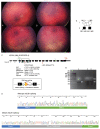
WGS and focused assays identify opsin gene hybrid involving within chromosome Xq28 (A) Ultra-wide-field pseudo colour fundus images from the proband shows a normal appearance. (B) Wide-field fundus autofluorescence (UWF-FAF) showed some irregular hyperautofluorescence at the fovea with the remaining fundus having a normal WF-FAF pattern. (C) OCT assessment shows focal ellipsoid zone outer retinal discontinuity; (D) Pedigree analysis across four generations, suggestive of X-linked inheritance (pedigree abbreviated for illustrative purposes), the proband and twin sibling both had visual acuities of 6/60 in each eye, M1 = mutant hybrid opsin allele, + = wild type allele; (E) Illustration of the genomic arrangement of the tandem opsin genes within chromosome Xq28. Wild type and the complex allele identified in Family 1 are shown, with primer locations for the focused assay indicated by green arrows; (F) Table highlighting the differences in single nucleotide (c.) and amino acid (p.) sequence between the OPN1LW (NM_020061) and OPN1MW (NM_000513.2) genes (Table modified from source [13]). Unique coding OPN1LW sequence detected in our family highlighted in green, and OPN1MW coding sequence is highlighted in orange/yellow (LCR: Locus Control Region). OCT Optical Coherence tomography, UWF-FAF Ultra-wide field fundus autofluorescence.
Figure 2
Figure 2
WGS identifies loss of function variant within the RPGR mutation hotspot ORF15 (A) Ultra-wide-field pseudo colour fundus images showing widespread retinal pigment migration in a “bone spicule” pattern with associated outer retinal atrophy; (B) UWF-FAF showing a central area hyper autofluorescence surrounded by a ring of incomplete hypo autofluorescence. Beyond the vascular arcades there are speckled areas of hypo autofluorescence of varying density; (C) OCT assessment shows preservation of an attenuated ellipsoid zone (EZ) and outer retinal structures at the fovea. In the para macular regions there is significant attenuation to loss of the EZ line representing outer retinal structures. (D) Pedigree of Family 2, M1 = RPGR ORF15 mutant allele, + = wild type allele; (E) IGV screenshot showing coverage of the RPGR(NM_001034853.2):c.2898_2899del p.(Glu967Argfs * 111) variant detected on WGS (dashed vertical lines), located within the highly repetitive GC-rich ORF15 locus; (F) Illustration of the RPGR ORF15 gene structure depicting the location of the variant identified within the low complexity region of the ORF15 mutation hotspot. OCT Optical Coherence tomography, UWF-FAF Ultra-wide field fundus autofluorescence, EZ ellipsoid zone. WGS identifies loss of function variant within the RPGR mutation hotspot ORF15 (A) Ultra-wide-field pseudo colour fundus images showing widespread retinal pigment migration in a “bone spicule” pattern with associated outer retinal atrophy; (B) UWF-FAF showing a central area hyper autofluorescence surrounded by a ring of incomplete hypo autofluorescence. Beyond the vascular arcades there are speckled areas of hypo autofluorescence of varying density; (C) OCT assessment shows preservation of an attenuated ellipsoid zone (EZ) and outer retinal structures at the fovea. In the para macular regions there is significant attenuation to loss of the EZ line representing outer retinal structures. (D) Pedigree of Family 2, M1 = RPGR ORF15 mutant allele, + = wild type allele; (E) IGV screenshot showing coverage of the RPGR(NM_001034853.2):c.2898_2899del p.(Glu967Argfs * 111) variant detected on WGS (dashed vertical lines), located within the highly repetitive GC-rich ORF15 locus; (F) Illustration of the RPGR ORF15 gene structure depicting the location of the variant identified within the low complexity region of the ORF15 mutation hotspot. OCT Optical Coherence tomography, UWF-FAF Ultra-wide field fundus autofluorescence, EZ ellipsoid zone.
Figure 3
Figure 3
WGS and RNA studies identify a novel deep intronic IQCB1 variant which activates a pseudoexon. (A) Right superior fundus, (B) Left superior fundus, (C) Right inferior fundus, and (D) Left inferior fundus shown for proband II.1 with widefield Retcam fundus images (RetCam, Natus, Pleasanton, CA, USA). The main clinical changes are seen in the inferior fundus of each eye (C,D). There is retinal atrophy with granular pigmentary changes involving the inferior fundus in each eye. There are also fine speckled pigmentary changes in the superior fundus on clinical exam, which is difficult to appreciate in the Retcam photos. (E) Pedigree of Family 3, M1 = IQCB1:c.897_900CTTGdup, M2 = IQCB1:c.263 + 201delA, + = wild type; (F) Illustration of IQCB1 gene structure, with exon numbering and the location of the variants identified in this study indicated by red arrows. Splicing prediction algorithm data for the novel intronic variant is shown, suggesting the activation of a cryptic acceptor splice site. Coloured arrows underneath indicate primer pair locations in exons 3 to 6 and 4 to 5. Orange box illustrates the location of the pseudoexon introduced between exons 4 and 5; (G) Agarose gel image showing the presence of the wild type band [WT, 161bp] in both the control (A) and proband (B), with the larger sized mutant band [Mut, 314bp] also present in the proband samples; (H) Sanger sequencing traces showing the retention of 153bp of intron 4 sequence into the mutant band of IQCB1 RNA. Note: only flanking 5 and 3′ ends of the pseudoexon are pictured and not all 153bp.
Figure 4
Figure 4
Homozygous deletion identified involving KCNV2. (A) Pedigree of family with two siblings with cone dystrophy, suggestive of autosomal recessive inheritance. M1 = KCNV2 heterozygous deletion allele, + = wild type allele; (B) Location of the genomic deletion identified by WGS including IGV screenshots of reads spanning the 5′ and 3′ breakpoints. Proband (II.1) [top] & affected sibling (II.2) [middle] showing no reads (homozygous deletion). Parental carrier (I.1) [bottom] showing reduced read depth (heterozygous deletion) compared to flanking adjacent genomic segments.
Figure 5
Figure 5
WGS resolves single exon CNV in ABCA4. (A) Ultra-Wide field pseudo colour fundus images show significant atrophy involving the posterior pole in each eye. There was blotchy retinal atrophy extending to the equator in each eye; (B) UWF-FAF shows dense loss of autofluorescence in the posterior pole of both eyes extending beyond the vascular arcades with coalescing paving stone like areas of dense hypo autofluorescence. In the periphery there is patchy areas of hyper and hypo autofluorescence; (C) OCT showed almost complete loss of the outer retinal layers at the macular with significant disruption and loss of the EZ in the para macular region; (D) Pedigree of Family 6. M1 = ABCA4:c.5461-10T>C, M2 = ABCA4 exon 18 deletion; (E) IGV screenshot of the 1029bp deletion involving ABCA4 exon 18, plus flanking intronic segments. Genomic deletion is describable as [hg19]chr1:94514389_94515418del. OCT Optical Coherence tomography, UWF-FAF Ultra-wide field fundus autofluorescence, EZ ellipsoid zone.

References

    1. Hanany M., Rivolta C., Sharon D. Worldwide carrier frequency and genetic prevalence of autosomal recessive inherited retinal diseases. Proc. Natl. Acad. Sci. USA. 2020;117:2710–2716. doi: 10.1073/pnas.1913179117. - DOI - PMC - PubMed
    1. Liew G., Michaelides M., Bunce C. A comparison of the causes of blindness certifications in England and Wales in working age adults (16–64 years), 1999–2000 with 2009–2010. BMJ Open. 2014;4:e004015. doi: 10.1136/bmjopen-2013-004015. - DOI - PMC - PubMed
    1. Heath Jeffery R.C., Mukhtar S.A., McAllister I.L., Morgan W.H., Mackey D.A., Chen F.K. Inherited retinal diseases are the most common cause of blindness in the working-age population in Australia. Ophthalmic Genet. 2021;42:431–439. doi: 10.1080/13816810.2021.1913610. - DOI - PMC - PubMed
    1. Nash B.M., Wright D.C., Grigg J.R., Bennetts B., Jamieson R.V. Retinal dystrophies, genomic applications in diagnosis and prospects for therapy. Transl. Pediatr. 2015;4:139–163. doi: 10.3978/j.issn.2224-4336.2015.04.03. - DOI - PMC - PubMed
    1. Maquire A.M., Russell S., Wellman J.A., Chung D.C., Yu Z., Tillman A., Wittes J., Pappas J., Elci O., Marshall K.A., et al. Efficacy, Safety, and Durability of Voretigene Neparvovec-rzyl in RPE65 Mutations Associated Inherited Retinal Dystrophy. Ophthalmology. 2019;126:1273–1285. doi: 10.1016/j.ophtha.2019.06.017. - DOI - PubMed

LinkOut - more resources