. 2022 Apr 1;23(7):3936.
doi: 10.3390/ijms23073936.

Glycoproteomic and Phenotypic Elucidation of B4GALNT2 Expression Variants in the SID Histo-Blood Group System

Affiliations

Glycoproteomic and Phenotypic Elucidation of B4GALNT2 Expression Variants in the SID Histo-Blood Group System

Linn Stenfelt et al. Int J Mol Sci. .

Abstract

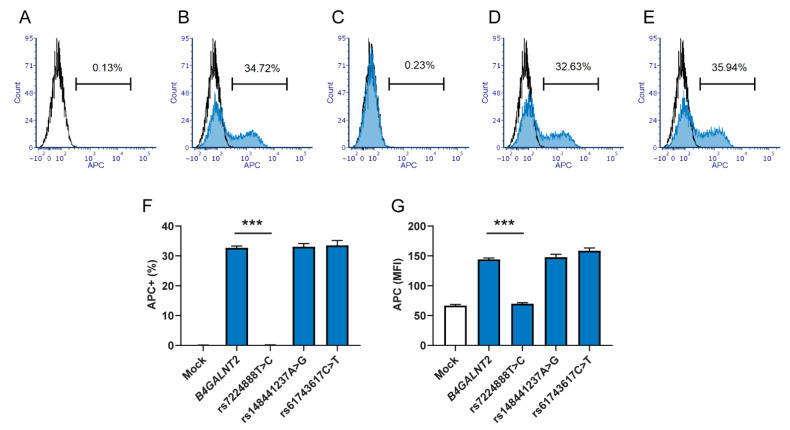
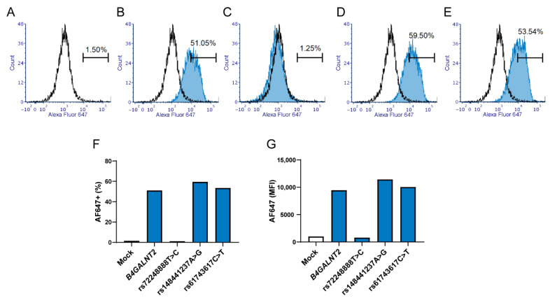
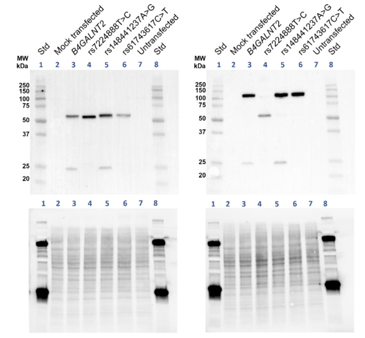
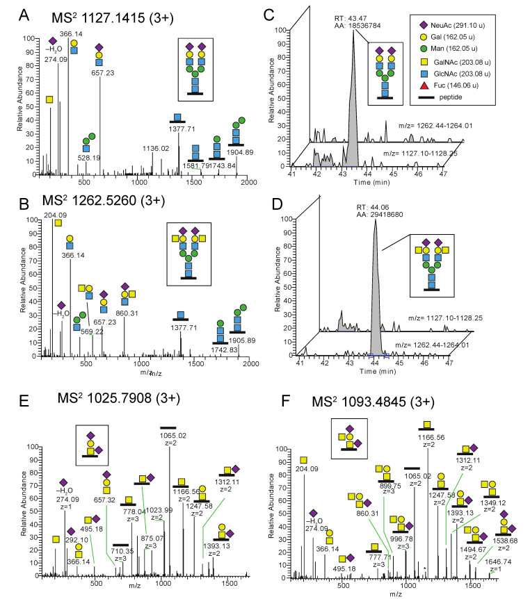
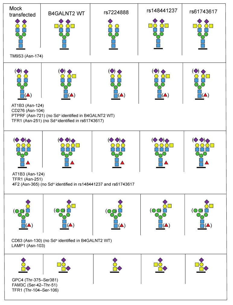
The Sda histo-blood group antigen (GalNAcβ1-4(NeuAcα2-3)Galβ-R) is implicated in various infections and constitutes a potential biomarker for colon cancer. Sd(a−) individuals (2−4% of Europeans) may produce anti-Sda, which can lead to incompatible blood transfusions, especially if donors with the high-expressing Sd(a++)/Cad phenotype are involved. We previously reported the association of B4GALNT2 mutations with Sd(a−), which established the SID blood-group system. The present study provides causal proof underpinning this correlation. Sd(a−) HEK293 cells were transfected with different B4GALNT2 constructs and evaluated by immunostaining and glycoproteomics. The predominant SIDnull candidate allele with rs7224888:T>C (p.Cys406Arg) abolished Sda synthesis, while this antigen was detectable as N- or O-glycans on glycoproteins following transfection of wildtype B4GALNT2. Surprisingly, two rare missense variants, rs148441237:A>G and rs61743617:C>T, found in a Sd(a−) compound heterozygote, gave results similar to wildtype. To elucidate on whether Sd(a++)/Cad also depends on B4GALNT2 alterations, this gene was sequenced in five individuals. No Cad-specific changes were identified, but a detailed erythroid Cad glycoprotein profile was obtained, especially for glycophorin-A (GLPA) O-glycosylation, equilibrative nucleoside transporter 1 (S29A1) O-glycosylation, and band 3 anion transport protein (B3AT) N-glycosylation. In conclusion, the p.Cys406Arg β4GalNAc-T2 variant causes Sda-deficiency in humans, while the enigmatic Cad phenotype remains unresolved, albeit further characterized.

Keywords: SID blood group system; carbohydrate biosynthesis; erythrocyte; flow cytometry; gene expression; glycobiology; glycopeptide; glycoprotein; glycosyltransferase; mass spectrometry (MS).


Conflict of interest statement

The authors have no conflict of interest in relation to this study.

Figures

Figure 1
Figure 1
Lectin analysis by flow cytometry of terminal GalNAc residues on HEK293 cells transfected with B4GALNT2 constructs. HEK293 cells transfected with the bicistronic GFP-expressing vector (CS-2719-M61) carrying B4GALNT2 constructs were stained with biotinylated DBA lectin followed by APC-conjugated streptavidin. Flow cytometry was used to record the level of GalNAc expression in GFP-positive cells. Cells transfected with the vector carrying (A) no construct (Mock) (B) B4GALNT2 wt (GenBank accession number AJ517771), (C) B4GALNT2 with Sd(a−)-associated SNP rs7224888:T > C, (D) rs148441237:A > G, or (E) rs61743617:C > T. Graphs present data from three separate experiments giving (F) the percentage of cells positive for the APC signal and (G) the mean MFI of the APC signal, with error bars that represent the standard error of the mean (SEM) and asterisks (***) indicate p < 0.001.
Figure 2
Figure 2
Antibody analysis of Sda surface expression in HEK293 cells transfected with B4GALNT2 constructs. One set of transfected cells were stained with human plasma containing anti-Sda (diluted in PBS) followed by staining with Alexa Fluor 647 conjugated Goat anti-human:IgM. The GFP positive population in HEK293 cells transfected with the vector CS-2719-M61 carrying (A) no construct (mock), (B) B4GALNT2 wt (GenBank accession number AJ517771), (C) B4GALNT2 with Sd(a−) associated SNPs rs7224888:T > C, (D) rs148441237:A > G or (E) rs61743617:C > T. Graphs present (F) the percentage of cells positive for the Alexa Fluor 647 signal and (G) the MFI of the Alexa Fluor 647 signal. Due to the small amounts of human anti-Sda available, only one set of transfected samples was run, not allowing for statistical evaluation.
Figure 3
Figure 3
Immunoblot of the B4GALNT2-expressed glycosyltransferase in transfected HEK293 cells. Western blot displaying the immunoblotted band corresponding to β4GalNAc-T2 in protein samples from transfected HEK293 cells. In the left blot, samples are dissolved in Laemmli buffer with reducing agent β-mercaptoethanol, while Laemmli buffer without reducing agent has been used for samples of the right blot. The standards (Lane 1 and 8) specify the molecular weights (kDa). Mock-transfected and untransfected cells are negative controls (Lane 2 and 7) with the B4GALNT2 wt transfected serves as the positive control (Lane 3). The glycosyltransferase is detected in all cell preparations transfected with either of the B4GALNT2-mutated constructs, rs7224888:T > C (Lane 4), rs148441237:A > G (Lane 5) or rs61743617:C > T (Lane 6). Colorimetric and chemiluminescent images of the membrane have been merged. The lower panel displays stain-free images of total protein of each blot, ChemiDoc imaging system, Bio-Rad.
Figure 4
Figure 4
Glycoproteomic analysis of N- and O-glycopeptides carrying the Sda epitope or its precursor structure in transfected HEK293 cells. MS2 spectra obtained at NCE 20% for (A) a complex type disialo biantennary N-glycopeptide with the amino acid sequence IVDVNLTSEGK including the Asn-174 glycosite (underlined), from transmembrane 9 superfamily member 3 (TM9S3) and for (B) a glycopeptide with the same amino acid sequence carrying one Sda epitope on each of the two antennae. The measured monoisotopic masses for precursor ions are provided in the panel headings (C,D) Extracted ion chromatograms (XICs) of the two precursor ions demonstrate that (C) the mock-transfected sample contains the complex biantennary glycopeptide but not the Sda epitope glycopeptide, and (D) vice versa is true for the wt B4GALNT2-transfected sample. (E) MS2 spectrum obtained at NCE 20% of a glycopeptide, with the amino acid sequence LAGTESPVREEPGEDFPAAR from transferrin receptor protein 1 (TFR1), carrying the disialo core 1 O-glycan, and (F) a glycopeptide with the same sequence carrying the Sda epitope. The glycosite is at Thr-104 or Ser-106. It should be observed that the measured monoisotopic masses for precursor ions at four decimals are provided in the headings of the MS2 spectra. Displayed m/z values of fragment ions are from the largest isotope peaks, not always from the monoisotopic ion. Thus, delta masses in the figures occasionally differ by ±1 u from calculated values.
Figure 5
Figure 5
Summary of identified sialic acid containing N- and O-glycopeptides from the transfected HEK293 cell preparations. The transfected HEK293 cells are organized in columns and the identified glycoproteins with their corresponding glycan isoforms in rows. UniProt abbreviations are used and unambiguous Asn- and candidate Ser/Thr-glycosites are indicated by amino acid numbers. The protein names and glycopeptide identities are provided in Table 1.
Figure 6
Figure 6
Analysis of Sda expression on erythrocyte surfaces by flow cytometry. Fixed erythrocytes were stained with human plasma containing anti-Sda (diluted in PBS) followed by PE-conjugated Goat anti-human:IgM. Erythrocytes from blood donors of (A) Sd(a–) phenotype (black line), also seen in the following graphs for comparison with other phenotypes. (BD) Blue curve represents the common Sd(a+) phenotype (B) and Cad phenotypes, as shown by the Cad-a sample (C) and Cad-b (D).
Figure 7
Figure 7
Glycoproteomic analysis of erythrocyte glycoproteins of a Cad sample. (A) MS2 spectrum of a glycopeptide DTYAATPR (residues 51–58) of GLPA carrying an O-glycan Sda epitope from the Cad-a sample. M denotes remaining precursor ions at m/z 1022.92. The MS2 spectrum of the corresponding non-Sda O-glycopeptide is shown in Supplementary Figure S2A. (B) MS2 spectrum of a glycopeptide LSVPDGFKVSNSSAR.G (residues 632–645) of B3AT with an N-glycan with the Sda epitope on one of the antennae obtained from the Cad-a individual. (C) Extracted MS2 ion chromatogram of the diagnostic ion at m/z 860.31 (occasionally measured as m/z 860.32, the exact mass is 860.3143 u). The major peaks (red encircled a.–d.) are indicated and identified in panels A and B and in the Supplementary Figure S1C–E. (D) Extracted MS1 ions of the GLPA glycopeptide (residues 51–58) demonstrated a relative peak intensity of 12% of the Sda epitope for the Cad-a sample. * Not glycopeptide related.

References

    1. Stenfelt L., Hellberg Å., Möller M., Thornton N., Larson G., Olsson M.L. Missense mutations in the C-terminal portion of the B4GALNT2-encoded glycosyltransferase underlying the Sd(a-) phenotype. Biochem. Biophys. Rep. 2019;19:100659. doi: 10.1016/j.bbrep.2019.100659. - DOI - PMC - PubMed
    1. Veldhuisen B., Ligthart P., van der Mark-Zoet J., Javadi A., Tissoudali A., Dengerink I., Folman C., van der Shoot C.E. Identification of a single homozygous mutation in the B4GALNT2 gene in individuals lacking the Sd(a) (SID) antigen on red blood cells. Vox Sang. 2019;114:193.
    1. Macvie S.I., Morton J.A., Pickles M.M. The reactions and inheritance of a new blood group antigen, Sda. Vox Sang. 1967;13:485–492. doi: 10.1111/j.1423-0410.1967.tb03795.x. - DOI
    1. Renton P.H. Anti-Sda new blood group antibody. Vox Sang. 1967;13:493–501. doi: 10.1111/j.1423-0410.1967.tb03796.x. - DOI
    1. Morton J.A., Pickles M.M., Vanhegan R.I. The Sda antigen in the human kidney and colon. Immunol. Investig. 1988;17:217–224. doi: 10.3109/08820138809052961. - DOI - PubMed

MeSH terms

Substances

LinkOut - more resources