. 2022 Apr 2;23(7):3966.
doi: 10.3390/ijms23073966.

In Silico and In Vitro Screening of 50 Curcumin Compounds as EGFR and NF-κB Inhibitors

Affiliations

In Silico and In Vitro Screening of 50 Curcumin Compounds as EGFR and NF-κB Inhibitors

Mohamed E M Saeed et al. Int J Mol Sci. .

Abstract

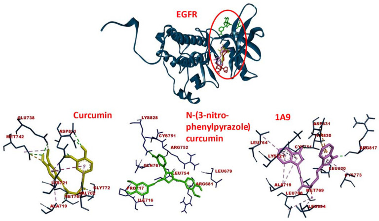
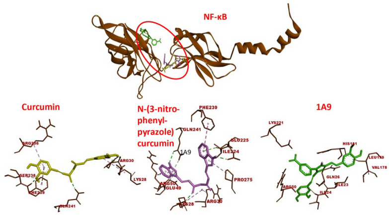
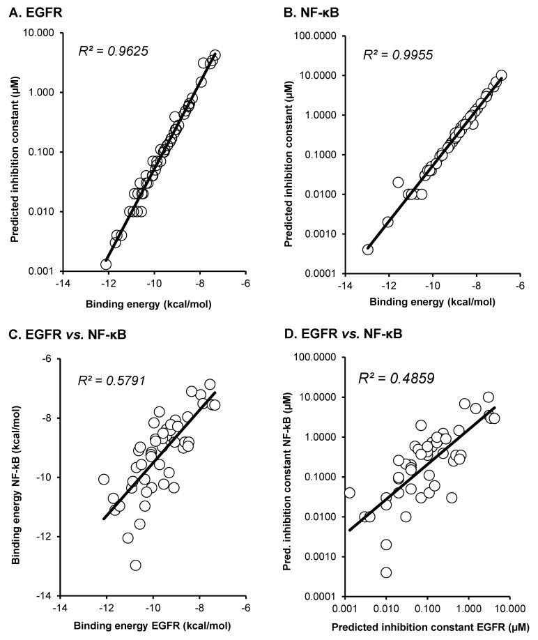
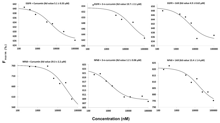
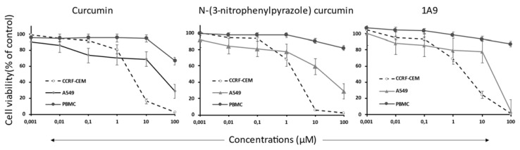
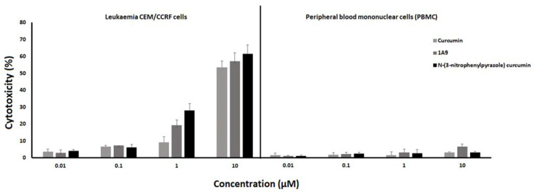
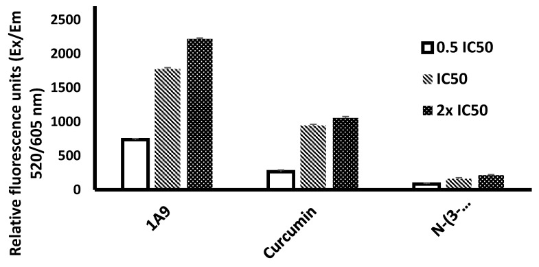
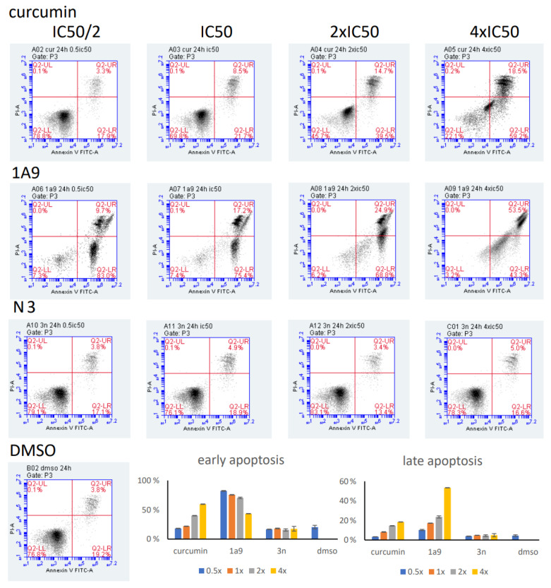
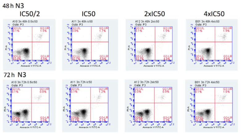
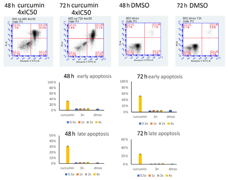
The improvement of cancer chemotherapy remains a major challenge, and thus new drugs are urgently required to develop new treatment regimes. Curcumin, a polyphenolic antioxidant derived from the rhizome of turmeric (Curcuma longa L.), has undergone extensive preclinical investigations and, thereby, displayed remarkable efficacy in vitro and in vivo against cancer and other disorders. However, pharmacological limitations of curcumin stimulated the synthesis of numerous novel curcumin analogs, which need to be evaluated for their therapeutic potential. In the present study, we calculated the binding affinities of 50 curcumin derivatives to known cancer-related target proteins of curcumin, i.e., epidermal growth factor receptor (EGFR) and nuclear factor κB (NF-κB) by using a molecular docking approach. The binding energies for EGFR were in a range of −12.12 (±0.21) to −7.34 (±0.07) kcal/mol and those for NF-κB ranged from −12.97 (±0.47) to −6.24 (±0.06) kcal/mol, indicating similar binding affinities of the curcumin compounds for both target proteins. The predicted receptor-ligand binding constants for EGFR and curcumin derivatives were in a range of 0.00013 (±0.00006) to 3.45 (±0.10) µM and for NF-κB in a range of 0.0004 (±0.0003) to 10.05 (±4.03) µM, indicating that the receptor-ligand binding was more stable for EGFR than for NF-κB. Twenty out of 50 curcumin compounds showed binding energies to NF-κB smaller than −10 kcal/mol, while curcumin as a lead compound revealed free binding energies of >−10 kcal/mol. Comparable data were obtained for EGFR: 15 out of 50 curcumin compounds were bound to EGFR with free binding energies of <−10 kcal/mol, while the binding affinity of curcumin itself was >−10 kcal/mol. This indicates that the derivatization of curcumin may indeed be a promising strategy to improve targe specificity and to obtain more effective anticancer drug candidates. The in silico results have been exemplarily validated using microscale thermophoresis. The bioactivity has been further investigated by using resazurin cell viability assay, lactate dehydrogenase assay, flow cytometric measurement of reactive oxygen species, and annexin V/propidium iodide assay. In conclusion, molecular docking represents a valuable approach to facilitate and speed up the identification of novel targeted curcumin-based drugs to treat cancer.

Keywords: bioinformatics; cancer; natural products; phytochemicals; synthetic derivatives; virtual drug screening.


Conflict of interest statement

The authors declare that there is no conflict of interest.

Figures

Figure 1
Figure 1
Molecular docking of curcumin-type compounds to EGFR. Top: The compounds were bound to the same domain of EGFR. Bottom: curcumin, N-(3-nitrophenylpyrazole) curcumin, and the derivative 1A9 were bound to different amino acids in this domain. The red circle indicates the binding site of the three compounds.
Figure 2
Figure 2
Molecular docking of curcumin-type compounds to NF-κB. Top: The compounds were bound to the same domain of EGFR. Bottom: curcumin, N-(3-nitrophenylpyrazole) curcumin, and the derivative 1A9 were bound to different amino acids in this domain. The red circle indicates the binding site of the three compounds.
Figure 3
Figure 3
Correlation of binding energies (kcal/mol) and predicted inhibition constants (pKi, µM) of 50 curcumin compounds were calculated using the Pearson correlation test. Correlation of binding energies and pKi values for (A) EGFR and (B) NF-κB. Correlation of (C) binding energies or (D) pKi values between EGFR and NF-κB.
Figure 4
Figure 4
Analysis of the interaction between curcumin derivatives with recombinant EGFR and NF-κB by microscale thermophoresis (MST). The recombinant proteins were used at a concentration of 200 nM, while the concentration of curcumin, N-(3-nitrophenylpyrazole) curcumin, and the curcumin derivative 1A9 ranged from 100 to 100,000 nM. The migration of the fluorescent proteins was determined upon local heating using a Monolith NT.115Pico with 40% LED power and 80% MST power for EGFR and with 20% LED power and 20% MST power for NF-κB at room temperature.
Figure 5
Figure 5
Cell viability dose-response curves of CCRF-CEM leukemia cells, A549 lung carcinoma cells, and healthy peripheral blood mononuclear cells treated with curcumin, 1A9, and N-(3-nitrophenylpyrazole) curcumin as determined by the resazurin assay. Cells were incubated with concentrations from 10−3 to 100 µM and incubated at 37 °C for 72 h. DMSO was used as vehicle control. Mean ± SD of three independent measurements.
Figure 6
Figure 6
Cytotoxicity in CCRF-CEM leukemia cells (left) and peripheral blood mononuclear cells (PBMCs) of a healthy donor (right) by curcumin, 1A9, and N-(3-nitrophenylpyrazole) curcumin as determined by the release of lactate dehydrogenase. Cells were incubated with concentrations from 0.01 to 10 µM and incubated at 37 °C for 48 h. DMSO was used as vehicle control. Mean ± SD of three independent measurements.
Figure 7
Figure 7
Generation of reactive oxygen species in CCRF-CEM cells by curcumin, N-(3-nitrophenylpyrazole) curcumin, and 1A9. Cells were incubated with 0.5×, 1×, 2×, or 4× IC50 and incubated at 37 °C for 24 h. DMSO was used as vehicle control. Mean ± SD of three independent measurements.
Figure 8
Figure 8
Induction of apoptosis in CCRF-CEM cells upon exposure at 37 °C for 24 h with curcumin, N-(3-nitrophenylpyrazole) curcumin, and 1A9 as determined by the annexin V/propidium iodide assay and flow cytometry. Cells were incubated with 0.5×, 1×, 2×, or 4× IC50. DMSO was used as vehicle control. Mean ± SD of three independent measurements.
Figure 9
Figure 9
Induction of apoptosis in CCRF-CEM cells upon exposure at 37 °C for 48 or 72 h with N-(3-nitrophenylpyrazole) curcumin as determined by the annexin V/propidium iodide assay and flow cytometry. Cells were incubated with 0.5×, 1×, 2×, or 4× IC50 N-(3-nitrophenylpyrazole) curcumin. Positive control: curcumin (4× IC50); negative control: DMSO. Mean ± SD of three independent measurements.
Figure 9
Figure 9
Induction of apoptosis in CCRF-CEM cells upon exposure at 37 °C for 48 or 72 h with N-(3-nitrophenylpyrazole) curcumin as determined by the annexin V/propidium iodide assay and flow cytometry. Cells were incubated with 0.5×, 1×, 2×, or 4× IC50 N-(3-nitrophenylpyrazole) curcumin. Positive control: curcumin (4× IC50); negative control: DMSO. Mean ± SD of three independent measurements.

References

    1. Siegel R.L., Miller K.D., Jemal A. Cancer statistics 2018. CA Cancer J. Clin. 2018;68:7–30. doi: 10.3322/caac.21442. - DOI - PubMed
    1. Toyoda Y., Tabuchi T., Nakayama T., Hojo S., Yoshioka S., Maeura Y. Past trends and future estimation of annual breast cancer incidence in Osaka, Japan. Asian Pac. J. Cancer Prev. 2016;17:52. - PubMed
    1. Mattiuzzi C., Lippi G. Current cancer epidemiology. J. Epidemiol. Glob. Health. 2019;9:217–222. doi: 10.2991/jegh.k.191008.001. - DOI - PMC - PubMed
    1. Newman D.J., Cragg G.M. Natural products as sources of new drugs over the last 25 years. J. Nat. Prod. 2007;70:461–477. doi: 10.1021/np068054v. - DOI - PubMed
    1. Kuete V., Efferth T. African flora has the potential to fight multidrug resistance of cancer. BioMed Res. Int. 2015;2015:914813. doi: 10.1155/2015/914813. - DOI - PMC - PubMed