. 2022 Apr 3;23(7):3986.
doi: 10.3390/ijms23073986.

Anti-Inflammatory Activities of an Anti-Histamine Drug, Loratadine, by Suppressing TAK1 in AP-1 Pathway

Affiliations

Anti-Inflammatory Activities of an Anti-Histamine Drug, Loratadine, by Suppressing TAK1 in AP-1 Pathway

Jiwon Jang et al. Int J Mol Sci. .

Abstract

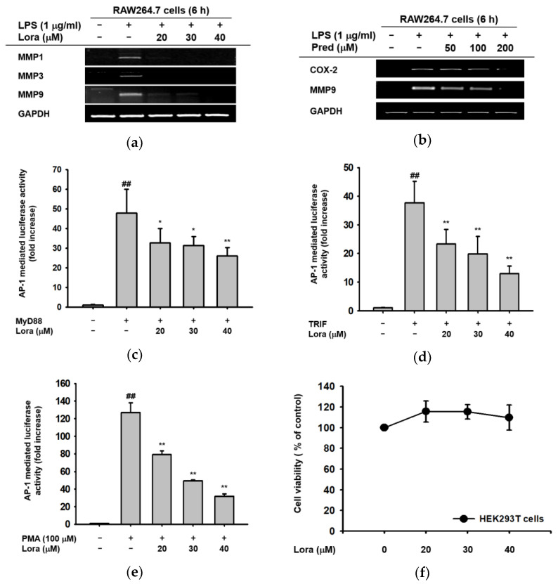
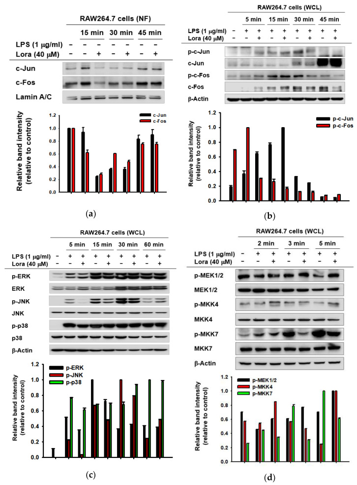
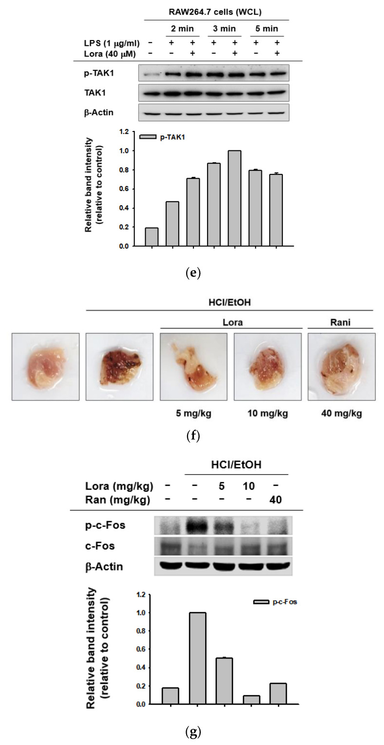
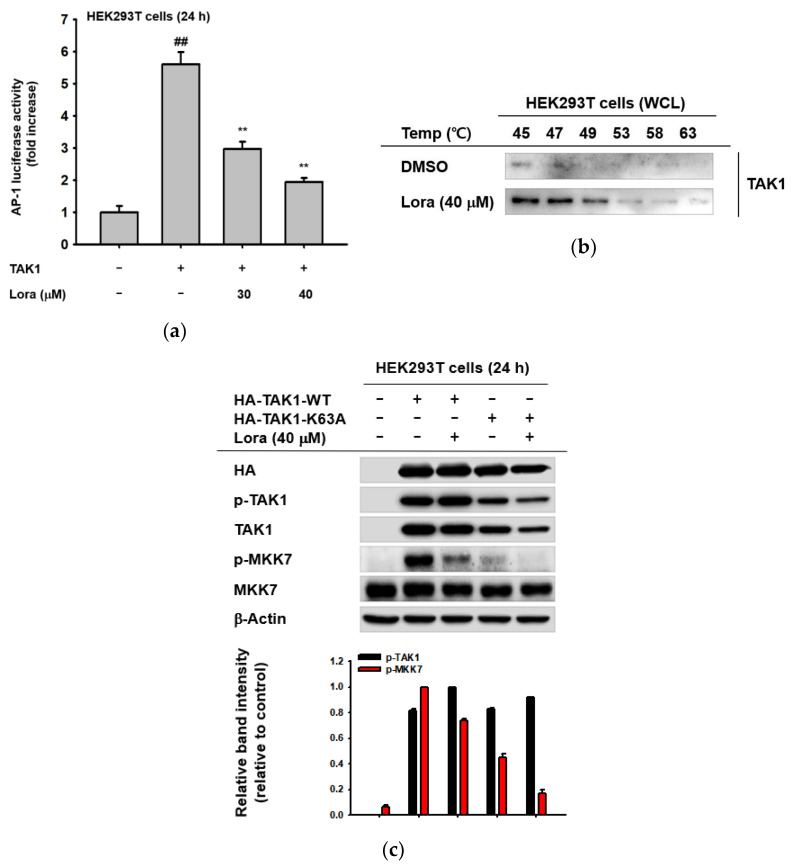
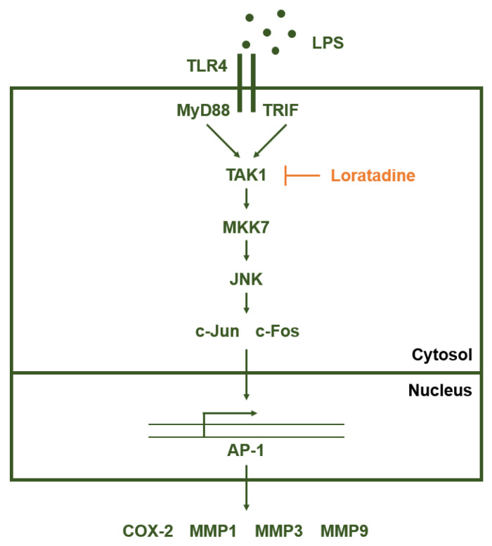
Loratadine is an anti-histamine routinely used for treating allergies. However, recent findings have shown that Loratadine may also have anti-inflammatory functions, while their exact mechanisms have not yet been fully uncovered. In this paper, we investigated whether Loratadine can be utilized as an anti-inflammatory drug through a series of in vitro and in vivo experiments using a murine macrophage cell line and an acute gastritis mouse model. Loratadine was found to dramatically reduce the expression of pro-inflammatory genes, including MMP1, MMP3, and MMP9, and inhibit AP-1 transcriptional activation, as demonstrated by the luciferase assay. Therefore, we decided to further explore its role in the AP-1 signaling pathway. The expression of c-Jun and c-Fos, AP-1 subunits, was repressed by Loratadine and, correspondingly, the expression of p-JNK, p-MKK7, and p-TAK1 was also inhibited. In addition, Loratadine was able to reduce gastric bleeding in acute gastritis-induced mice; Western blotting using the stomach samples showed reduced p-c-Fos protein levels. Loratadine was shown to effectively suppress inflammation by specifically targeting TAK1 and suppressing consequent AP-1 signaling pathway activation and inflammatory cytokine production.

Keywords: AP-1; Loratadine; TAK1; anti-inflammatory effect.


Conflict of interest statement

The authors have no conflicts of interest to declare.

Figures

Figure 1
Figure 1
Inhibitory effects of Loratadine on the mRNA expression of pro-inflammatory genes and the transcriptional activation of inflammatory transcription factors. (a,b) RAW264.7 cells were pre-treated with the indicated concentrations of Loratadine (20–40 μM) or prednisolone (50–200 μM) for 30 min. The cells were treated with LPS (1 μg/mL) for 6 h, and the mRNA expression levels of MMP1, MMP3, MMP9, and GAPDH (control) were measured by reverse-transcription polymerase chain reaction (RT-PCR) and agarose gel electrophoresis. Visualization of DNA bands was achieved by exposing the gel to UV irradiation. (cf) HEK293T cells were transfected with AP-1-luc, MyD88, TRIF, and β-galactosidase (control) using PEI for 24 h. Cells were treated with the indicated concentrations of Loratadine (20–40 μM) for 6 h before cell harvesting. The expression of AP-1 was measured by luciferase activity. (f) HEK293T cells were treated with the indicated doses of Loratadine (20–40 μM) for 24 h, and viability was measured using the MTT cell viability assay. ## p < 0.01 compared to normal group; * p < 0.05 and ** p < 0.01 compared to control group. All data are presented (af) as mean ± standard deviation of the experiments performed with 4–6 samples.
Figure 2
Figure 2
Anti-inflammatory effects of Loratadine on the constituent kinases of the AP-1 signaling pathway. (a) RAW264.7 cells were pre-treated with Loratadine (40 μM) for 30 min following LPS treatment (1 μg/mL) for the indicated time points (15–45 min). The expression levels of c-Jun, c-Fos, and Lamin A/C (control) within the nuclear fraction were assessed by Western blotting analysis. (be) RAW264.7 cells were pre-treated with Loratadine (40 μM) for 30 min following LPS treatment (1 μg/mL) for the indicated time points (5–60 min). The levels of total or phosphorylated c-Jun, c-Fos, ERK, JNK, p-38, MEK1/2, MKK4, MKK7, TAK-1, and β-actin (control) within the whole lysate were measured by Western blotting analysis. NF: nuclear fraction; WCL: whole cell lysate. (f) Acute gastritis was induced using HCl/EtOH after vehicle, Loratadine, or ranitidine injection. Stomach samples were obtained and cut open after sacrifice to assess stomach bleeding levels. (g) Stomach samples were ground in liquid nitrogen and lysed using cell lysis buffer. Western blotting was performed to assess the expression levels of total and phosphorylated c-Fos and β-Actin (control). NF: nuclear fraction; WCL: whole cell lysate; Ran: Ranitidine. (Bottom panels of ae,g). Relative intensity of these proteins was calculated by ImageJ.
Figure 2
Figure 2
Anti-inflammatory effects of Loratadine on the constituent kinases of the AP-1 signaling pathway. (a) RAW264.7 cells were pre-treated with Loratadine (40 μM) for 30 min following LPS treatment (1 μg/mL) for the indicated time points (15–45 min). The expression levels of c-Jun, c-Fos, and Lamin A/C (control) within the nuclear fraction were assessed by Western blotting analysis. (be) RAW264.7 cells were pre-treated with Loratadine (40 μM) for 30 min following LPS treatment (1 μg/mL) for the indicated time points (5–60 min). The levels of total or phosphorylated c-Jun, c-Fos, ERK, JNK, p-38, MEK1/2, MKK4, MKK7, TAK-1, and β-actin (control) within the whole lysate were measured by Western blotting analysis. NF: nuclear fraction; WCL: whole cell lysate. (f) Acute gastritis was induced using HCl/EtOH after vehicle, Loratadine, or ranitidine injection. Stomach samples were obtained and cut open after sacrifice to assess stomach bleeding levels. (g) Stomach samples were ground in liquid nitrogen and lysed using cell lysis buffer. Western blotting was performed to assess the expression levels of total and phosphorylated c-Fos and β-Actin (control). NF: nuclear fraction; WCL: whole cell lysate; Ran: Ranitidine. (Bottom panels of ae,g). Relative intensity of these proteins was calculated by ImageJ.
Figure 3
Figure 3
Loratadine specifically targets TAK-1 to exert anti-inflammatory activity. (a) HEK293T cells were transfected with AP-1-luc, HA-TAK-1, and β-galactosidase (control) using PEI for 24 h. Cells were treated with the indicated doses of Loratadine (30–40 μM) for 1 h. The expression level of AP-1 was assessed by measuring luciferase activity. (b) HEK293T cells were transfected with TAK1 using PEI for 24 h and treated with Loratadine (40 μM) for 1 h after transfection. The binding affinity of TAK-1 and Loratadine was evaluated using the cellular thermal shift assay at the indicated temperatures. (c) HEK293T cells were transfected with HA-TAK-1-WT or HA-TAK-1-K63A constructs using PEI for 24 h and treated with Loratadine (40 μM) for 1 h and whole-cell lysates were prepared. The expression levels of total or phosphorylated TAK-1, MKK7, HA, and β-actin (control) were measured through Western blotting analysis. (Bottom panel of c) Relative intensity of these proteins was calculated by ImageJ. ## p < 0.01 compared to the normal group; ** p < 0.01 compared to the control group. Data are presented as mean ± the standard deviation of experiments performed with 4–6 samples. WCL: whole cell lysate.
Figure 4
Figure 4
Schematic representation of Loratadine targeting the AP-1 signaling pathway. Loratadine specifically inhibits TAK1 in the AP-1 signaling pathway upon LPS induction, thereby suppressing the constituent kinases of the AP-1 pathway and eventually reducing the production of pro-inflammatory cytokines.

Similar articles

Cited by

References

    1. Hawiger J. Innate immunity and inflammation: A transcriptional paradigm. Immunol. Res. 2001;23:99–110. doi: 10.1385/IR:23:2-3:099. - DOI - PubMed
    1. Medzhitov R. Origin and physiological roles of inflammation. Nature. 2008;454:428–435. doi: 10.1038/nature07201. - DOI - PubMed
    1. Larsen G.L., Henson P.M. Mediators of inflammation. Annu. Rev. Immunol. 1983;1:335–359. doi: 10.1146/annurev.iy.01.040183.002003. - DOI - PubMed
    1. Gupta S.C., Kunnumakkara A.B., Aggarwal S., Aggarwal B.B. Inflammation, a double-edge sword for cancer and other age-related diseases. Front. Immunol. 2018;9:2160. doi: 10.3389/fimmu.2018.02160. - DOI - PMC - PubMed
    1. Chen L., Deng H., Cui H., Fang J., Zuo Z., Deng J., Li Y., Wang X., Zhao L. Inflammatory responses and inflammation-associated diseases in organs. Oncotarget. 2018;9:7204–7218. doi: 10.18632/oncotarget.23208. - DOI - PMC - PubMed

MeSH terms