

Aquaporins Are One of the Critical Factors in the Disruption of the Skin Barrier in Inflammatory Skin Diseases
- PMID: 35409378
- PMCID: PMC8999368
- DOI: 10.3390/ijms23074020
Aquaporins Are One of the Critical Factors in the Disruption of the Skin Barrier in Inflammatory Skin Diseases
Abstract
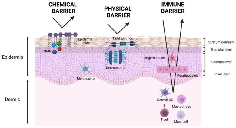
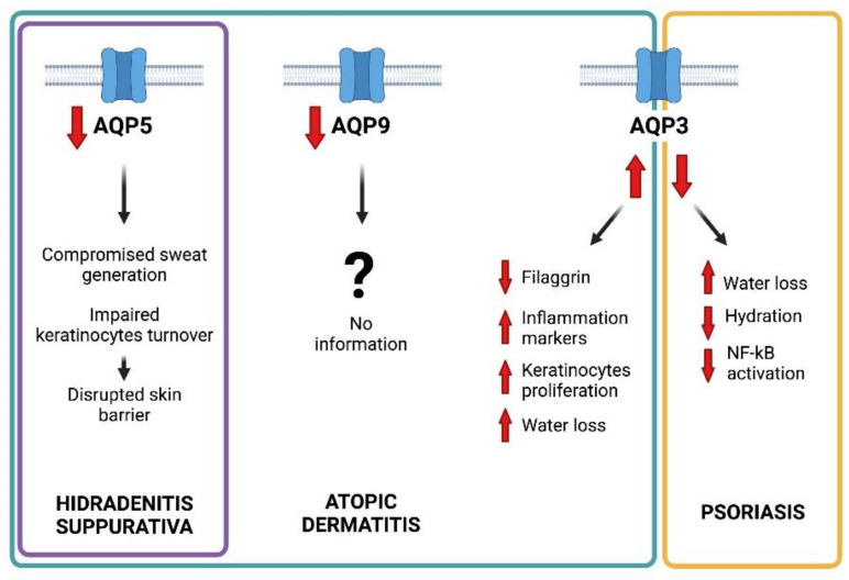
The skin is the largest organ of the human body, serving as an effective mechanical barrier between the internal milieu and the external environment. The skin is widely considered the first-line defence of the body, with an essential function in rejecting pathogens and preventing mechanical, chemical, and physical damages. Keratinocytes are the predominant cells of the outer skin layer, the epidermis, which acts as a mechanical and water-permeability barrier. The epidermis is a permanently renewed tissue where undifferentiated keratinocytes located at the basal layer proliferate and migrate to the overlying layers. During this migration process, keratinocytes undertake a differentiation program known as keratinization process. Dysregulation of this differentiation process can result in a series of skin disorders. In this context, aquaporins (AQPs), a family of membrane channel proteins allowing the movement of water and small neutral solutes, are emerging as important players in skin physiology and skin diseases. Here, we review the role of AQPs in skin keratinization, hydration, keratinocytes proliferation, water retention, barrier repair, wound healing, and immune response activation. We also discuss the dysregulated involvement of AQPs in some common inflammatory dermatological diseases characterised by skin barrier disruption.
Keywords: AQP3; aquaporin channels; atopic dermatitis; hidradenitis suppurativa; membrane transport; psoriasis.
Conflict of interest statement
The authors declare no conflict of interest.
Figures
References
-
- Patel R., Kevin Heard L., Chen X., Bollag W.B. Aquaporins in the Skin. Adv. Exp. Med. Biol. 2017;969:173–191. - PubMed
-
- Coates M., Mariottoni P., Corcoran D.L., Kirshner H.F., Jaleel T., Brown D.A., Brooks S.R., Murray J., Morasso M.I., MacLeod A.S. The skin transcriptome in hidradenitis suppurativa uncovers an antimicrobial and sweat gland gene signature which has distinct overlap with wounded skin. PLoS ONE. 2019;14:e0216249. doi: 10.1371/journal.pone.0216249. - DOI - PMC - PubMed
Publication types
MeSH terms
Substances
Grants and funding
LinkOut - more resources
Full Text Sources
Other Literature Sources
