. 2022 Apr 5;23(7):4026.
doi: 10.3390/ijms23074026.

More Prominent Inflammatory Response to Pachyman than to Whole-Glucan Particle and Oat-β-Glucans in Dextran Sulfate-Induced Mucositis Mice and Mouse Injection through Proinflammatory Macrophages

Affiliations

More Prominent Inflammatory Response to Pachyman than to Whole-Glucan Particle and Oat-β-Glucans in Dextran Sulfate-Induced Mucositis Mice and Mouse Injection through Proinflammatory Macrophages

Pratsanee Hiengrach et al. Int J Mol Sci. .

Abstract

(1→3)-β-D-glucans (BG) (the glucose polymers) are recognized as pathogen motifs, and different forms of BGs are reported to have various effects. Here, different BGs, including Pachyman (BG with very few (1→6)-linkages), whole-glucan particles (BG with many (1→6)-glycosidic bonds), and Oat-BG (BG with (1→4)-linkages), were tested. In comparison with dextran sulfate solution (DSS) alone in mice, DSS with each of these BGs did not alter the weight loss, stool consistency, colon injury (histology and cytokines), endotoxemia, serum BG, and fecal microbiome but Pachyman-DSS-treated mice demonstrated the highest serum cytokine elicitation (TNF-α and IL-6). Likewise, a tail vein injection of Pachyman together with intraperitoneal lipopolysaccharide (LPS) induced the highest levels of these cytokines at 3 h post-injection than LPS alone or LPS with other BGs. With bone marrow-derived macrophages, BG induced only TNF-α (most prominent with Pachyman), while LPS with BG additively increased several cytokines (TNF-α, IL-6, and IL-10); inflammatory genes (iNOS, IL-1β, Syk, and NF-κB); and cell energy alterations (extracellular flux analysis). In conclusion, Pachyman induced the highest LPS proinflammatory synergistic effect on macrophages, followed by WGP, possibly through Syk-associated interactions between the Dectin-1 and TLR-4 signal transduction pathways. Selection of the proper form of BGs for specific clinical conditions might be beneficial.

Keywords: DSS-induced mucositis; extracellular flux; mice; microbiome; proinflammatory macrophages; β-glucans.


Conflict of interest statement

The authors declare no conflict of interest.

Figures

Figure 1
Figure 1
Schema of the experiments (A, upper part) and characteristics of mice with drinking water (Control) with or without oral β-glucan gavage using whole-glucan particles (WGP), Pachyman or Oat glucans (Oat-BG), which are combined into the BG (mixed) group, or dextran sulfate solution with or without several glucans. Survival analysis (A), bodyweight alteration (B), stool consistency index (C), colon injury score from Hematoxylin and Eosin (H&E) staining (D), and colon cytokines (TNF-α and IL-6) (E,F) are shown (n = 6–8/group). #, p < 0.05 vs. Control as determined by ANOVA with Tukey’s analysis. Data from oral glucan-administered mice using different types of glucans were combined as BG (mixed) due to the nonsignificant difference between groups.
Figure 2
Figure 2
Characteristics of mice with drinking water (Control) with or without oral β-glucan gavage using whole-glucan particles (WGP), Pachyman, or Oat glucans (Oat-BG), which are combined into the BG (mixed) group, or dextran sulfate solution with or without several glucans, as indicated by serum cytokines (TNF-α, IL-6, and IL-10) (AC); gut permeability (FITC-dextran assay) (D); endotoxemia (E); and serum (1→3)-β-D-glucan (BG) (F), which are shown (n = 6–8/group). #, p < 0.05 vs. Control; *, p < 0.05 vs. other groups as determined by ANOVA with Tukey’s analysis. Data from oral glucan-administered mice using different types of glucans were combined as BG (mixed) due to the nonsignificant differences between groups.
Figure 3
Figure 3
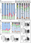
Fecal microbiome analysis from mice. Dextran sulfate solution (DSS) with or without several glucans using whole-glucan particles (WGP), Pachyman, or Oat glucans (Oat-BG) was performed. Meanwhile, in nondrinking water (Control) with oral β-glucans gavage, only one of these glucans was used in each mouse, but the data are combined into the BG (mixed) group. Phylum and genus level determination (A), average value of the analysis (B), graph presentation in the phylum analysis (C), and bacterial diversity (D) are shown (n = 3–4/group). #, p < 0.05 vs. Control; *, p < 0.05 vs. other groups; ϕ, p < 0.05 vs. the indicated group as determined by ANOVA with Tukey’s analysis. Data from oral glucan-administered mice using different types of glucans were combined as BG (mixed) due to the nonsignificant differences between these groups.
Figure 4
Figure 4
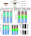
Fecal microbiome analysis of the feces of healthy mice after 24 h of incubation with whole-glucan particles (WGP), Pachyman, or Oat glucans (Oat), as presented in the schema of the experiments, characterized by phylum and genus level determination (A,B), the average value of the analysis (C), and the bacterial diversity (D) (n = 3/group).
Figure 5
Figure 5
Characteristics of mice with drinking water (Control) with or without oral β-glucans gavage using whole-glucan particles (WGP), Pachyman, or Oat glucans (Oat-BG), which are combined into the BG (mixed) group or dextran sulfate solution with or without several glucans. The alteration in serum cytokines (TNF-α, IL-6, and IL-10); gut permeability (FITC-dextran assay); and liver enzyme (alanine transaminase) (AE), together with liver cytokines (TNF-α, IL-6, and IL-10), (FH) are shown (n = 6–8/group). #, p < 0.05 vs. Control; *, p < 0.05 vs. other groups; ϕ, p < 0.05 vs. the indicated group as determined by ANOVA with Tukey’s analysis. Data from oral glucan-administered mice using different types of glucans were combined as the BG (mixed) due to the nonsignificant different values among the groups with glucan injection alone.
Figure 6
Figure 6
Characteristics of bone marrow-derived macrophages after 24 h of incubation in the control medium (Control) or control medium supplemented with whole-glucan particles (WGP), Pachyman, Oat glucans (Oat-BG), lipopolysaccharide (LPS), and LPS with the different glucans. Supernatant cytokines (TNF-α, IL-6, and IL-10) with gene expression (AF) are shown (n = 6–8/group). #, p < 0.05 vs. Control; *, p < 0.05 vs. other groups; ϕ, p < 0.05 vs. the indicated group as calculated by ANOVA with Tukey’s analysis. Independent triplicate experiments were performed.
Figure 7
Figure 7
Gene expression characteristics of bone marrow-derived macrophages after 24 h of incubation in the control medium (Control) or supplemented with whole-glucan particles (WGP), Pachyman, Oat glucans (Oat-BG), lipopolysaccharide (LPS), and LPS with the different glucans. Pro- (iNOS and IL-1β) and anti- inflammatory responses (Arginase-1, TGF-β, and Fizz-1) (AE) with the signaling genes (TLR-4, Dectin-1, Syk, and NF-κB) (FI) are shown (n = 6–8/group). #, p < 0.05 vs. Control; *, p < 0.05 vs. other groups; ϕ, p < 0.05 vs. the indicated group as calculated by ANOVA with Tukey’s analysis. Independent triplicate experiments were performed.
Figure 8
Figure 8
Characteristics of bone marrow-derived macrophages after 24 h of incubation in the control medium (Control) or with or without different forms of β-glucans using whole-glucan particles (WGP), Pachyman, or Oat glucans (Oat-BG), which are combined into the BG (mixed) group, or lipopolysaccharide (LPS) with or without these glucans. Extracellular metabolite flux analysis patterns, including oxygen consumption rate (OCR) of the mitochondrial function (A) and extracellular acidification rate (ECAR) of glycolysis activity (B) with the graph presentation of mitochondrial (basal respiration, maximal respiration, and respiratory reserve) (CE) and glycolysis (glycolysis capacity and glycolysis reserve) (F,G) parameters, which are shown (n = 6–8/group). #, p < 0.05 vs. Control; *, p < 0.05 vs. other groups; ϕ, p < 0.05 vs. the indicated group. ANOVA with Tukey’s analysis was used. Data from oral glucan-administered mice using different types of glucans were combined as BG (mixed) due to the nonsignificant differences between groups and independent triplicate experiments were performed.
Figure 9
Figure 9
The proposed working hypothesis demonstrates the possible additional proinflammatory impact with lipopolysaccharides (LPS) plus the different forms of (1→3)-β-D-glucan (BGs), including whole-glucan particles (WGP; BGs with 1,6 linkage), Pachyman (BGs without 1→6 nor 1→4 linkages), and Oat β-glucans (Oat-BG; BGs with 1,4 linkage), through the crosslink between Toll-like receptor-4 (TLR-4) and Dectin-1, the pattern recognition receptors for LPS and BGs, respectively, with Spleen tyrosine kinase (Syk), an inhibitor of nuclear factor kappa-B kinase (IKK), and nuclear factor kappa B (NF-κB) as the downstream signals (thickness of the arrows represents the intensity of the activation). While Myeloid differentiation primary response 88 (MyD88) and Syk-Caspase recruitment domain-containing protein 9 (CARD-9) are the main downstream signals of TLR-4 and Dectin-1, respectively [48,64,82], the crosslink of Dectin-1 with TLR-4, by a proper form of glucan (Pachyman), induced stronger signaling through Syk and NF-κB [64]. TRAP-6, Thrombin Receptor Activator for Peptide 6; IκB, I kappa B kinase. The picture is created by BioRender.com (accessed on 1 March 2022).

Similar articles

Cited by

References

    1. Chaiyasut C., Pengkumsri N., Sivamaruthi B., Sirilun S., Kesika P., Saelee M., Chaiyasut K., Peerajan S. Extraction of Β-glucan of Hericium erinaceus, Avena sativa, L., and Saccharomyces cerevisiae and in vivo evaluation of their immunomodulatory effects. Food Sci. Technol. 2018;38:138–146. doi: 10.1590/fst.18217. - DOI
    1. Vetvicka V., Vannucci L., Sima P., Richter J. Beta Glucan: Supplement or Drug? From Laboratory to Clinical Trials. Molecules. 2019;24:1251. doi: 10.3390/molecules24071251. - DOI - PMC - PubMed
    1. Jayachandran M., Chen J., Chung S.S.M., Xu B. A critical review on the impacts of beta-glucans on gut microbiota and human health. J. Nutr. Biochem. 2018;61:101–110. doi: 10.1016/j.jnutbio.2018.06.010. - DOI - PubMed
    1. Driscoll M., Hansen R., Ding C., Cramer D.E., Yan J. Therapeutic potential of various beta-glucan sources in conjunction with anti-tumor monoclonal antibody in cancer therapy. Cancer Biol. Ther. 2009;8:218–225. doi: 10.4161/cbt.8.3.7337. - DOI - PubMed
    1. Karp S., Wyrwisz J., Kurek M.A. The impact of different levels of oat beta-glucan and water on gluten-free cake rheology and physicochemical characterisation. J. Food Sci. Technol. 2020;57:3628–3638. doi: 10.1007/s13197-020-04395-5. - DOI - PMC - PubMed

LinkOut - more resources