. 2022 Mar 22;19(7):3782.
doi: 10.3390/ijerph19073782.

Sclerocarya birrea (Marula) Extract Inhibits Hepatic Steatosis in db/db Mice

Affiliations

Sclerocarya birrea (Marula) Extract Inhibits Hepatic Steatosis in db/db Mice

Lawrence Mabasa et al. Int J Environ Res Public Health. .

Abstract

Non-alcoholic fatty liver disease (NAFLD) is a spectrum of hepatic metabolic perturbations ranging from simple steatosis to steatohepatitis, cirrhosis and hepatocellular carcinoma. Currently, lifestyle modifications to reduce weight gain are considered the most effective means of preventing and treating the disease. The aim of the present study was to determine the therapeutic benefit of Sclerocarya birrea (Marula leaf extract, MLE) on hepatic steatosis. Obese db/db mice were randomly stratified into the obese control, metformin (MET) or MLE-treated groups. Mice were treated daily for 29 days, at which point all mice were euthanized and liver samples were collected. Hematoxylin and eosin staining was used for histological assessment of the liver sections, while qRT-PCR and Western blot were used to determine hepatic mRNA and protein expression, respectively. Thereafter, the association between methylenetetrahydrofolate reductase (Mthfr a key enzyme in one-carbon metabolism and DNA-methylation-induced regulation of gene transcription) and lipogenic genes was evaluated using Pearson's correlation coefficient. Mice treated with MLE presented with significantly lower body and liver weights as compared with the obese control and MET-treated mice (p ≤ 0.05). Further, MLE treatment significantly inhibited hepatic steatosis as compared with the obese control and MET-treated mice (p ≤ 0.05). The reduced lipid accumulation was associated with low expression of fatty acid synthase (Cpt1; p ≤ 0.05) and an upregulation of the fatty acid oxidation gene, carnitine palmitoyltransferase (Cpt1; p ≤ 0.01), as compared with the obese control mice. Interestingly, MLE treatment improved the correlation between Mthfr and Cpt1 mRNA expression (r = 0.72, p ≤ 0.01). Taken together, the results suggest that Marula leaf extracts may inhibit hepatic steatosis by influencing the association between Mthfr and genes involved in hepatic lipid metabolism. Further studies are warranted to assess DNA methylation changes in lipid metabolism genes.

Keywords: DNA methylation; Marula leaf extract; methylenetetrahydrofolate reductase; non-alcoholic fatty liver disease; β-oxidation.


Conflict of interest statement

There is no conflict of interest reported by the authors. The funding sponsors had no role in the design of the study; in the collection, analyses, or interpretation of data; in the writing of the manuscript, and in the decision to publish the results.

Figures

Figure 1
Figure 1
Bodyweight (A) and non-fasted blood glucose (B) results for db/db mice. Data are represented as means ± SEM, with a sample size of 5 animals/group. GraphPad Prism 6 was used to test for statistical significance at a p-value ≤ 0.05.
Figure 2
Figure 2
Hepatocellular steatosis, liver weight and percent lipid accumulation results. Representative photomicrographs of liver cross-sections of the obese C (A), obese + MET (B) and obese + MLE (C) mice. Histologically steatosis severity was classified by the number and distribution of hypertrophied hepatocytes (dotted circle) with either macrovesicular (bold arrows) or microvesicular (dotted arrows) steatosis. All photomicrographs were stained with H&E at 40× magnification. Liver weight (D), % lipid accumulation (E), and liver to body weight ratio (F) results are represented as means ± SEM, with a sample size of 5/group (10 images per slide were taken). GraphPad Prism 9 was used to test for statistical significance at a p-value ≤ 0.05. Note: * p ≤ 0.05 and ** p ≤ 001 vs. obese C, # p ≤ 0.05 and ### p ≤ 0.0001 vs. obese + MET.
Figure 3
Figure 3
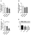
The effects of Marula leaf extract (MLE) on mRNA expression of lipogenic genes, Acc (A), Srebf1 (B), and Fasn (C), as well as protein levels of FASN (D). Data are represented as means ± SEM, with a sample size of 5–6/group. GraphPad Prism 9 was used to test for statistical significance at a p-value ≤ 0.05. Note: * p ≤ 0.05 vs. obese C, *** p ≤ 0.001 vs. obese C. Acc; acetyl-CoA carboxylase, Srebf1; sterol regulatory element binding transcription factor 1, Fasn; fatty acid synthase.
Figure 4
Figure 4
The effects of Marula leaf extract (MLE) on mRNA expression of β-oxidation genes, Pparα (A), Ampk (B) and Cpt1 (C). Data are represented as means ± SEM, with a sample size of 5-6/group. One-way ANOVA was used to test for statistical significance at a p-value ≤ 0.05. Note: * p ≤ 0.05 vs. obese C, ** p ≤ 0.01 vs. obese C. Pparα; peroxisome proliferator-activated receptor alpha, Ampk; AMP-activated protein kinase, Cpt1; carnitine palmitoyltransferase I.
Figure 5
Figure 5
The effects of Marula leaf extract (MLE) on mRNA expression of insulin signaling genes, Akt (A), Pi3k (B), Irs2 (C) and Glut2 (D). Data are represented as means ± SEM, with a sample size of 5-6/group. GraphPad Prism 9 was used to test for statistical significance at a p-value ≤ 0.05. Note: * p ≤ 0.05 vs. obese C, # p ≤ 0.05 vs. obese + MET and #### p ≤ 0.0001 vs. obese + MET. Akt; protein kinase B, PI3k; phosphatidyl-inositol-3-kinases, Irs2; insulin receptor substrate 2, Glut2; glucose transporter 2.
Figure 6
Figure 6
The effects of Marula leaf extract (MLE) on mRNA expression Dnmt1 (A), Mthfr (B) and Cbs (C) levels of genes involved in the control of DNA methylation. Data are represented as means ± SEM, with a sample size of 5-6/group. GraphPad Prism 9 was used to test for statistical significance at a p-value ≤ 0.05. Note: ## p ≤ 0.01 vs. obese + MET. Dnmt1; DNA methyltransferase 1, Mthfr; methylene tetrahydrofolate reductase, Cbs; cystathionine-β-synthase.
Figure 7
Figure 7
Schematic representation of the potential mechanism through which Marula leaf extract lowers hepatic lipid accumulation. Marula leaf extract can significantly influence the association between MTHFR and CPT1, which impacts the DNA methylation profile of Cpt1, and in turn triggers the activation of the β-oxidation pathway.

References

    1. Fabbrini E., Sullivan S., Klein S. Obesity and nonalcoholic fatty liver disease: Biochemical, metabolic, and clinical implications. Hepatology. 2010;51:679–689. doi: 10.1002/hep.23280. - DOI - PMC - PubMed
    1. Sayiner M., Koenig A., Henry L. Epidemiology of Nonalcoholic Fatty Liver Disease and Nonalcoholic Steatohepatitis in the United States and the Rest of the World. Clin. Liver Dis. 2016;20:22042. doi: 10.1016/j.cld.2015.10.001. - DOI - PubMed
    1. Geisler C.E., Renquist B.J. Hepatic lipid accumulation: Cause and consequence of dysregulated glucoregulatory hormones. J. Endocrinol. 2017;234:R1–R21. doi: 10.1530/JOE-16-0513. - DOI - PubMed
    1. Jarvis H., Craig D., Barker R., Spiers G., Stow D., Anstee Q.M., Hanratty B. Metabolic risk factors and incident advanced liver disease in non-alcoholic fatty liver disease (NAFLD): A systematic review and meta-analysis of population-based observational studies. PLoS Med. 2020;17:e1003100. doi: 10.1371/journal.pmed.1003100. - DOI - PMC - PubMed
    1. Xia H.M., Wang J., Xie X.J., Xu L.J., Tang S.Q. Green tea polyphenols attenuate hepatic steatosis, and reduce insulin resistance and inflammation in high-fat diet-induced rats. Int. J. Mol. Med. 2019;44:1523–1530. doi: 10.3892/ijmm.2019.4285. - DOI - PubMed