. 2022 Mar 15;12(3):1102-1115.
eCollection 2022.

PIK-75 overcomes venetoclax resistance via blocking PI3K-AKT signaling and MCL-1 expression in mantle cell lymphoma

Affiliations

PIK-75 overcomes venetoclax resistance via blocking PI3K-AKT signaling and MCL-1 expression in mantle cell lymphoma

Shengjian Huang et al. Am J Cancer Res. .

Abstract

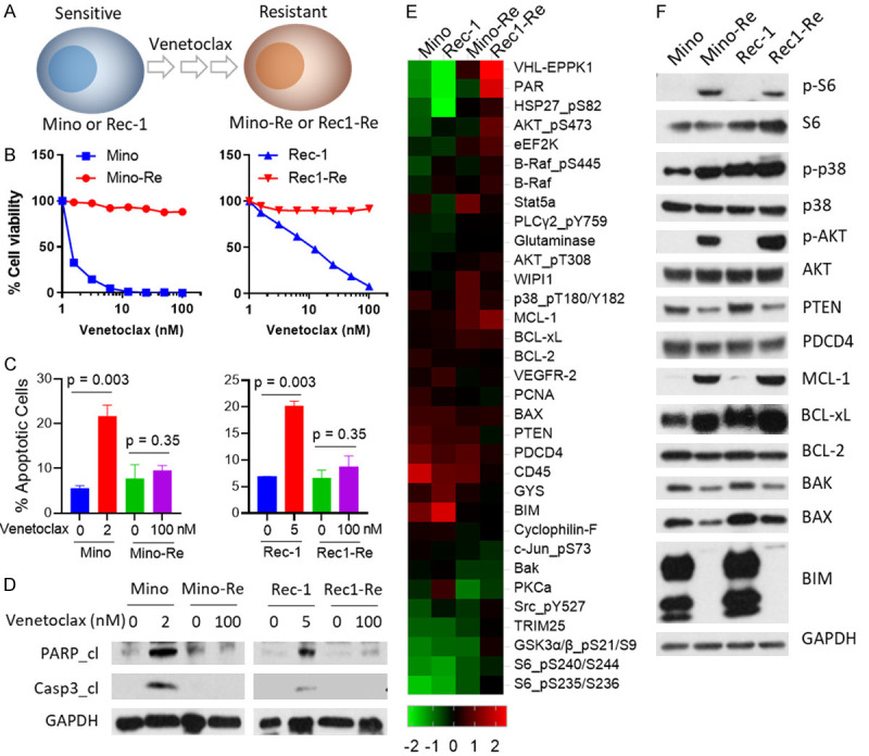
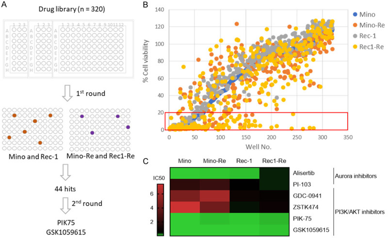
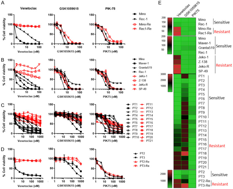
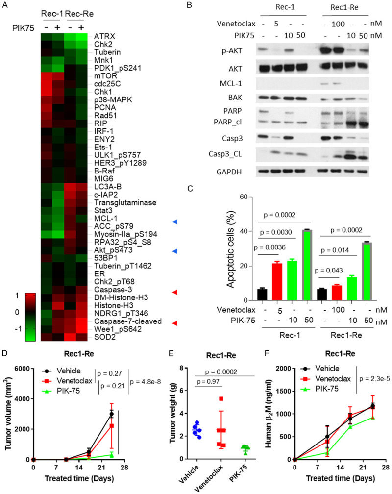
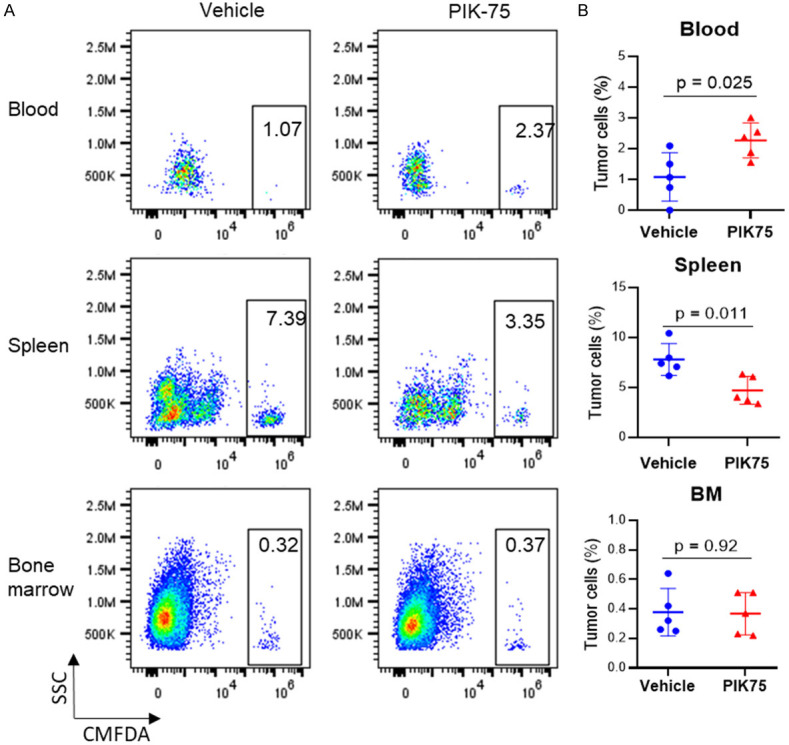
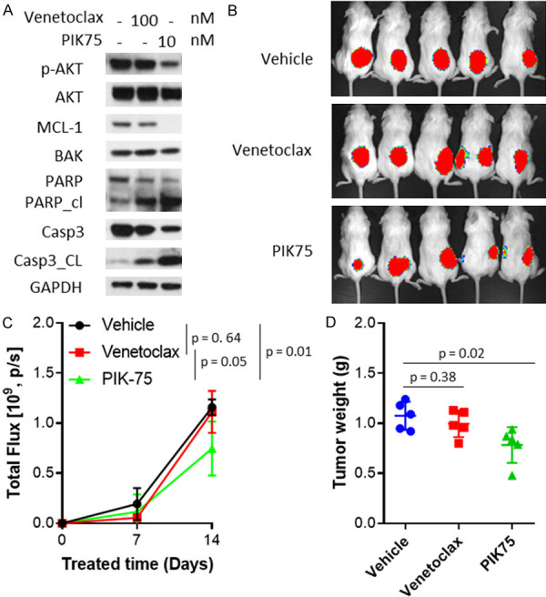
Therapeutic resistance is the major challenge in clinic for patients with mantle cell lymphoma (MCL), an aggressive subtype of B-cell lymphoma. In addition to the FDA-approved Bruton's tyrosine kinase (BTK) inhibitors, multiple clinical trials have demonstrated clinical benefits in targeting BCL-2 by venetoclax and reported to greatly improve clinical outcome for refractory/relapsed patients with MCL alone or in combination with BTK inhibitors. However, resistance to venetoclax is no exception and marks as a new clinic challenge. To decode the underlying mechanisms driving venetoclax resistance, we established two MCL cell lines, Mino-Re and Rec1-Re, with acquired resistance to venetoclax from sensitive Mino and Rec-1. Using reverse phase protein assay (RPPA), an agnostic proteomic approach, we identified targetable signaling pathways that are associated with acquired venetoclax resistance in Mino-Re and Rec1-Re cells. A panel of pro-survival signals was identified to correlate well with venetoclax-resistance, including increased expression of MCL-1, BCL-xL and AKT phosphorylation, and decreased expression of BIM, BAX and PTEN. Based on a high throughput drug screening of over 320 FDA-approved/investigational drugs in the paired venetoclax-sensitive and -resistant cell lines Mino-Re and Rec1-Re, we identified the top candidates that are capable to overcome acquired venetoclax resistance in these cells. The best candidate is PIK-75, a dual inhibitor targeting both PI3K and CDK9. Its action to overcome venetoclax resistance was further confirmed in additional cell lines with primary venetoclax resistance (n=4) and primary patient samples (n=21). Mechanistically, PIK75 treatment potently diminished the elevated MCL-1 expression and AKT activation in cells with acquired or primary venetoclax resistance and resulted in potent anti-MCL activity to overcome these resistances. In addition, PIK75 is also potent in overcoming tumor microenvironment (TME)-associated venetoclax resistance. Furthermore, PIK-75 treatment is efficacious in overcoming primary and acquired venetoclax resistance in xenograft models and inhibited tumor cell dissemination to spleen in mice. Altogether, our data demonstrated that PIK-75 is highly potent in overcoming primary, acquired, or stromal cells-induced venetoclax resistances in MCL cells and revealed a new tumor vulnerability that can be exploited clinically in difficult to treat MCL cases, especially those with venetoclax resistance.

Keywords: AKT; MCL-1; PI3K; PIK75; mantle cell lymphoma; venetoclax resistance.


Conflict of interest statement

M.W. is a consultant for: InnoCare, Loxo Oncology, Juno, Oncternal, CStone, AstraZeneca, Janssen, VelosBio, Pharmacyclics, Genentech, Bayer Healthcare. His research is funded by: Pharmacyclics, Janssen, AstraZeneca, Celgene, Loxo Oncology, Kite Pharma, Juno, BioInvent, VelosBio, Acerta Pharma, Oncternal, Verastem, Molecular Templates, Lilly, Innocare. All other authors declare no competing financial interests.

Figures

Figure 1
Figure 1
Establishment of venetoclax-resistant cell lines and differential gene expression associated with acquired venetoclax resistance. (A) Two sensitive MCL cell lines (Mino and Rec-1) were exposed to increasing doses of venetoclax stepwise up to 1000 nM. (B) 8-dose cell viability was performed in both parental and resistant cell lines for 72 h. Each treatment for the cell viability assay was set up in triplicate. (C) Mino cells were treated with 2 nM venetoclax, Rec-1 cells were treated with 5 nM venetoclax, the corresponding resistant cells were treated with 100 nM venetoclax for 24 h, and apoptosis was measured by annexin-V/PI assay. Annexin-V+PI- subpopulation and annexin-V+PI+ subpopulations were summed up as total apoptosis. Each treatment for the cell apoptosis assay was set up in triplicate. (D) Cells with the same treatments as described in (C) were collected for western blot. Total PARP, cleaved PARP, total caspase-3, and cleaved caspase-3 were detected in these cell lines. GAPDH was used as internal loading control. Each experiment in (B-D) was repeated three times. (E) Heatmaps of dysregulated proteins. 5×10⁶ cells of each cell line (Mino, Mino-Re, Rec-1 and Rec1-Re) were collected and subjected to RPPA analysis. 425 proteins in total were detected, and proteins with at least two-fold change were selected for generating the heatmap. High-expressing proteins are shown in red, and low-expressing proteins are shown in green. Each sample for RPPA analysis was set up in triplicate. (F) Western blotting was used to confirm dysregulated protein expression comparing parental and resistant cell lines.
Figure 2
Figure 2
High throughput drug screening to identify candidates for overcoming venetoclax resistance. (A) Schematic illustration of the high-throughput drug screen. The drug screen was performed over a library consisting of 320 drugs in two parental and venetoclax-resistant paired cell lines by a 72 h cell viability test with one dose (5 μM) as first round screen followed by 2nd round drug validation via 4-dose viability assay in the same cell lines to identify the top candidates in overcoming venetoclax resistance. (B) 1st round drug screen at 5 μM by a cell viability inhibition assay in Mino, Mino-Re, Rec-1 and Rec1-Re cells. Each treatment for the cell viability assay was set up in triplicate. Relative cell viability was normalized to DMSO control. Cell viability less than 20% was considered as positive (red box). (C) 2nd round drug validation for the top candidates (six drugs) was performed by a four-dose (three-fold dilution) cell viability assay at 72 h in the same cell lines. The heatmap shown represents the IC50 values (µM). Each treatment for the cell viability assay was set up in triplicate and the experiment was repeated three times.
Figure 3
Figure 3
PIK-75 is potent in overcoming a variety of venetoclax resistances. (A, B) In vitro efficacy of BCL2i venetoclax, PI3Ki PIK-75 and GSK1059615 in paired venetoclax-sensitive (Mino and Rec-1) and -resistant (Mino-Re and Rec1-Re) cell lines (A), and additional 8 MCL cell line panel (B) by eight-dose cell viability assay at 72 hours upon treatment. (C) In vitro efficacy of venetoclax, PIK-75 and GSK1059615 in 21 primary MCL patient samples at 24 hours upon treatment. (D) Two venetoclax-sensitive (ex vivo) patient samples (PT2 and PT3) were co-cultured with HS-5 stromal cells and supplemental cytokines for two weeks. An eight-dose 24-hour cell viability assay was conducted. Each treatment for the cell viability assay was set up in triplicate. (E) Heatmaps of IC50 of venetoclax, GSK1059615 and PIK-75 in MCL cell lines and primary patient samples with/without coculture with HS-5 cells based on the data (A-D). Sensitivity to venetoclax was indicated in the right for each cell line or patient sample.
Figure 4
Figure 4
PIK-75 inhibits PI3K-AKT activity and overcomes acquired venetoclax resistance in vitro and in vivo. (A) Rec-1 and Rec1-Re cells were treated with 50 nM PIK-75 for 24 h and subjected to RPPA analysis. A heatmap was made with NormLog2_MedianCentered values that were < and >0.5 times between untreated and treated samples. (B, C) Rec-1 and Rec1-Re cells were treated with designated venetoclax or PIK-75 concentrations for 24 h. Cells were harvested and analyzed by western blotting (B) and cell apoptosis assay (C) to confirm the RPPA data and biological effects. (D-F) 5×10⁶ Rec1-Re cells were injected subcutaneously into each NSG mouse and treated with vehicle, 50 mg/kg venetoclax, or 10 mg/kg PIK-75, daily. Tumor volumes were measured weekly (D), and mouse blood was collected weekly; human B2M in mouse serum was measured by ELISA assay (F). Tumor weight was measured at the end of experiment (E). Student t test was used for statistical analysis, n=5.
Figure 5
Figure 5
PI3K inhibitor PIK-75 blocks in vivo homing of MCL cells to mouse spleen. (A) CMFDA-labelled patient cells (PT2) were pre-treated with vehicle (DMSO) or PIK-75 at 100 nM for 30 min before tail vein inoculation into NSG mice. Blood, spleen, and bone marrow were collected at 16 hours post inoculation for tumor cell detection by flow cytometry based on CMFDA labeling (x-axis). (B) Percentage of tumor cells was quantified based on flow cytometry analysis. Student t test was used for statistical analysis, n=5.
Figure 6
Figure 6
PIK-75 overcomes primary venetoclax resistance in mouse xenograft models. (A) JeKo-1 cells were treated with venetoclax at 100 nM or PIK-75 at 10 nM for 24 hours. Cells were harvested and analyzed by western blotting. (B-D) 5×10⁶ luciferase-expressing JeKo-luc cells were injected subcutaneously into each NSG mouse and treated with vehicle, 50 mg/kg venetoclax, or 10 mg/kg PIK-75 daily. Tumor load was measured weekly by bioluminescence imaging (B) and total flux was calculated based on live imaging data (C). Tumor weight was measured at the end of the experiment (D). Student t test was used for statistical analysis, n=5.

References

    1. Roberts AW, Davids MS, Pagel JM, Kahl BS, Puvvada SD, Gerecitano JF, Kipps TJ, Anderson MA, Brown JR, Gressick L, Wong S, Dunbar M, Zhu M, Desai MB, Cerri E, Heitner Enschede S, Humerickhouse RA, Wierda WG, Seymour JF. Targeting BCL2 with venetoclax in relapsed chronic lymphocytic leukemia. N Engl J Med. 2016;374:311–322. - PMC - PubMed
    1. Khan N, Kahl B. Targeting BCL-2 in hematologic malignancies. Targeted Oncol. 2018:257–267. - PubMed
    1. Davids MS. Targeting BCL-2 in B-cell lymphomas. Blood. 2017;130:1081–1088. - PubMed
    1. Davids MS, Roberts AW, Seymour JF, Pagel JM, Kahl BS, Wierda WG, Puvvada S, Kipps TJ, Anderson MA, Salem AH, Dunbar M, Zhu M, Peale F, Ross JA, Gressick L, Desai M, Kim SY, Verdugo M, Humerickhouse RA, Gordon GB, Gerecitano JF. Phase I first-in-human study of venetoclax in patients with relapsed or refractory non-Hodgkin lymphoma. J. Clin. Oncol. 2017;35:826–833. - PMC - PubMed
    1. Tam C, Roberts AW, Anderson M, Dawson S, Hicks R, Burbury K, Turner G, Di Iulio J, Bressel M, Westerman D. Combination ibrutinib (Ibr) and venetoclax (Ven) for the treatment of mantle cell lymphoma (MCL): primary endpoint assessment of the phase 2 AIM study. Hematol Oncol. 2017;35:144–145.

LinkOut - more resources