

Visualization of energy-based device-induced thermal tissue alterations using bimodal ex-vivo confocal microscopy with digital staining. A proof-of-concept study
- PMID: 35411961
- PMCID: PMC9907604
- DOI: 10.1111/srt.13155
Visualization of energy-based device-induced thermal tissue alterations using bimodal ex-vivo confocal microscopy with digital staining. A proof-of-concept study
Abstract
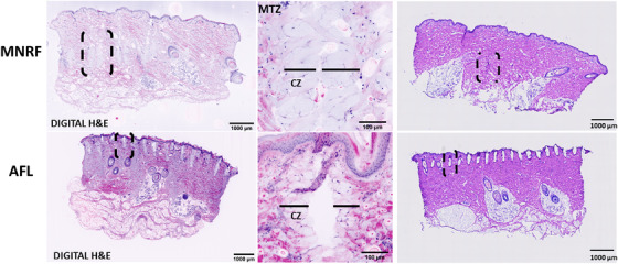
Background: Ex-vivo confocal microscopy (EVCM) enables examination of tissue alterations immediately after treatment with energy-based devices (EBDs). This proof-of-concept study aimed to describe EBD-induced tissue effects in ex-vivo porcine skin after treatment with microneedle radiofrequency (MNRF) and ablative fractional CO2 -laser (AFL) using EVCM.
Materials and methods: Ex-vivo porcine skin was treated with MNRF and AFL. Three cryosections from each intervention were stained with acridine orange (AO) and scanned with EVCM. Reflectance confocal microscopy (RCM, 638 nm) and fluorescence confocal microscopy (FCM, 488 nm) images were captured and evaluated individually, after image fusion, and after digital hematoxylin and eosin (H&E) staining.
Results: Bimodal EVCM was able to visualize EBD-induced thermal alterations in porcine skin. In RCM mode, the full width and depth of the vertically aligned microscopic treatment zones (MTZs) were displayed with clear demarcation to surrounding intact skin. In FCM mode, the ablation of the epidermis after AFL was prominent in contrast with the almost intact epidermis observed in MNRF treated skin. In fusion mode, fluorescence signal from AO marked the surrounding coagulation zone (CZ) from both interventions, with enhanced discrimination between ablation and coagulation. Digitally H&E-stained images closely resembled conventional histopathology but proved superior in terms of visualization of the CZ.
Conclusion: Bimodal EVCM with digital H&E-staining facilitates the identification and qualitative evaluation of thermal alterations induced by treatment with EBD. By providing high-resolution images comparable to standard histology, EVCM is a useful tool in the research and development of EBD to visualize and evaluate device-tissue interactions.
Keywords: acridine orange; coagulation zone; ex-vivo confocal microscopy; fractional CO2-laser; microscopic ablation zone; radiofrequency microneedling.
© 2022 The Authors. Skin Research and Technology published by John Wiley & Sons Ltd.
Figures

References
-
- Shin M‐K, Choi JH, Ahn SB, Lee MH. Histologic comparison of microscopic treatment zones induced by fractional lasers and radiofrequency. J Cosmet Laser Ther Off Publ Eur Soc Laser Dermatol. 2014;16(6):317‐23. - PubMed
-
- Malvehy J, Pérez‐Anker J, Toll A, Pigem R, Garcia A, Alos LL, et al. Ex vivo confocal microscopy: revolution in fast pathology in dermatology. Br J Dermatol. 2020;183(6):1011‐25. - PubMed
-
- Karmisholt KE, Taudorf EH, Wulff CB, Wenande E, Philipsen PA, Haedersdal M. Fractional CO2 laser treatment of caesarean section scars‐a randomized controlled split‐scar trial with long term follow‐up assessment. Lasers Surg Med. 2017;49(2):189‐97. - PubMed
-
- Mahar PD, Spinks AB, Cleland H, Bekhor P, Waibel JS, Lo C, et al. Improvement of burn scars treated with fractional ablative CO2 lasers ‐ a systematic review and meta‐analysis using the Vancouver Scar Scale. J Burn Care Res Off Publ Am Burn Assoc. 2020;42:200‐6. - PubMed
MeSH terms
LinkOut - more resources
Full Text Sources
