. 2022 May 25;14(646):eabn1252.
doi: 10.1126/scitranslmed.abn1252. Epub 2022 May 25.

Multivalent designed proteins neutralize SARS-CoV-2 variants of concern and confer protection against infection in mice

Affiliations

Multivalent designed proteins neutralize SARS-CoV-2 variants of concern and confer protection against infection in mice

Andrew C Hunt et al. Sci Transl Med. .

Abstract

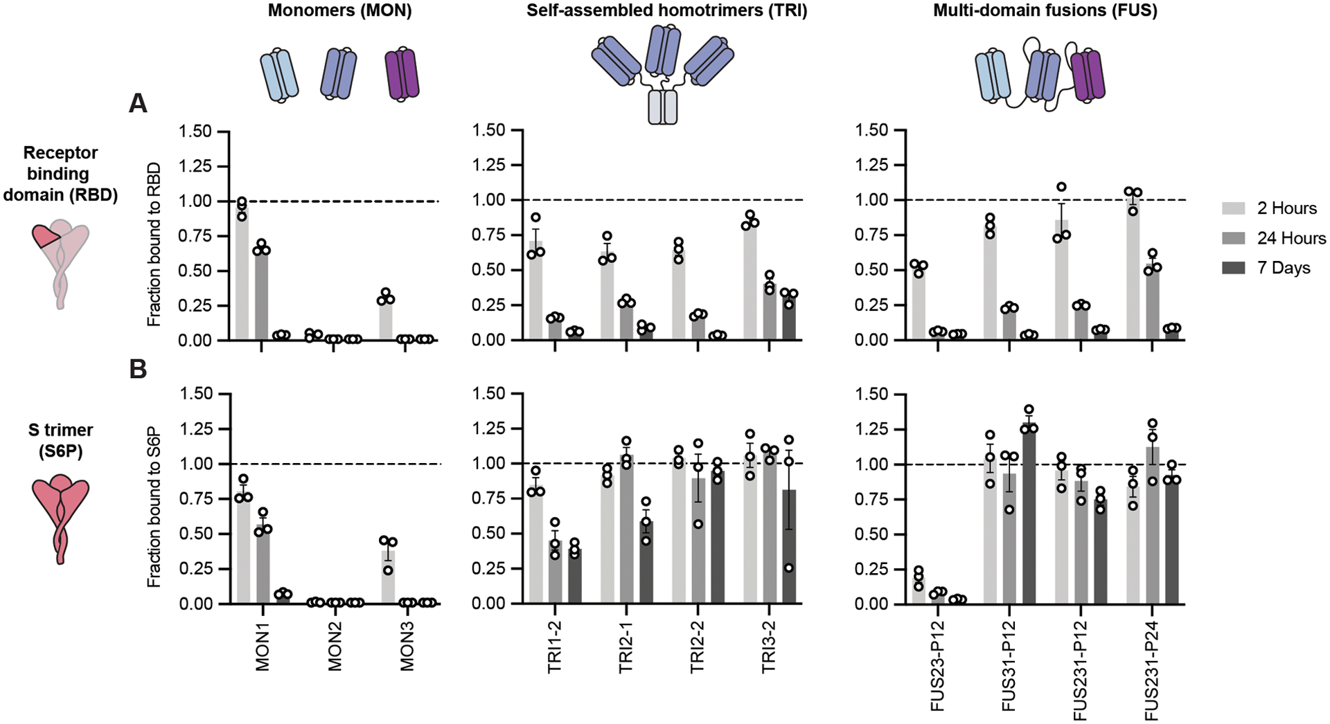
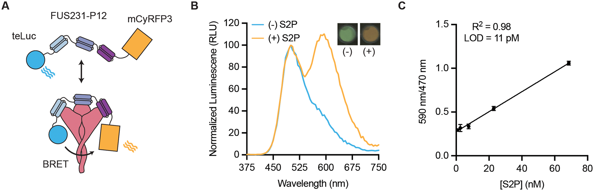
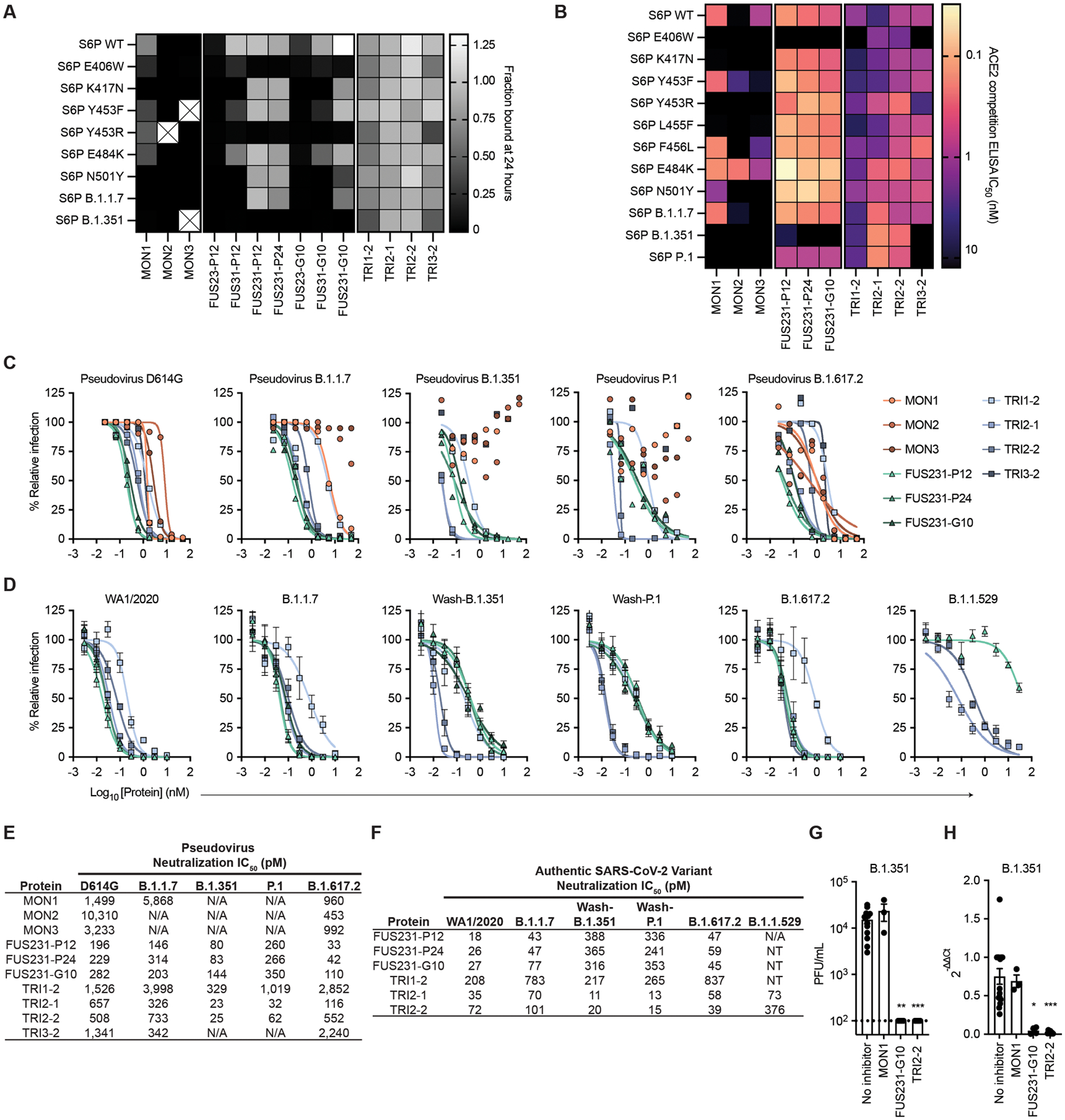
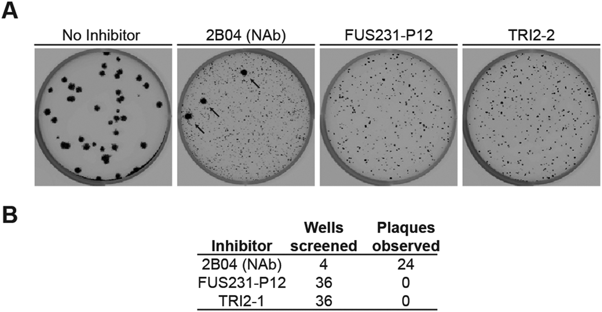
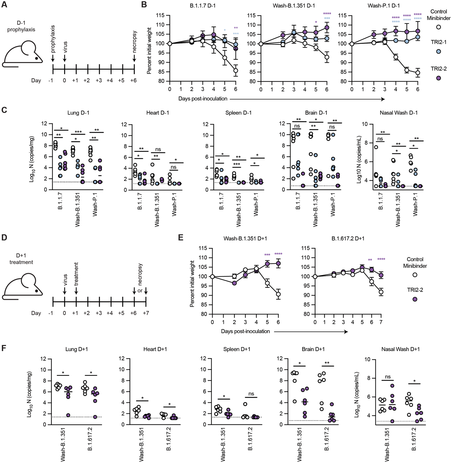
New variants of severe acute respiratory syndrome coronavirus 2 (SARS-CoV-2) continue to arise and prolong the coronavirus disease 2019 (COVID-19) pandemic. Here, we used a cell-free expression workflow to rapidly screen and optimize constructs containing multiple computationally designed miniprotein inhibitors of SARS-CoV-2. We found the broadest efficacy was achieved with a homotrimeric version of the 75-residue angiotensin-converting enzyme 2 (ACE2) mimic AHB2 (TRI2-2) designed to geometrically match the trimeric spike architecture. Consistent with the design model, in the cryo-electron microscopy structure TRI2-2 forms a tripod at the apex of the spike protein that engaged all three receptor binding domains simultaneously. TRI2-2 neutralized Omicron (B.1.1.529), Delta (B.1.617.2), and all other variants tested with greater potency than the monoclonal antibodies used clinically for the treatment of COVID-19. TRI2-2 also conferred prophylactic and therapeutic protection against SARS-CoV-2 challenge when administered intranasally in mice. Designed miniprotein receptor mimics geometrically arrayed to match pathogen receptor binding sites could be a widely applicable antiviral therapeutic strategy with advantages over antibodies in greater resistance to viral escape and antigenic drift, and advantages over native receptor traps in lower chances of autoimmune responses.


Figures

Fig 1.
Fig 1.. Multivalent minibinders exhibit very slow dissociation rates upon binding to the prefusion SARS-CoV-2-S glycoprotein trimer.
Dissociation of the minibinder construct was monitored by competition with 100-fold molar excess of untagged MON1 using AlphaLISA (Mean ± SEM, n = 3 technical replicates from a single experiment). (A) Dissociation was measured for indicated minibinder constructs complexed with the receptor-binding domain of SARS-CoV-2 (RBD). (B) Dissociation was measured for the indicated minibinder constructs complexed with the S trimer (S6P).
Fig 2.
Fig 2.. CryoEM structures of multivalent minibinders in complex with the SARS-CoV-2 S6P glycoprotein.
TRI2–2 is a homotrimer of MON2 using the SB175 homotrimerization domain, FUS31-G10 is a fusion of MON3 to MON1 with a 10 amino acid glycine-serine linker, FUS213-P24 is a fusion of MON2 to MON1 to MON 3 with a 24 amino acid proline-alanine-serine linker, and FUS213-G10 is a fusion of MON2 to MON1 to MON 3 with a 10 amino acid glycine-serine linker. (A) A CryoEM map of TRI2–2 in complex with the S6P in two orthogonal orientations is shown. (B) A zoomed-in view of the TRI2–2 and RBD complex was obtained using focused 3D classification and local refinement. The RBD and MON2 built in the 2.9 Å resolution cryoEM map are shown in yellow and blue, respectively. (C) A cryoEM map of FUS31-G10 bound to S6P is shown. (D) A cryoEM map of FUS231-P24 bound to S6P is shown. (E) A negative-stain EM map of FUS231-G10 in complex with S6P is shown. S and minibinder models were docked in the whole map by rigid body fitting for visualization. In all panels, the EM density is shown as a transparent gray surface, S protomers (PDB 7JZL) are rendered in yellow, cyan, and pink and minibinders (PDB 7JZU, 7JZM, and MON2 structure was determined in this study) are shown in orange.
Fig 3.
Fig 3.. FUS231-P12 enables detection of SARS-CoV-2 S trimer through BRET.
(A) A schematic representation of the BRET sensor, teluc-FUS231-P12-mCyRFP3, to detect S trimer is shown. (B) Luminescence emission spectra and image of the BRET sensor (100 pM) in the presence (orange trace, 100 pM) and absence (blue trace) of S2P are shown. Emission color change was observed using a mobile phone camera (inset top right). RLU, relative light units. (C) Titration of S2P with 100 pM sensor protein is shown. LOD indicates limit of detection. R2 value is shown on the graph. Data are presented as mean ± SEM with n = 3 technical replicates from a single experiment.
Fig 4.
Fig 4.. Multivalency enhances both the breadth and potency of neutralization against SARS-CoV-2 variants by minibinders.
(A) Dissociation of minibinder constructs from S6P variants after 24 hours was measured by competition with untagged TRI2–1 using AlphaLISA. Means are shown with n = 3 technical replicates from a single experiment. Cells containing an X indicate insufficient signal in the absence of a competitor to quantify the fraction of protein bound. (B) Competition of minibinder constructs with ACE2 for binding S6P were measured by ELISA. Data are presented as mean values for n = 2 technical replicates representative of two independent experiments. (C) Neutralization of SARS-CoV-2 pseudovirus variants by minibinder constructs are shown. Data are presented as means of n = 2 technical replicates representative of two independent experiments. (D) Neutralization of authentic SARS-CoV-2 by minibinder constructs was measured. Data are presented as mean ± SEM with n = 4 technical replicates from two independent experiments for all but B.1.1.529, where n = 8 technical replicates from four independent experiments. (E) Summary of neutralization potencies of multivalent minibinder constructs against SARS-CoV-2 pseudovirus variants are shown. N/A indicates an IC50 value above the tested concentration range and an IC50 greater than 50,000 pM. (F) Summary of neutralization potencies of multivalent minibinder constructs against authentic SARS-CoV-2 variants are shown. N/A indicates an IC50 value above the tested concentration range and an IC50 greater than 30,000 pM. NT indicates not tested. (G) Replicating authentic B.1.351 virus in the presence of minibinder constructs (0.3 μM) was quantified in human kidney organoids. Data are presented as mean ± SEM, n = 4 biological replicates with 2 to 3 technical replicates per experiment. Data were compared to the no inhibitor control by a Kruskal-Wallis test with Dunn’s post-hoc analysis; ** P < 0.01, *** P < 0.001. Dashed line indicates lower limit of detection of plaque assay. (H) Relative gene expression of SARS-CoV-2 envelope protein (SARS-CoV2-E) was measured in kidney organoids post viral infection with and without multivalent minibinders (0.3 μM). Data are presented as mean ± SEM of n = 4 biological replicates with 2 to 3 technical replicates per experiment. Data were compared to the no inhibitor control by a Kruskal-Wallis test with Dunn’s post-hoc analysis; * P < 0.05, *** P < 0.001.
Fig 5.
Fig 5.. Top multivalent minibinder candidates are resistant to viral escape.
(A) Plaque assays were performed to isolate VSV-SARS-CoV-2 S chimera virus escape mutants against a control neutralizing antibody (2B04) and the FUS231-P12 and TRI2–2 multivalent minibinders. For each inhibitor tested, Vero CCL-81 cells were incubated with VSV-SARS-CoV-2 S chimera virus for one hour, followed by addition of the inhibitor protein at a fully neutralizing concentration and further incubation to allow for replication and spread of resistant viruses. Thirty-six independent selections were carried out for each minibinder compound in a single experiment; representative examples are shown in the images. Small plaques are indicative of inhibited viral spreading and large plaques, highlighted by black arrows, are indicative of viral escape mutants spreading. (B) A summary of the results of the viral escape screen are shown. NAb, neutralizing antibody.
Fig 6.
Fig 6.. Top multivalent minibinder candidates protect mice from SARS-CoV-2 challenge.
(A) K18-hACE2-transgenic mice (n = 6 from two independent experiments) were dosed with 50 μg of the indicated minibinder by intranasal (i.n.) administration (50 μl total) 24 hours prior (D-1) to infection with 103 focus forming units (FFU) of SARS-CoV-2 variants B.1.1.7, Wash-B.1.351, or Wash-P.1 i.n. on Day 0. (B) Daily weight change following inoculation was measured. Data are presented as mean ± SEM. Data were analyzed by a two-way ANOVA with Sidak’s post-test; * P < 0.05, ** P < 0.01, *** P < 0.001, **** P < 0.0001 as compared to the control minibinder. (C) At 6 days post infection (dpi), animals (n = 6 from two independent experiments) were euthanized and analyzed for SARS-CoV-2 viral RNA by RT-qPCR in the lung, heart, spleen, brain, and nasal wash. Horizontal bars indicate median; dashed lines represent the limit of detection. Data were analyzed by a Kruskal-Wallis test with Dunn’s post-hoc analysis; ns, not significant, * P < 0.05, ** P < 0.01, *** P < 0.001. (D) K18-hACE2-transgenic mice (n = 6 from two independent experiments) were dosed with 50 μg of the indicated minibinder by i.n. administration (50 μl total) 24 hours after (D+1) infection with 103 FFU of the SARS-CoV-2 Wash-B.1.351 or B.1.617.2 variant on Day 0. (E) Daily weight change following inoculation was measured. Data are presented as mean ± SEM. Data were analyzed by two-way ANOVA with Sidak’s post-test; * P < 0.05, ** P < 0.01, *** P < 0.001, **** P < 0.0001). (F) At 6 dpi (B.1.351) or 7 dpi (B.1.617.2), animals (n = 6 from two independent experiments) were euthanized and analyzed for SARS-CoV-2 viral RNA by RT-qPCR in the lung, heart, spleen, brain, and nasal wash. Horizontal bars indicate median; dashed lines represent the limit of detection. Data were analyzed by a two-tailed Mann-Whitney test; ns, not significant, * P < 0.05, ** P < 0.01.

Update of

  • Multivalent designed proteins protect against SARS-CoV-2 variants of concern.
    Hunt AC, Case JB, Park YJ, Cao L, Wu K, Walls AC, Liu Z, Bowen JE, Yeh HW, Saini S, Helms L, Zhao YT, Hsiang TY, Starr TN, Goreshnik I, Kozodoy L, Carter L, Ravichandran R, Green LB, Matochko WL, Thomson CA, Vögeli B, Krüger-Gericke A, VanBlargan LA, Chen RE, Ying B, Bailey AL, Kafai NM, Boyken S, Ljubetič A, Edman N, Ueda G, Chow C, Addetia A, Panpradist N, Gale M Jr, Freedman BS, Lutz BR, Bloom JD, Ruohola-Baker H, Whelan SPJ, Stewart L, Diamond MS, Veesler D, Jewett MC, Baker D. Hunt AC, et al. bioRxiv [Preprint]. 2021 Jul 7:2021.07.07.451375. doi: 10.1101/2021.07.07.451375. bioRxiv. 2021. Update in: Sci Transl Med. 2022 May 25;14(646):eabn1252. doi: 10.1126/scitranslmed.abn1252. PMID: 34268509 Free PMC article. Updated. Preprint.

References

    1. Walls AC, Park YJ, Tortorici MA, Wall A, McGuire AT, Veesler D, Structure, Function, and Antigenicity of the SARS-CoV-2 Spike Glycoprotein. Cell 181, 281–292.e6 (2020). - PMC - PubMed
    1. Dougan M, Nirula A, Azizad M, Mocherla B, Gottlieb RL, Chen P, Hebert C, Perry R, Boscia J, Heller B, Morris J, Crystal C, Igbinadolor A, Huhn G, Cardona J, Shawa I, Kumar P, Adams AC, Van Naarden J, Custer KL, Durante M, Oakley G, Schade AE, Holzer TR, Ebert PJ, Higgs RE, Kallewaard NL, Sabo J, Patel DR, Dabora MC, Klekotka P, Shen L, Skovronsky DM, BLAZE-1 Investigators, Bamlanivimab plus Etesevimab in Mild or Moderate Covid-19. N. Engl. J. Med 385, 1382–1392 (2021). - PMC - PubMed
    1. Gupta A, Gonzalez-Rojas Y, Juarez E, Crespo Casal M, Moya J, Falci DR, Sarkis E, Solis J, Zheng H, Scott N, Cathcart AL, Hebner CM, Sager J, Mogalian E, Tipple C, Peppercorn A, Alexander E, Pang PS, Free A, Brinson C, Aldinger M, Shapiro AE, COMET-ICE Investigators, Early Treatment for Covid-19 with SARS-CoV-2 Neutralizing Antibody Sotrovimab. N. Engl. J. Med 385, 1941–1950 (2021). - PubMed
    1. Weinreich DM, Sivapalasingam S, Norton T, Ali S, Gao H, Bhore R, Xiao J, Hooper AT, Hamilton JD, Musser BJ, Rofail D, Hussein M, Im J, Atmodjo DY, Perry C, Pan C, Mahmood A, Hosain R, Davis JD, Turner KC, Baum A, Kyratsous CA, Kim Y, Cook A, Kampman W, Roque-Guerrero L, Acloque G, Aazami H, Cannon K, Simón-Campos JA, Bocchini JA, Kowal B, DiCioccio AT, Soo Y, Geba GP, Stahl N, Lipsich L, Braunstein N, Herman G, Yancopoulos GD, Trial Investigators REGEN -COV Antibody Combination and Outcomes in Outpatients with Covid-19. N. Engl. J. Med 385, e81 (2021). - PMC - PubMed
    1. Corti D, Purcell LA, Snell G, Veesler D, Tackling COVID-19 with neutralizing monoclonal antibodies. Cell 184, 4593–4595 (2021). - PMC - PubMed

Publication types

Substances

Supplementary concepts