. 2022 Apr 12;17(4):e0266073.
doi: 10.1371/journal.pone.0266073. eCollection 2022.

Sera from women with different metabolic and menopause states differentially regulate cell viability and Akt activation in a breast cancer in-vitro model

Affiliations

Sera from women with different metabolic and menopause states differentially regulate cell viability and Akt activation in a breast cancer in-vitro model

Laura C Flores-García et al. PLoS One. .

Abstract

Obesity is associated with an increased incidence and aggressiveness of breast cancer and is estimated to increment the development of this tumor by 50 to 86%. These associations are driven, in part, by changes in the serum molecules. Epidemiological studies have reported that Metformin reduces the incidence of obesity-associated cancer, probably by regulating the metabolic state. In this study, we evaluated in a breast cancer in-vitro model the activation of the IR-β/Akt/p70S6K pathway by exposure to human sera with different metabolic and hormonal characteristics. Furthermore, we evaluated the effect of brief Metformin treatment on sera of obese postmenopausal women and its impact on Akt and NF-κB activation. We demonstrated that MCF-7 cells represent a robust cellular model to differentiate Akt pathway activation influenced by the stimulation with sera from obese women, resulting in increased cell viability rates compared to cells stimulated with sera from normal-weight women. In particular, stimulation with sera from postmenopausal obese women showed an increase in the phosphorylation of IR-β and Akt proteins. These effects were reversed after exposure of MCF-7 cells to sera from postmenopausal obese women with insulin resistance with Metformin treatment. Whereas sera from women without insulin resistance affected NF-κB regulation. We further demonstrated that sera from post-Metformin obese women induced an increase in p38 phosphorylation, independent of insulin resistance. Our results suggest a possible mechanism in which obesity-mediated serum molecules could enhance the development of luminal A-breast cancer by increasing Akt activation. Further, we provided evidence that the phenomenon was reversed by Metformin treatment in a subgroup of women.


Conflict of interest statement

The authors have declared that no competing interests exist.

Figures
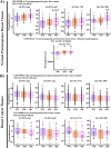
Fig 1
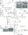
Fig 1. Akt phosphorylation landscape evaluated through gene expression signatures in non-tumoral or tumor breast tissues of women with different BMI.
Bioinformatic analysis of the mRNA gene expression signatures that mirrored the phosphorylation of Akt in Ser 473 and Thr 308 in public datasets recovered from the GEO database in breast tissue of normal-weight (NW), overweight (OW) and obese (OB) women. A) RNA gene signature scores describing Akt activation (Akt sig 1 –in-house signature- and sig 2) or Akt phosphorylation in non-tumoral breast tissue from premenopausal women. B) RNA gene signature scores describing Akt activation (Akt sig 1–in-house signature- and sig 2) or Akt phosphorylation in tumors of pre and postmenopausal women (upper panel) or breast tumors from premenopausal women (lower panel).*<0.05 **<0.005 comparisons between groups.
Fig 2
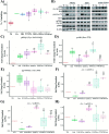
Fig 2. Effect of human sera on MCF-7 line.
A) Viability of MCF-7 cells stimulated with sera from women with different BMI. B) Western Blot of the time course of IR-β/Akt/p70S6K pathway activation under stimulation with sera from women with different BMI. C) Group analysis of densitometric quantification stratified by sera condition of pIR-β (Tyr 1162–1163). D) Group analysis of densitometric quantification stratified by sera condition of pAkt (Ser 473). E) Group analysis of densitometric quantification stratified by sera condition of pp70S6K (Thr 389). F) Western Blot of Inhibition of Akt activation by stimulation with sera from women with different BMI. G) Group analysis of densitometric quantification of Inhibition of Akt activation by stimulation with sera from women with different BMI. For viability assay, the MCF-7 cells were treated with 10% inactivated fetal bovine serum (Ctr), 5% FBS or 5% sera from normal-weight premenopausal women (NWSPre) or sera from obese premenopausal women (OSPre). T0 corresponds to the viability at the time the different sera were added, and normalized against Ctr. For Western Blot, the cells were stimulated for 10 min with recombinant human insulin (Ins) (0.5 UI/ml) (positive control for activation), 5% NWSPre or 5% OSPre. T0 represents the phosphorylation level before stimulation with Ins or human sera. All experiments were carried out in triplicate (n = 9) for each serum evaluated. Comparison of means ** P <0.005, * P <0.05. ** Ctr or T0, ## 5% FBS or Ins, ++ NWSPre, @@ Without inhibitors.
Fig 3
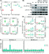
Fig 3. Effect on the viability and activation of the IR-β/Akt/p70S6K pathway of MCF-7 cells by stimulation with human sera from normal-weight and obese women with different hormonal states.
A) Viability of MCF-7 cells exposed to sera from women with different BMIs and hormonal states. B) Western Blot of IR-β/Akt/p70S6K pathway members on MCF-7 cells exposed to sera from women with different BMIs and hormonal states. C) Group analysis of densitometric quantification stratified by serum condition of pIR-β (Tyr 1162–1163). D) Group analysis of densitometric quantification stratified by serum condition of pAkt (Ser 473). E) Group analysis of densitometric quantification stratified by serum condition of pp70S6K (Thr 389). F) Akt-1 expression levels on MCF-7 cells stimulated with human sera. G) Akt-2 expression levels on MCF-7 cells stimulated with human sera. H) Akt-3 expression levels on MCF-7 cells stimulated with human sera. For viability assay, the MCF-7 cells were treated with 10% inactivated fetal bovine serum (Ctr), 5% FBS, sera from normal-weight premenopausal women (NWSPre), sera from obese premenopausal women (OSPre), sera from obese postmenopausal women (OSPost) or sera from normal-weight postmenopausal women (NWSPost). For Western Blot test the cells were stimulated with recombinant human insulin (Ins) (0.5 UI/ml) (positive control for activation of the IR-β/Akt pathway) or 5% NWSPre, OSPre, OSPost or NWSPost. Labels of Western Blot as described in Fig 2. RT-PCR for Akt isoforms: Akt-1, Akt-2, Akt-3, and peptidylprolyl isomerase A (PPIA) as constitutive control. Western blot armed image. All experiments were carried out in triplicate (n = 9) for each serum evaluated. Comparison of means ** P <0.005, * P <0.05. ** Ctr or T0, ## 5% FBS or Ins, ++ NWSPre, $ $ OSPre, && OSPost.
Fig 4
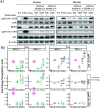
Fig 4. Effect of sera from Metformin-treated postmenopausal obese women on MCF-7 cells.
A) Viability of MCF-7 cells exposed to OSPost serum without insulin resistance (HOMA<3) and OSPost serum with insulin resistance (HOMA>3). B) Western Blot of MCF-7 cells exposed to OSPost serum without insulin resistance (HOMA<3) and OSPost serum with insulin resistance (HOMA>3). C) Western Blot’s Densitometry of pErk (Thr 202-Tyr204) and pAkt (Ser 473) of MCF-7 divided by the presence of insulin resistance. D) Akt Isoforms expression levels on MCF-7 cells stimulated with OSPost at 0 and 10 weeks of Metformin treatment. E) Determination of serum molecule levels in postmenopausal obese women treated at 0 and 10 weeks of treatment with Metformin. Labels of Western Blot as described in Fig 2. Labels of RT-PCR as described in Fig 3. Western blot armed image. The plotted data correspond to three independent experiments (n = 3) for each serum evaluated. Comparison of means ** P <0.005, * P <0.05. ** Ctr or T0, ## 5% FBS or Ins, !! W0, ^^ respect HOMA<3 vs HOMA>3.
Fig 5
Fig 5. Effect of sera from Metformin-treated postmenopausal obese women on NF-κB signaling in MCF-7 cells.
A) Western Blot of pp38 (Tyr 182), IKBα and pp65 (Ser 536) under stimulation with OSPost serum without insulin resistance (HOMA<3) and OSPost serum with insulin resistance (HOMA>3) at 10 and 30 min of stimulation. B) Group analysis of densitometric quantification of pp38 (Tyr 138), IKBα and pp65 (Ser 536) by stimulation with OSPost serum without insulin resistance (HOMA<3) and OSPost serum with insulin resistance (HOMA>3) at 10 min and 30 min. Labels of Western Blot as described in Fig 2. Western blot armed image. The plotted data correspond to three independent experiments (n = 3) for each serum evaluated. Comparison of means ** P <0.005, * P <0.05). ** T0, ¡¡ TNF-α, ## Ins, !! W0, ^^ respect HOMA<3 vs HOMA>3.
Fig 6
Fig 6. Schematic representation of the activation of the IR-β/Akt/p70S6K pathway in MCF-7 cells exposed to human sera with different metabolic characteristics and effect of Metformin treatment in obese patients.

References

    1. Soto T and Lagos E. Obesidad y cáncer: un enfoque epidemiológico. Revista Médica de Costa Rica y Centroamérica (2009) LXVI(587), 27–32.
    1. Barone I, Giordano C, Bonofiglio D, Andò S, Catalano S. The weight of obesity in breast cancer progression and metastasis: Clinical and molecular perspectives. Semin Cancer Biol. 2020. Feb;60:274–284. doi: 10.1016/j.semcancer.2019.09.001 - DOI - PubMed
    1. Aguilar MJ, Neri M, Padilla CA, Pimentel ML, García A, Muy N. Sobrepeso/obesidad en mujeres y su implicación en el cáncer de mama; edad de diagnóstico. Nutr Hosp (2012) 27(5):1643–1647. 10.3305/nh.2012.27.5.5998 - DOI - PubMed
    1. GLOBOCAN (2018) https://gco.iarc.fr/
    1. Bowers L, Cavazos D, Maximo I, Brenner A, Hursting S, deGrafferenried L. Obesity enhances nongenomic estrogen receptor crosstalk with the PI3K/Akt and MAPK pathways to promote in vitro measures of breast cancer progression. Breast Cancer Research (2013) 15:R59. doi: 10.1186/bcr3453 - DOI - PMC - PubMed

Publication types