. 2022 Apr 12;22(1):67.
doi: 10.1186/s12880-022-00792-8.

A novel technique using ultrasonography in upper airway management after anterior cervical decompression and fusion

Affiliations

A novel technique using ultrasonography in upper airway management after anterior cervical decompression and fusion

Shizumasa Murata et al. BMC Med Imaging. .

Abstract

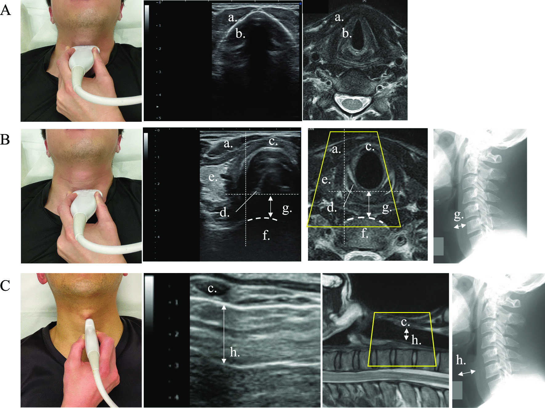
Background: Airway complications are the most serious complications after anterior cervical decompression and fusion (ACDF) and can have devastating consequences if their detection and intervention are delayed. Plain radiography is useful for predicting the risk of dyspnea by permitting the comparison of the prevertebral soft tissue (PST) thickness before and after surgery. However, it entails frequent radiation exposure and is inconvenient. Therefore, we aimed to overcome these problems by using ultrasonography to evaluate the PST and upper airway after ACDF and investigate the compatibility between X-ray and ultrasonography for PST evaluation.

Methods: We included 11 radiculopathy/myelopathy patients who underwent ACDF involving C5/6, C6/7, or both segments. The condition of the PST and upper airway was evaluated over 14 days. The Bland-Altman method was used to evaluate the degree of agreement between the PST values obtained using radiography versus ultrasonography. The Pearson correlation coefficient was used to determine the relationship between the PST measurement methods. Single-level and double-level ACDF were performed in 8 and 3 cases, respectively.

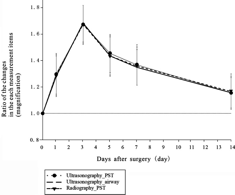
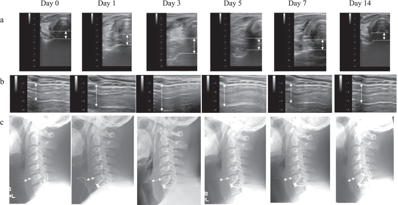
Results: PST and upper airway thickness peaked on postoperative day 3, with no airway complications. The Bland-Altman bias was within the prespecified clinically nonsignificant range: 0.13 ± 0.36 mm (95% confidence interval 0.04-0.22 mm). Ultrasonography effectively captured post-ACDF changes in the PST and upper airway thickness and detected airway edema.

Conclusions: Ultrasonography can help in the continuous assessment of the PST and the upper airway as it is simple and has no risk of radiation exposure risk. Therefore, ultrasonography is more clinically useful to evaluate the PST than radiography from the viewpoint of invasiveness and convenience.

Keywords: Airway complication; Anterior cervical decompression and fusion; Cervical spine; Prevertebral soft tissue evaluation; Spine surgery; Ultrasonography.


Conflict of interest statement

The authors declare that they have no competing interests.

Figures

Fig. 1
Fig. 1
Illustration of the anatomical features around the cervical spine Left (A): bones around cervical spine at C5/6. Right (B): soft tissues. a: hyoid bone, b: thyroid cartilage, c: cricoid cartilage
Fig. 2
Fig. 2
Macro photograph, ultrasound image, and magnetic resonance image A, B: Thyroid cartilage and cricoid cartilage in the transverse plane. C: Cricoid cartilage and tracheal cartilage in the sagittal plane a: sternohyoid muscle, b: thyroid cartilage, c: cricoid cartilage, d; arytenoid cartilage, e: thyroid gland, f: C6 vertebral body, g: the thickness of the prevertebral soft tissue, h: upper airway
Fig. 3
Fig. 3
Thickness of the PST and airway in the pilot study. The data are expressed as mean ± standard deviation. PST: prevertebral soft tissue
Fig. 4
Fig. 4
Statistical analysis results of the pilot study. a: Bland–Altman plot for mean thickness of prevertebral soft tissue (PST) obtained using radiography versus ultrasonography. b: Relationship between PST thickness obtained using radiography (R-PST) and ultrasonography (U-PST) (r = 0.9, P < 0.0001)
Fig. 5
Fig. 5
Postoperative course of the prevertebral soft tissue (PST) and upper airway in the representative case. a: Postoperative changes in the thickness of the PST measured via ultrasonography. b: Postoperative changes in the upper airway measured via ultrasonography. c: Postoperative changes in the thickness of the PST measured via lateral radiography of the cervical spine
Fig. 6
Fig. 6
Summary of the information presented in Fig. 4. It can be seen that ultrasonography (upper airway) or both lateral radiography of the cervical spine and ultrasonography (prevertebral soft tissue ) followed a similar course. PST: prevertebral soft tissue

References

    1. Fountas KN, Kapsalaki EZ, Nikolakakos LG, Smisson HF, Johnston KW, Grigorian AA, et al. Anterior cervical discectomy and fusion associated complications. Spine. 2007;32:2310–7. doi: 10.1097/BRS.0b013e318154c57e. - DOI - PubMed
    1. Debkowska MP, Butterworth JF, Moore JE, Kang S, Appelbaum EN, Zuelzer WA. Acute post-operative airway complications following anterior cervical spine surgery and the role for cricothyrotomy. J Spine Surg. 2019;5:142–54. doi: 10.21037/jss.2019.03.01. - DOI - PMC - PubMed
    1. Li H, Huang Y, Shen B, Ba Z, Wu D. Multivariate analysis of airway obstruction and reintubation after anterior cervical surgery: a retrospective cohort study of 774 patients. Int J Surg. 2017;41:28–33. doi: 10.1016/j.ijsu.2017.03.014. - DOI - PubMed
    1. Suk KS, Kim KT, Lee SH, Park SW. Prevertebral soft tissue swelling after anterior cervical discectomy and fusion with plate fixation. Int Orthop. 2006;30:290–4. doi: 10.1007/s00264-005-0072-9. - DOI - PMC - PubMed
    1. Lim S, Kesavabhotla K, Cybulski GR, Dahdaleh NS, Smith ZA. Predictors for airway complications following single- and multilevel anterior cervical discectomy and fusion. Spine. 2017;42:379–84. doi: 10.1097/BRS.0000000000001737. - DOI - PubMed