

Case Reports
doi: 10.7759/cureus.22986.
eCollection 2022 Mar.
Spontaneous Rupture of the Renal Pelvis Due to Extrinsic Obstruction by Metastatic Retroperitoneal Lymphadenopathy
Affiliations
- PMID: 35415055
- PMCID: PMC8994016
- DOI: 10.7759/cureus.22986
Item in Clipboard
Case Reports
Spontaneous Rupture of the Renal Pelvis Due to Extrinsic Obstruction by Metastatic Retroperitoneal Lymphadenopathy
Cureus.
.
Abstract
Spontaneous rupture of the renal pelvis due to metastatic disease is a rare complication. Renal pelvis rupture often goes undiagnosed in cases of non-traumatic origin due to its vague abdominal and flank symptoms. We present a case of an 81-year-old male with primary non-small cell lung cancer who had renal pelvis rupture due to extrinsic compression of the ureter by retroperitoneal lymphadenopathy secondary to metastatic disease.
Keywords: forniceal rupture; renal pelvis; retroperitoneal lymphadenopathy; spontaneous rupture; ureteric obstruction.
Copyright © 2022, Fink et al.
Conflict of interest statement
The authors have declared that no competing interests exist.
Figures
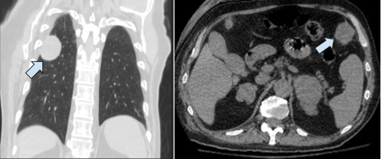
CT cross-sectional imaging in the coronal plane (left panel) of a well-circumscribed peripheral nodularity measuring 4.3 x 3.3 x 4 cm (arrow). Cross-sectional CT imaging in transverse section (right panel) of an enlarged nodularity in left anterior upper abdomen measuring 3.5 x 3.1 cm (arrow).
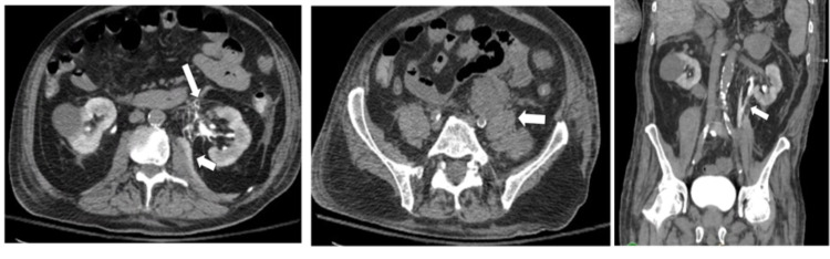
CT abdomen and pelvis (CTAP) in transverse plane (left panel) showing extravasation of contrast at the anteromedial aspect of the left renal pelvis (long arrow) with extension along the left iliopsoas muscle (short arrow). Transverse plane CTAP without contrast (middle panel) displaying compression of the left distal ureter by enlarged retroperitoneal lymph nodes measuring 10 x 4 cm (arrow). Coronal plane view of CTAP without contrast (right panel) showing extravasation of retained contrast medial to the renal pelvis outlining the iliopsoas (arrow).
Similar articles
-
Causes of renal forniceal rupture.BJU Int. 2011 Dec;108(11):1909-11; discussion 1912. doi: 10.1111/j.1464-410X.2011.10164.x. Epub 2011 Jul 8. BJU Int. 2011. PMID: 21736690
-
Spontaneous bilateral renal pelvis rupture during CT in the absence of urinary tract obstruction: case report.BMC Urol. 2020 Jul 13;20(1):98. doi: 10.1186/s12894-020-00669-4. BMC Urol. 2020. PMID: 32660460 Free PMC article.
-
Urothelial carcinoma of the proximal ureter revealed by spontaneous forniceal rupture: A case report.Urol Case Rep. 2022 May 2;43:102098. doi: 10.1016/j.eucr.2022.102098. eCollection 2022 Jul. Urol Case Rep. 2022. PMID: 35573088 Free PMC article.
-
[Spontaneous rupture of the ureter caused by metastatic ureteric tumor: a case report].Hinyokika Kiyo. 1995 Jan;41(1):57-60. Hinyokika Kiyo. 1995. PMID: 7900570 Review. Japanese.
-
Spontaneous renal rupture during pregnancy.Mayo Clin Proc. 1991 Feb;66(2):179-82. doi: 10.1016/s0025-6196(12)60490-x. Mayo Clin Proc. 1991. PMID: 1994136 Review.
References
-
- Causes of renal forniceal rupture. Gershman B, Kulkarni N, Sahani DV, Eisner BH. BJU Int. 2011;108:1909–1911. - PubMed
-
- Lescay HA, Jiang J, Tuma F. Treasure Island, FL: StatPearls Publishing; 2021. Anatomy, abdomen and pelvis, ureter. - PubMed
-
- Urinary system: ureter. http://www.histology.leeds.ac.uk/urinary/ureter.php 2003
-
- Evaluation of the tensile strength of the human ureter - preliminary results. Shilo Y, Pichamuthu JE, Averch TD, Vorp DA. J Endourol. 2014;28:1470–1473. - PubMed
Publication types
LinkOut - more resources
Full Text Sources