

Nipple Adenoma: Case Report of a Rare Entity
- PMID: 35415057
- PMCID: PMC8992876
- DOI: 10.7759/cureus.22996
Nipple Adenoma: Case Report of a Rare Entity
Abstract
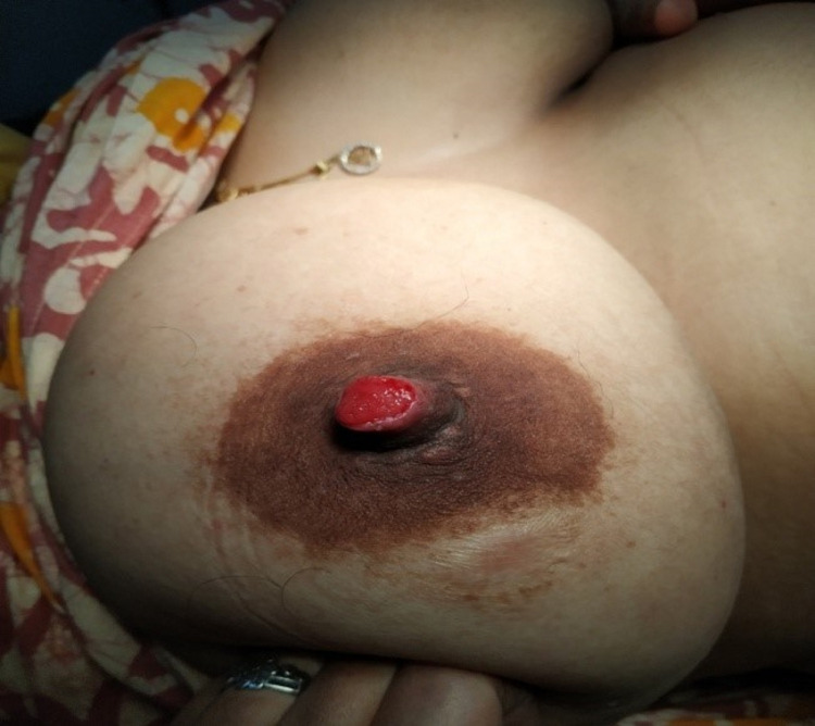
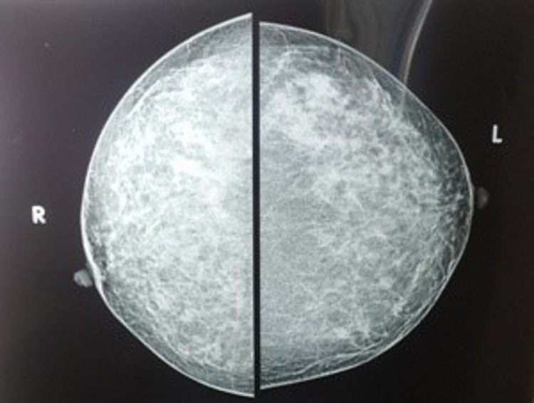
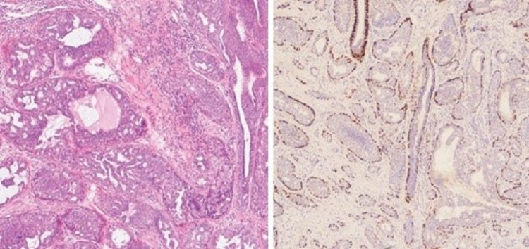
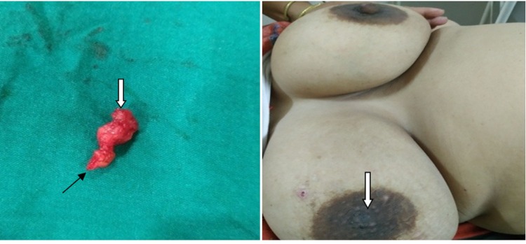
A nipple adenoma is a rare benign breast tumor. The commonest presentation of this rare entity is nipple erosion, serosanguinous discharge, induration, or tumor formation at the nipple. It often mimics malignant breast lesions or nipple eczema and is mistaken for Paget's disease of the nipple or dermatological pathology. It may be misdiagnosed pathologically as ductal carcinoma of the breast. This may cause a diagnostic delay or a faulty diagnosis. Treatment is the excision of the tumor with or without nipple excision. Here, we report a case of nipple adenoma that projected out of the nipple along with nipple erosion, serosanguinous discharge, and occasional bleeding from the adenoma. A 37- year-old woman presented with a tumor on her right nipple for eight months, with the erosion of the nipple and serosanguinous discharge. The patient gave a history of a small amount of bleeding occasionally. Axilla was normal. The patient was advised to have a mammosonography. It showed an oval-shaped, well-demarcated, hypoechoic, uniformly solid nodule in the right nipple. There was no microcalcification seen on mammography. A punch biopsy was done to establish the diagnosis. It showed ductal hyperplasia and papillary proliferation of glandular structures suggestive of nipple adenoma. Complete resection of the tumor with partial excision of the nipple was done with a satisfactory cosmetic result. Though very uncommon, the possibility of nipple adenoma should be thought of when a patient presents with nipple erosion and discharge with or without a clinically obvious tumor. Timely diagnosis with histopathological correlation is important since it allows for less invasive surgical methods. In our case, we could attain a cosmetically satisfactory outcome without a remnant tumor. Paget's disease of the nipple also has a similar clinical presentation, and it is a premalignant condition. The objective of presenting this case is to highlight the possibility of this rare benign condition, which may be easily missed clinically and also demands careful histopathological examination for its correct diagnosis.
Keywords: erosion; lactiferous duct; myoepithelial cells; nipple adenoma; paget’s disease.
Copyright © 2022, Dalal et al.
Conflict of interest statement
The authors have declared that no competing interests exist.
Figures
Similar articles
-
Nipple adenoma in a female patient presenting with persistent erythema of the right nipple skin: case report, review of the literature, clinical implications, and relevancy to health care providers who evaluate and treat patients with dermatologic conditions of the breast skin.BMC Dermatol. 2016 May 20;16(1):4. doi: 10.1186/s12895-016-0041-6. BMC Dermatol. 2016. PMID: 27206635 Free PMC article.
-
Unusual presentation of intraductal papilloma on the nipple: A case report.Int J Surg Case Rep. 2024 Apr;117:109483. doi: 10.1016/j.ijscr.2024.109483. Epub 2024 Mar 8. Int J Surg Case Rep. 2024. PMID: 38493616 Free PMC article.
-
Adenoma of the Nipple: A Case Report.Cureus. 2024 Jul 8;16(7):e64105. doi: 10.7759/cureus.64105. eCollection 2024 Jul. Cureus. 2024. PMID: 39114234 Free PMC article.
-
Florid papillomatosis of the nipple: a rare presentation and review of the literature.Breast Dis. 2015;35(2):153-6. doi: 10.3233/BD-150397. Breast Dis. 2015. PMID: 25585841 Review.
-
Adenoma of the nipple in an adolescent.Breast Cancer. 2002;9(3):254-6. doi: 10.1007/BF02967598. Breast Cancer. 2002. PMID: 12185338 Review.
Cited by
-
Mammary Paget's Disease Mimicking Benign and Malignant Dermatological Conditions: Clinical Challenges and Diagnostic Considerations.Cureus. 2024 Jul 25;16(7):e65378. doi: 10.7759/cureus.65378. eCollection 2024 Jul. Cureus. 2024. PMID: 39188449 Free PMC article. Review.
References
-
- Nipple adenoma: a review of the literature. Tatterton MR, Fiddes R. Ann Breast Surg. 2019;3:29.
-
- Nipple adenoma in an adolescent. Tao W, Kai F, Yue Hua L. Pediatr Dermatol. 2010;27:399–401. - PubMed
Publication types
LinkOut - more resources
Full Text Sources
Molecular Biology Databases