. 2022 Mar 28:33:153-161.
doi: 10.1016/j.jot.2022.03.004. eCollection 2022 Mar.

Biodegradable magnesium implant enhances angiogenesis and alleviates medication-related osteonecrosis of the jaw in rats

Affiliations

Biodegradable magnesium implant enhances angiogenesis and alleviates medication-related osteonecrosis of the jaw in rats

Wang-Yong Zhu et al. J Orthop Translat. .

Abstract

Background: Medication-related osteonecrosis of the jaw (MRONJ) is a serious complication associated with antiresorptive and antiangiogenic medications, of which impaired angiogenesis is a key pathological alteration. Since Magnesium (Mg)-based implants possess proangiogenic effects, we hypothesized that the biodegradable Mg implant could alleviate the development of MRONJ via enhancing angiogenesis.

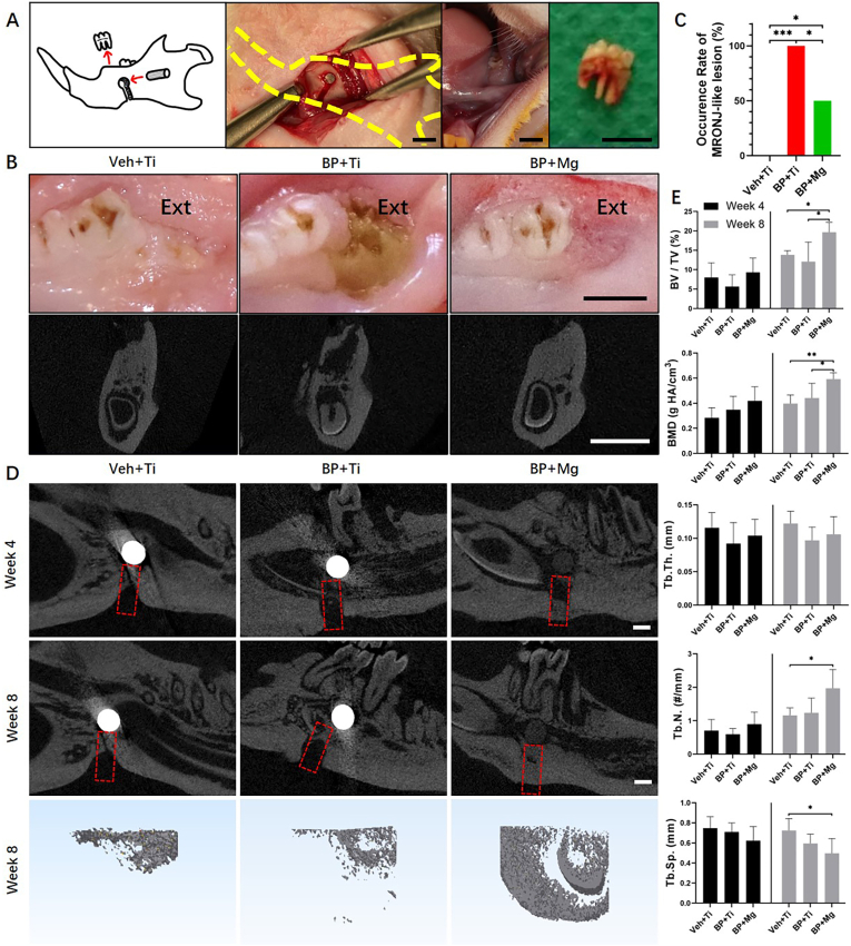
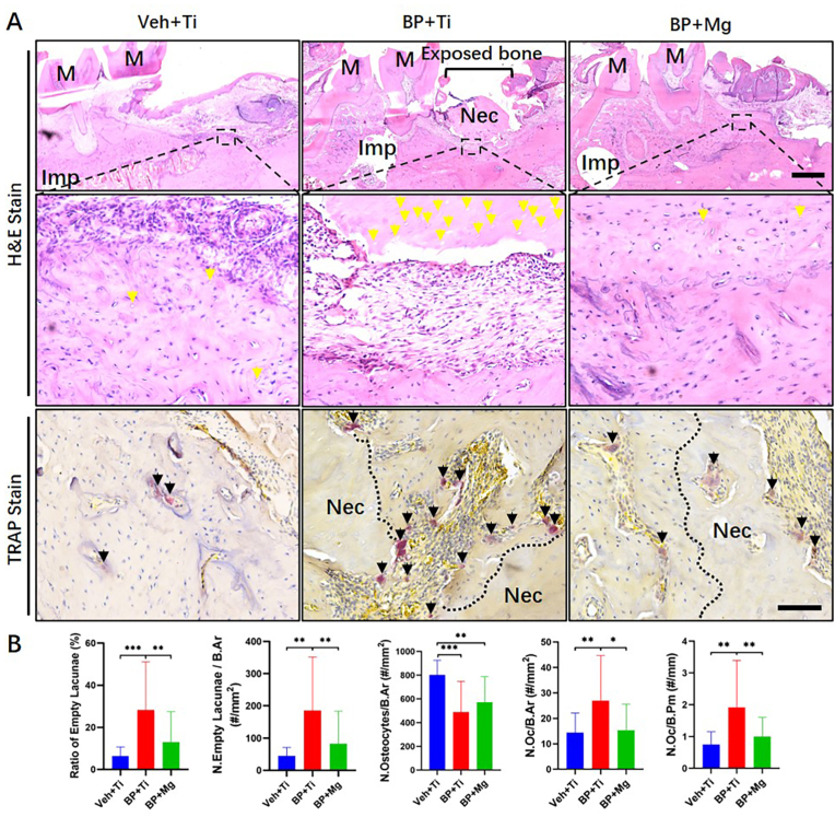
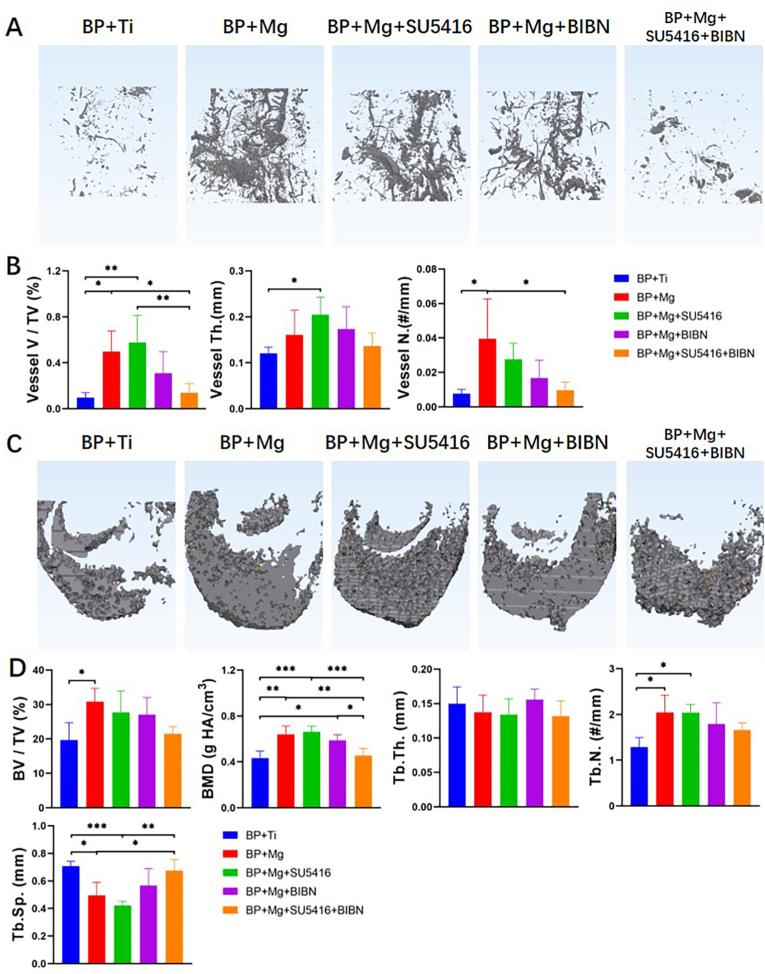
Methods: MRONJ model was established and divided into the Veh ​+ ​Ti group (Vehicle-treated rat, with Titanium (Ti) implant), BP ​+ ​Ti group (Bisphosphonate (BP)-treated rat, with Ti implant), BP ​+ ​Mg group (BP-treated rat, with Mg implant), BP ​+ ​Mg ​+ ​SU5416 group (BP-treated rat, with Mg implant and vascular endothelial growth factor (VEGF) receptor-2 inhibitor), BP ​+ ​Mg ​+ ​BIBN group (BP-treated rat, with Mg implant and calcitonin gene-related peptide (CGRP) receptor antagonist), and BP ​+ ​Mg ​+ ​SU5416+BIBN group (BP-treated rat, with Mg implant and VEGF receptor-2 inhibitor and CGRP receptor antagonist). The occurrence of MRONJ, alveolar bone necrosis, new bone formation and vessel formation were assessed by histomorphometry, immunohistochemistry, and micro-CT analysis.

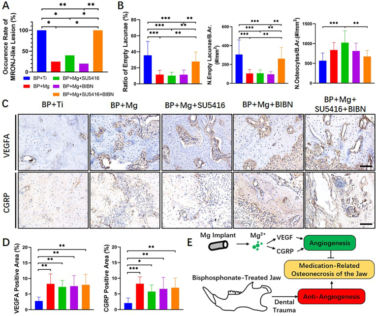
Results: Eight weeks after surgery, the BP ​+ ​Mg group had significantly reduced occurrence of MRONJ-like lesion and histological osteonecrosis, increased bone microstructural parameters, and increased expressions of VEGFA and CGRP, than the BP ​+ ​Ti group. By simultaneously blocking VEGF receptor-2 and CGRP receptor, the vessel volume and new bone formation in the BP ​+ ​Mg group were significantly decreased, meanwhile the occurrence of MRONJ-like lesion and histological bone necrosis were significantly increased.

Conclusion: Biodegradable Mg implant could alleviate the development of MRONJ-like lesion, possibly via upregulating VEGF- and CGRP-mediated angiogenesis. Mg-based implants have the translational potential to be developed as a novel internal fixation device for patients with the risk of MRONJ.

The translational potential of this article: This work reports a biodegradable Mg implant which ameliorates the development of MRONJ-like lesions possibly due to its angiogenic property. Mg-based implants have the potential to be developed as a novel internal fixation device for patients at the risk of MRONJ.

Keywords: Angiogenesis; Biodegradable implants; Bisphosphonate; Magnesium; Medication-related osteonecrosis of the jaw.


Conflict of interest statement

The author(s) have no conflicts of interest relevant to this article.

Figures

Fig. 1
Fig. 1
BP ​+ ​Mg group presented alleviated MRONJ occurrence and enhanced bone defect repair as compared to the BP ​+ ​Ti group (A) Schematic and intraoperative images of surgical procedures for establishing the MRONJ model with implant fixation, bone defect creation and dental extraction. A Mg rod (with a purity of 99.99%, 3 ​mm in length, and 1.3 ​mm in diameter) or a Ti rod (pure titanium, the same size with Mg rod) was implanted into the mandible; a 1 ​mm-wide bone defect was cut inferior to the implantation site; the mandibular 1st molar was extracted. Scale bar: 3 ​mm (B) Representative photos and coronal micro-CT images of extraction sites (Ext) at 8 weeks after surgery, showing complete healing mucosae (Left and Right) and a MRONJ-like lesion with exposure of necrotic bone and sequestrum (Middle). Scale bar: 3 ​mm (C) Occurrence rate of MRONJ-like lesion in three groups at 8 weeks after surgery. Veh ​+ ​Ti group n ​= ​5, BP ​+ ​Ti group n ​= ​9, and BP ​+ ​Mg group n ​= ​10, by Chi-square test (D) Representative sagittal micro-CT images and 3D reconstructed coronal images of mandibular bone defect (red dotted box) in three groups, 4 weeks and 8 weeks postoperatively. Scale bar: 1 ​mm (E) Bone micro-architectural morphometric assessment on the newly formed bone in the defect at 4 weeks and 8 weeks after surgery by micro-CT. Column and bar indicate mean and SD, respectively. n ​= ​5 for each group, by One-way ANOVA with Bonferroni post hoc test. ∗, p ​< ​0.05. ∗∗, p ​< ​0.01. ∗∗∗, p ​< ​0.001.
Fig. 2
Fig. 2
Comparison of histological osteonecrosis and TRAP-positive osteoclasts at MRONJ-like lesions (A) Representative H&E staining and TRAP staining images of extracted tooth socket 8 weeks after removal in three groups. Imp, implant. M, molar. Nec, necrotic bone. Yellow arrow points the empty lacunae. Black arrow points the TRAP-positive stained osteoclast. Scale bar in H&E stain: 1 ​mm. Scale bar in TRAP stain: 100 ​μm (B) Histomorphometric assessments of H&E staining on the degree of alveolar bone necrosis, and TRAP staining on the number of TRAP-positive cells among groups. Column and bar indicate mean and SD, respectively. Veh ​+ ​Ti group n ​= ​5, BP ​+ ​Ti group n ​= ​9, and BP ​+ ​Mg group n ​= ​10, by One-way ANOVA with Bonferroni post hoc test. ∗, p ​< ​0.05. ∗∗, p ​< ​0.01. ∗∗∗, p ​< ​0.001.
Fig. 3
Fig. 3
VEGFA and CGRP expression compared among Veh ​+ ​Ti, BP ​+ ​Ti and BP ​+ ​Mg groups (A) Representative images of IHC staining of CD31, VEGFA and CGRP in three groups, respectively. Black arrow points the positive stained cell. Scale bar: 400 ​μm (B) Corresponding quantitative data of VEGFA and CGRP. Column and bar indicate mean and SD, respectively. n ​= ​5 for each group, by One-way ANOVA with Bonferroni post hoc test. ∗∗∗, p ​< ​0.001.
Fig. 4
Fig. 4
Mg enhanced angiogenesis and coupled osteogenesis in MRONJ, which could be inhibited by blocking VEGF receptor-2 and CGRP receptor simultaneously, as indicated by micro-CT based angiography and bone histomorphometry analysis (A) Representative 3D reconstructed images of micro-CT based angiography showing the differences of vessel volume compared among the BP ​+ ​Ti, BP ​+ ​Mg, and BP ​+ ​Mg ​+ ​inhibitor/antagonist groups at 8 weeks after surgery. Scale bar: 5 ​mm (B) Quantitative analysis of angiography in different groups at 8 weeks after surgery (C) Representative 3D reconstructed images showing the differences of newly formed bone in the defect compared among groups at 8 weeks after surgery. Scale bar: 1 ​mm (D) Quantitative analysis of newly formed bone in different groups at 8 weeks after surgery. SU5416, VEGF receptor-2 inhibitor; BIBN, CGRP receptor antagonist. Column and bar indicate mean and SD, respectively. n ​= ​4–5 for each group, by One-way ANOVA with Bonferroni post hoc test. ∗, p ​< ​0.05; ∗∗, p ​< ​0.01; ∗∗∗, p ​< ​0.001.
Fig. 5
Fig. 5
Mg-alleviated MRONJ-like lesion development, possibly due to angiogenesis and osteogenesis (A) Occurrence rate of MRONJ-like lesion in the BP ​+ ​Ti, BP ​+ ​Mg, and BP ​+ ​Mg ​+ ​inhibitor/antagonist groups at 8 weeks after surgery (B) Histomorphometric assessments on the degree of alveolar bone necrosis among groups (H&E staining) (C) IHC staining of VEGFA (brown) and CGRP (brown) in the alveolar bone region in each group at 8 weeks after surgery. Scale bar: 100 ​μm (D) Corresponding quantitative data from (C). Column and bar indicate mean and SD, respectively. n ​= ​4–5 for each group, by Chi-square test (A), and One-way ANOVA with Bonferroni post hoc test (B and D). ∗∗, p ​< ​0.01; ∗∗∗, p ​< ​0.001 (E) Illustration of the underlying mechanism of biodegradable magnesium implant on attenuating the development of MRONJ via VEGF- and CGRP-mediated angiogenesis.

References

    1. Ruggiero S.L., Dodson T.B., Fantasia J., Goodday R., Aghaloo T., Mehrotra B., et al. American Association of Oral and Maxillofacial Surgeons position paper on medication-related osteonecrosis of the jaw--2014 update. J Oral Maxillofac Surg. 2014;72(10):1938–1956. - PubMed
    1. Otto S., Pautke C., Van den Wyngaert T., Niepel D., Schiodt M. Medication-related osteonecrosis of the jaw: prevention, diagnosis and management in patients with cancer and bone metastases. Cancer Treat Rev. 2018;69:177–187. - PubMed
    1. Coleman R., Woodward E., Brown J., Cameron D., Bell R., Dodwell D., et al. Safety of zoledronic acid and incidence of osteonecrosis of the jaw (ONJ) during adjuvant therapy in a randomised phase III trial (AZURE: BIG 01-04) for women with stage II/III breast cancer. Breast Cancer Res Treat. 2011;127(2):429–438. [eng] - PubMed
    1. Mauri D., Valachis A., Polyzos I.P., Polyzos N.P., Kamposioras K., Pesce L.L. Osteonecrosis of the jaw and use of bisphosphonates in adjuvant breast cancer treatment: a meta-analysis. Breast Cancer Res Treat. 2009;116(3):433–439. [eng] - PubMed
    1. Scagliotti G.V., Hirsh V., Siena S., Henry D.H., Woll P.J., Manegold C., et al. Overall survival improvement in patients with lung cancer and bone metastases treated with denosumab versus zoledronic acid: subgroup analysis from a randomized phase 3 study. J Thorac Oncol : Off Publ Int Assoc Stud Lung Cancer. 2012;7(12):1823–1829. [eng] - PubMed