. 2022 Feb 25:4:100091.
doi: 10.1016/j.fochms.2022.100091. eCollection 2022 Jul 30.

FIP-nha, a fungal immunomodulatory protein from Nectria haematococca, induces apoptosis and autophagy in human gastric cancer cells via blocking the EGFR-mediated STAT3/Akt signaling pathway

Affiliations

FIP-nha, a fungal immunomodulatory protein from Nectria haematococca, induces apoptosis and autophagy in human gastric cancer cells via blocking the EGFR-mediated STAT3/Akt signaling pathway

Shu-Ying Li et al. Food Chem (Oxf). .

Abstract

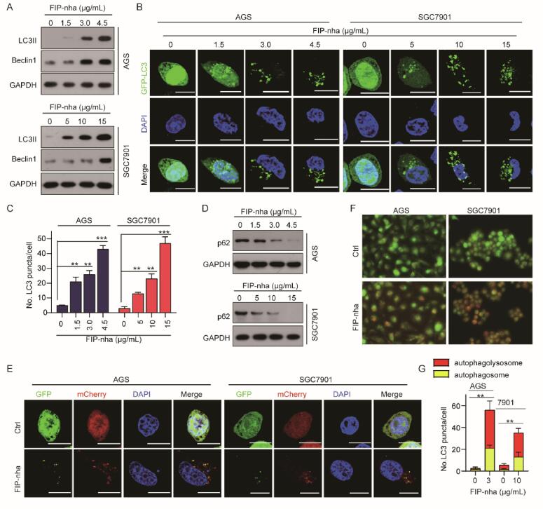
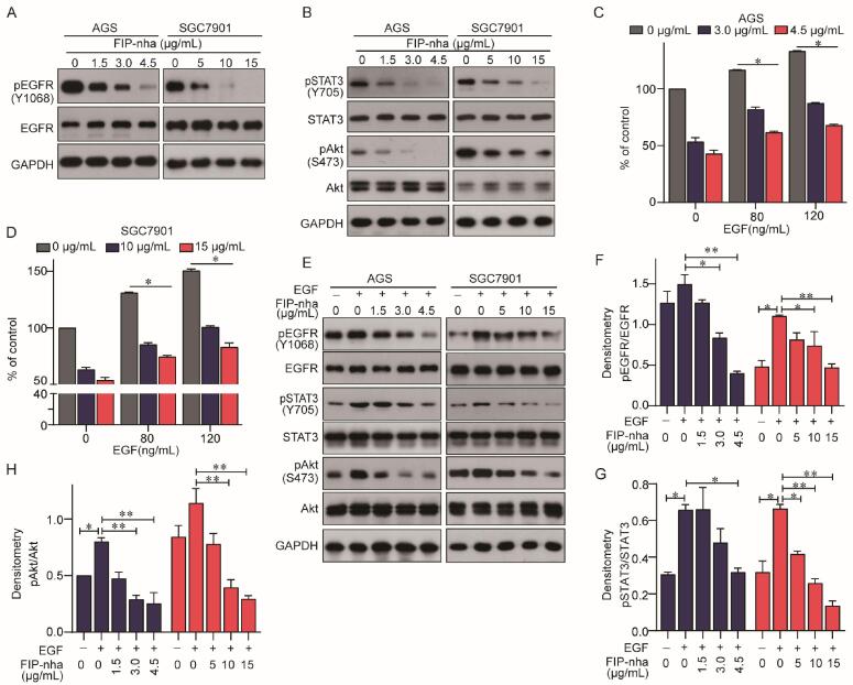
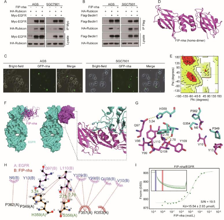
FIP-nha, a fungal immunomodulatory protein from Nectria haematococca, has been demonstrated a broad spectrum of antitumor activity and cell selectivity against human cancers in our previous study. However, the effect and mechanism of FIP-nha on gastric cancer remains unclear. In this study, we systematically observed the cytotoxicity, biological effect, regulatory mechanism and interaction target of FIP-nha on human gastric cancer cell lines, AGS and SGC7901. Our results demonstrated that FIP-nha inhibited the growth of AGS and SGC7901 cells in a dose-dependent manner and exerted proapoptotic effects on both cells as confirmed by flow cytometry, DAPI staining and western blot analysis. Additionally, the exposure of AGS and SGC7901 to FIP-nha induced autophagy as indicated by western blot analysis, GFP-LC3 and mCherry-GFP-LC3 transfection and acridine orange staining. Furthermore, we found that FIP-nha decreased the phosphorylation of EGFR, STAT3 and Akt and inhibited activation effect of ligand factor EGF to EGFR and its downstream signal molecule STAT3 and Akt. Finally, we proved that FIP-nha located on the surface of gastric cancer cells and bound directly to the transmembrane protein of EGFR by immunoprecipitation, cellular localization, molecular docking, microscale thermophoresis assay. The above findings indicated that FIP-nha inhibited the growth of gastric cancer and induced apoptosis and autophagy through competitively binding to EGFR with EGF to blocking the EGFR-mediated STAT3/Akt pathway. In summary, our study provided novel insights regarding the activity of FIP-nha against gastric cancer and contributed to the clinical application of FIP-nha as a potential chemotherapy drugs that targeted EGFR for human gastric cancer.

Keywords: Apoptosis; Autophagy; EGFR; FIP-nha; Gastric cancer; STAT3/Akt pathway.


Conflict of interest statement

The authors declare that they have no known competing financial interests or personal relationships that could have appeared to influence the work reported in this paper.

Figures

Fig. 1
Fig. 1
FIP-nha inhibited growth of GC cells. (A-C): The inhibitory effects of FIP-nha on AGS, SGC7901, and GES-1 cells analyzed by MTT assay. (D): The morphology images of AGS and SGC7901cells treated with FIP-nha at the indicated concentrations. (E, F): The cell viabilities of AGS and SGC7901 cells analyzed by MTS assay. (G, H): The colony formation assays of AGS and SGC7901 cells treated with FIP-nha atthe indicated concentrations. *, P < .05; **, P < .01.
Fig. 2
Fig. 2
FIP-nha induced apoptosis in GC cells. (A): AGS and SGC7901 cells were treated with increasing concentrations of FIP-nha for 24 h. Apoptotic induction was determined by flow cytometric analysis of Annexin V and PI-staining. The upper right quadrant (UR) represented late apoptotic cells stained with Annexin V and PI, and the lower right quadrant (LR) represented early apoptotic cells stained with Annexin V. (B): Thenuclear morphology evaluation ofAGS and SGC7901 cells, treated with the indicated concentrationof FIP-nha, was detected by DAPI staining. (C): AGS and SGC7901 cells were treated with increasing concentrations of FIP-nha for 24 h. Western blot was performed using antibodies indicated. GAPDH was used as the loading control.
Fig. 3
Fig. 3
FIP-nha induced autophagy in GC cells. (A, D): AGS and SGC7901 cells were treated with increasing concentrations of FIP-nha for 24 h. Western blot was performed using antibodies indicated. GAPDH was used as the loading control. (B): AGS and SGC7901 cells transfected with pQCXIP-GFP-LC3 plasmid were treated with increasing concentrations of FIP-nha for 24 h, and assessed by immunofluorescence analyses. Scale bar = 20 μm. (C): Graph shows quantification of LC3-positive punctate cells in (B). (E-F): AGS and SGC7901 cells transfected with pQCXIP-GFP-LC3 or pmCherry-GFP-LC3 plasmids were treated with indicated concentration of FIP-nha for 24 h, and assessed by immunofluorescence analyses. (G): Graph shows quantification of LC3-positive punctuate cells in (F). Scale bar = 20 μm. **, P < .01; ***, P < .001.
Fig. 4
Fig. 4
FIP-nha inhibited proliferation of GC cells through inactivating the EGFR/STAT3/Akt signaling. (A, B): AGS and SGC7901 cells were treated with increasing concentrations of FIP-nha for 24 h. Western blot assay was performed using antibodies indicated. (C, D): AGS and SGC7901 cells were treated with increasing concentrations of FIP-nha and EGF for 24 h. MTT assay was performed. (E): AGS and SGC7901 cells were treated with 120 ng/mL EGF and different concentrations of FIP-nha for 24 h. Western blot assay was performed using antibodies indicated.(F-H): Graph shows quantification of pEGFR/EGFR, pSTAT3/STAT3 and pAkt/Akt in (E). *, P < .05; **, P < .01.
Fig. 5
Fig. 5
FIP-nha activated autophagy in GC cells by directly binding to EGFR. (A,B): The effects of FIP-nha on the binding of EGFR to Rubicon or Rubicon to Beclin 1 were detected by immunoprecipitation. After cultured for 24 h, AGS and SGC7901 cells were treated with FIP-nha (AGS: 4.5 μg/mL, SGC 7901:15 μg/mL) for 24 h, respectively. Western blot assay was performed using antibodies indicated.(C): The localization analysis of FIP-nha. After cultured for 24 h, AGS and SGC7901 cells were treated with GFP-FIP-nha (AGS: 4.5 μg/mL, SGC 7901:15 μg/mL) for 24 h, respectively. The localization of GFP-FIP-nha was observed under fluorescence microscope, and the cell morphological changes were observed under microscopeat 20x lens. (D): FIP-nha structure was modeled by Modeller 9.20 software. FIP-nha mainly exists in the form of homodimer. Each monomer consists of an FNIII-like domain with an N-terminal a-helix and b-strand. (E): Analysis of pull conformation diagram. The proportion of amino acid residues in a reasonable range was more than 99% by Procheck Server evaluation.(F): The binding mode of FIP-nha docked into EGFR. EGFR is blue, FIP-nha is purple. (G, H) The interaction residues of FIP-nha with EGFR. (I): Binding affinity of FIP-nha with EGFR by MST in standard treated capillaries. (For interpretation of the references to colour in this figure legend, the reader is referred to the web version of this article.)

References

    1. Wang M., Zhao H., Hu J., Xu Z., Lin Y., Zhou S., Penicilazaphilone C. a New Azaphilone, Induces Apoptosis in Gastric Cancer by Blocking the Notch Signaling Pathway. Frontiers in Oncology. 2020;10:116. - PMC - PubMed
    1. Siegel R.L., Miller K.D., Jemal A. Cancer statistics, 2019. CA: A Cancer Journal for Clinicians. 2019;69(1):7–34. doi: 10.3322/caac.21551. Epub 2019 Jan 8. PMID: 30620402. - DOI - PubMed
    1. Chen W., Zheng R., Baade P.D., Zhang S., Zeng H., Bray F., Jemal A., Yu X.Q., He J. Cancer statistics in China, 2015. CA: A Cancer Journal for Clinicians. 2016;66(2):115–132. doi: 10.3322/caac.21338. Epub 2016 Jan 25. PMID: 26808342. - DOI - PubMed
    1. Zhang G., Li J., Li S., Wang Y. Exploring Spatial Trends and Influencing Factors for Gastric Cancer Based on Bayesian Statistics: A Case Study of Shanxi, China. International Journal of Environmental Research and Public Health. 2018;15(9):1824. - PMC - PubMed
    1. Giuppi M., La Salvia A., Evangelista J., Ghidini M. The Role and Expression of Angiogenesis-Related miRNAs in Gastric Cancer. Biology (Basel). 2021;10(2):146. - PMC - PubMed