. 2022 Apr;5(2):189-196.
doi: 10.1002/ame2.12228. Epub 2022 Apr 12.

A 3D-printable device allowing fast and reproducible longitudinal preparation of mouse intestines

Affiliations

A 3D-printable device allowing fast and reproducible longitudinal preparation of mouse intestines

Beckey DeLucia et al. Animal Model Exp Med. 2022 Apr.

Abstract

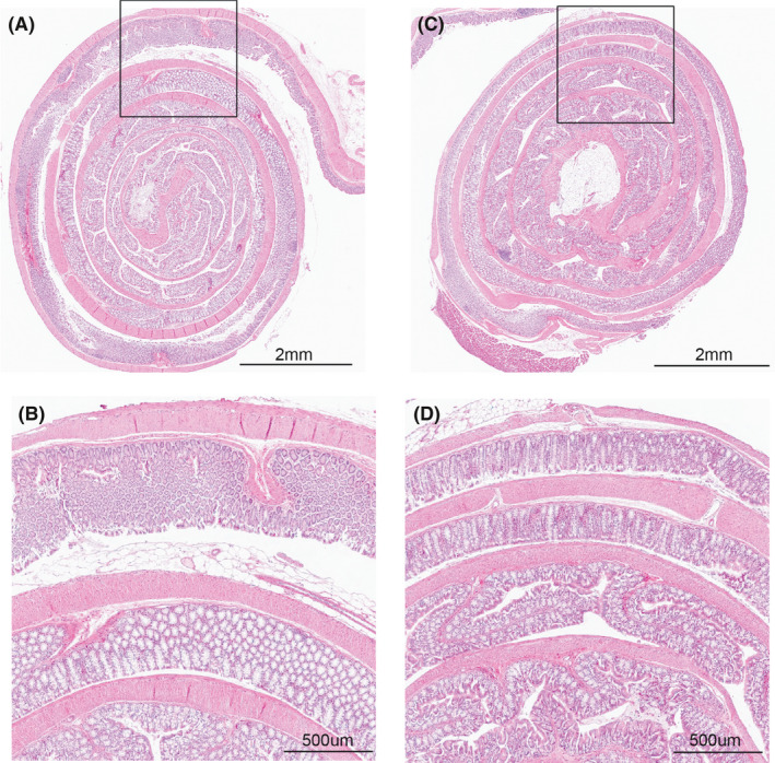
Accurate and reproducible analysis of murine small and large intestinal tissue is key for preclinical models involving intestinal pathology. Currently, there is no easily accessible, standardized method that allows researchers of different skill levels to consistently dissect intestines in a time-efficient manner. Here, we describe the design and use of the 3D-printed "Mouse Intestinal Slicing Tool" (MIST), which can be used to longitudinally dissect murine intestines for further analysis. We benchmarked the MIST against a commonly used procedure involving scissors to make a longitudinal cut along the intestines. Use of the MIST halved the time per mouse to prepare the intestines and outperformed alternative methods in smoothness of the cutting edge and overall reproducibility. By sharing the plans for printing the MIST, we hope to contribute a uniformly applicable method for saving time and increasing consistency in studies of the mouse gastrointestinal tract.

Keywords: adenoma; dissection; intestines; mice; printing, three-dimensional; reproducibility of results.


Conflict of interest statement

J.C. is a scientific advisor for Seed Health, Inc. Other authors do not have competing interests.

Figures

FIGURE 1
FIGURE 1
Schematic representations of the IPD and MIST. (A) Drawing of the IPD fully assembled with needles in place. (B) Cross‐section of the IPD showing the metal slanted cutting guides that are used to secure the intestinal segment onto the needle. (C) Front view of the MIST without needle, showing the forks that prevent the needle from rolling and the end bars that prevent the needle from sliding out. (D) Schematic cross‐section of the MIST technique. The intestinal tissue (orange) is loaded onto the needle (dark blue) and kept in place between the device and the wax surface (black) through the operator’s downward hand force. The tissue is cut with a scalpel along the slanted cutting guide
FIGURE 2
FIGURE 2
Overview of intestinal preparation methods. (A,B) Scissors method: the intestinal segment was cut open longitudinally (A), and the lumen was spread open using tweezers (B). (C–F) IPD method: loaded needles were placed into the IPD base (C) and secured in place by the lid (D). Metal cutting guides were inserted into the lid the tissue was cut with a scalpel (E). The IPD lid was removed, and the needles with cut intestines were transferred to the working surface (F). (G,H) Needle method: tissue was secured against the working surface by 2 fingers (G) and cut with a scalpel (H). (I,J) MIST method: tissue was secured against the working surface by uniform downward pressure from the MIST (I) and cut with a scalpel along the built‐in slanted cutting guide (J)
FIGURE 3
FIGURE 3
Comparison of preparation times across different methods. The preparation time for 4 intestinal segments per mouse was compared with the scissors method. Use of the IPD (*p = 0.0111), needle (**p = 0.0020), and MIST (**p = 0.0032) methods significantly improved prep times. Statistical analysis was performed using Brown‐Forsythe and Welch ANOVA tests uncorrected for multiple comparisons. N = 4–5 per group
FIGURE 4
FIGURE 4
The MIST method reproducibly yields the neatest cutting edges. (A–D) Representative photos of longitudinal intestine preparation using the scissors method (A), IPD method (B), needle method (C), and MIST method (D). (E) The neatness of the cutting edge was quantified by the ratio of the edge length to the total segment length, with a ratio of 1 representing a “perfect cut.” Compared with the scissors method, the cutting edge quality was significantly improved for the MIST (**p = 0.0054) and needle (*p = 0.0277). Statistical analysis was performed using Brown‐Forsythe and Welch ANOVA tests uncorrected for multiple comparisons. N = 4–5 per group
FIGURE 5
FIGURE 5
The MIST device allows for higher‐quality Swiss‐roll histology. (A–D) Representative hematoxylin and eosin (H&E)‐stained colonic Swiss‐roll preparations using the scissors method (A,B) and MIST method (C,D). The insets (B and D) give a higher‐magnification view. The even edge obtained by using the MIST (D) results in better and more consistent orientation of the tissue and visualization of the colonic crypts when compared with use of the scissors (B) for paraffin blocks that are sectioned at the same depth

References

    1. Kretser A, Murphy D, Bertuzzi S, et al. Scientific integrity principles and best practices: recommendations from a Scientific Integrity Consortium. Sci Eng Ethics. 2019;25:327‐355. - PMC - PubMed
    1. Begley CG, Buchan AM, Dirnagl U. Robust research: institutions must do their part for reproducibility. Nature. 2015;525:25‐27. - PubMed
    1. Casadevall A, Ellis LM, Davies EW, McFall‐Ngai M, Fang FC. A framework for improving the quality of research in the biological sciences. mBio. 2016;7:e01256‐16. - PMC - PubMed
    1. Freedman LP, Inglese J. The increasing urgency for standards in basic biologic research. Cancer Res. 2014;74:4024‐4029. - PMC - PubMed
    1. Taketo MM, Edelmann W. Mouse models of colon cancer. Gastroenterology. 2009;136:780‐798. - PubMed

Publication types