Case Reports
. 2022 Mar;56(2):152-156.
doi: 10.5152/j.aott.2022.21271.

Two-year follow-up after operative treatment of an osseous Bankart lesion with a flap-detached cartilage lesion of the glenoid: A case report

Affiliations
Case Reports

Two-year follow-up after operative treatment of an osseous Bankart lesion with a flap-detached cartilage lesion of the glenoid: A case report

Masayoshi Saito et al. Acta Orthop Traumatol Turc. 2022 Mar.

Abstract

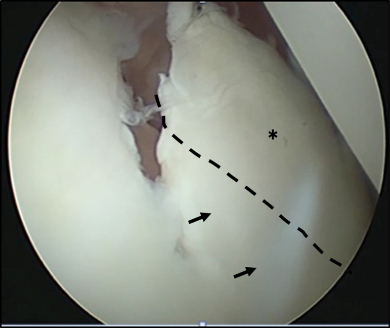
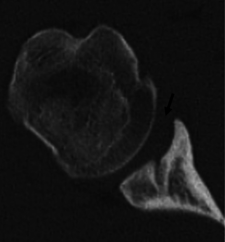
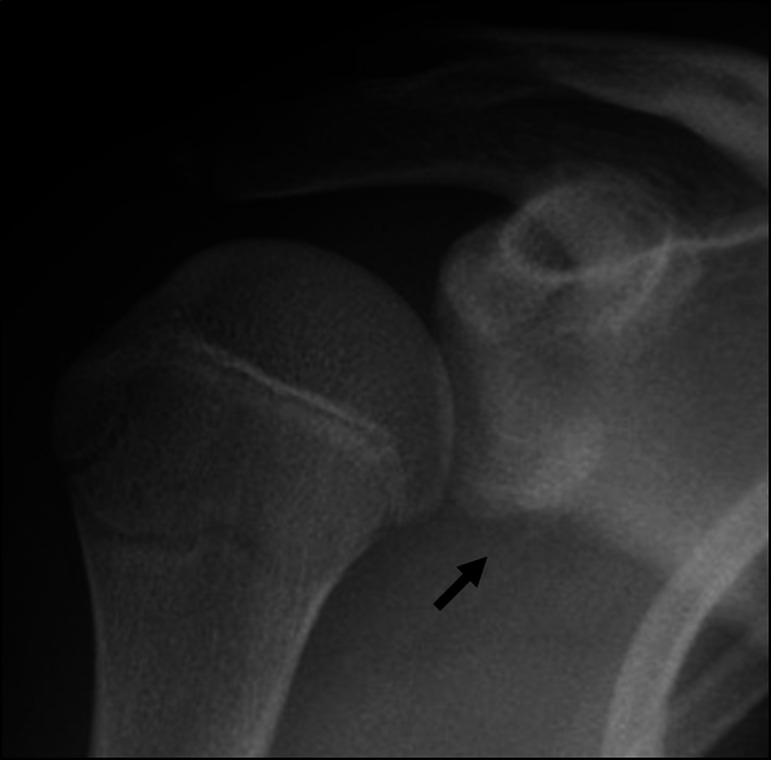
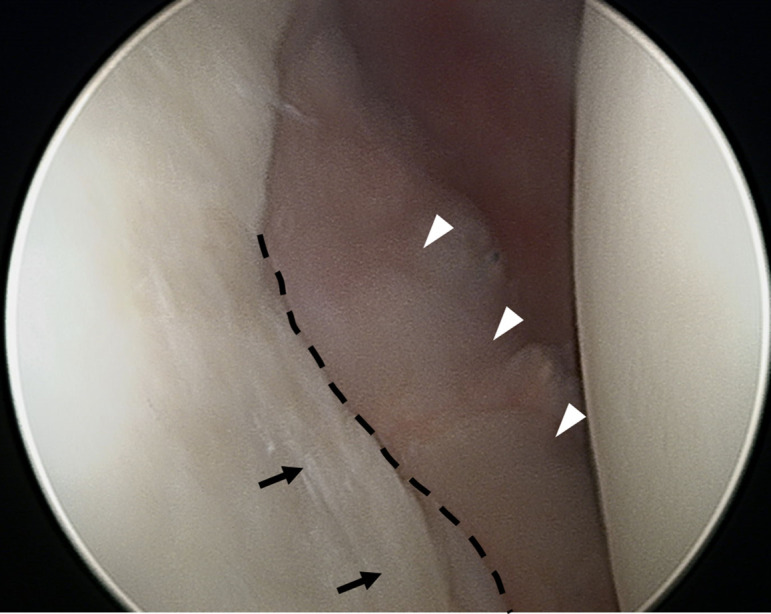
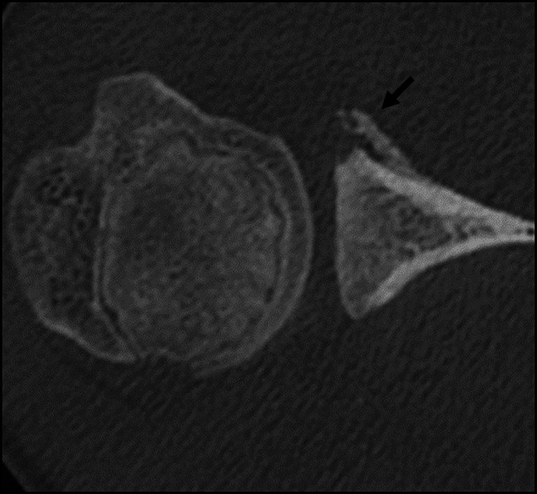
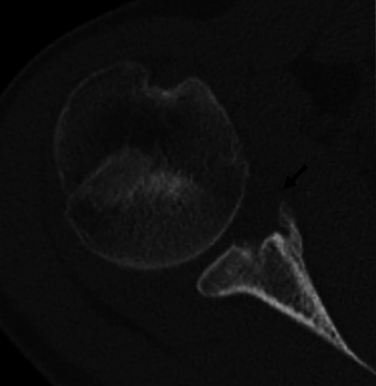
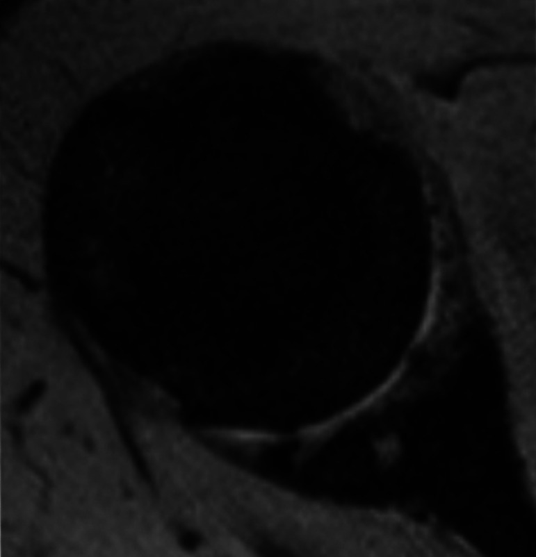
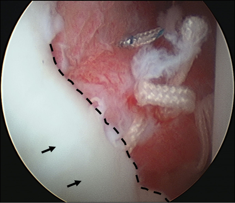
Glenoid articular cartilage lesion is a rare complication following traumatic anterior dislocation of the shoulder. We report the case of a 14-year-old male rugby player with traumatic anterior shoulder instability, an extensively flapped lesion on the glenoid articular cartilage, and an osseous Bankart lesion. Arthroscopic findings revealed that the glenoid cartilage was flap-detached, extending from the anteroinferior to the center. Repair of the osseous Bankart lesion using suture anchors and resection of the unstable peripheral part of the cartilage was performed arthroscopically. The main region of the injured articular surface was left untouched. During postoperative follow-up, absorption of the glenoid articular surface near the suture anchor holes was identified. Arthroscopic examination three months post-surgery showed that the flap detached lesion of the residual cartilage was stable and appeared adapted on the glenoid surface. The resected area was covered by fibrous tissue. A follow-up computed tomography scan revealed that the osseous lesion was united. The patient returned to his previous sports capacity eight months following the operation. At the 2-year-follow-up, magnetic resonance imaging revealed that the glenoid surface was remodeled to a flattened round shape with no signs of osteoarthritis, exhibiting proper conformity of the joint surfaces to the humeral head. Arthroscopic Bankart repair using suture anchors may cause bone resorption at the glenoid surface, leading to remodeling of the glenoid surface from the damaged glenoid cartilage lesion in young patients.


Figures

Figure 1.
Figure 1.
Preoperative images. Plain radiograph of the anteroposterior view; arrow shows osseous Bankart lesion.
Figure 2.
Figure 2.
CT image of axial view. An osseous bony lesion was observed in the anteroinferior part of the glenoid. CT, computed tomography.
Figure 3.
Figure 3.
Arthroscopic view of the anterior part from the posterior portal at the initial surgery. An extensively flap-detached cartilage lesion (asterisk) in the anteroinferior part of the glenoid. The dotted line indicates the trimming line of the anterior edge of the articular cartilage. Arrows show the unmoved cartilage lesion after partial resection.
Figure 4.
Figure 4.
Post-repair of the osseous Bankart lesion at the initial surgery. The dotted line and arrows represent the same locations as shown in Figure 3.
Figure 5.
Figure 5.
CT images 3 months post-operation at the axial view. The osseous Bankart lesion that had not been fully reduced was united after 3 months although osteolytic changes in the glenoid were observed. CT, computed tomography.
Figure 6.
Figure 6.
Arthroscopic view of the anterior part from the posterior portal at the second surgery. The dotted line and arrows represent the same locations as shown in Figures 3 and 4. The white arrowheads show the detached lesion of the residual cartilage, flattened and stable with fibrous soft tissue covering the area of the healed osseous Bankart lesion.
Figure 7.
Figure 7.
CT images at 2 years post-operation at the axial view. Arrows show the peripheral flattening of the osteolytic lesion as remodeling was noted. CT, computed tomography.
Figure 8.
Figure 8.
MRI at the 2-year follow-up at the proton axial view. Sclerotic changes in the subchondral bone and fibrocartilaginous tissue formation on top of it were noted. Proper conformity with the humeral head was observed. MRI, magnetic resonance imaging.

References

    1. Tasaki A, Morita W, Yamakawa A.et al. Combined arthroscopic Bankart repair and coracoid process transfer to anterior glenoid for shoulder dislocation in rugby players: evaluation based on ability to perform sport-specific movements effectively. Arthroscopy. 2015;31(9):1693 1701. 10.1016/j.arthro.2015.03.013) - DOI - PubMed
    1. Neviaser TJ. The GLAD lesion: another cause of anterior shoulder pain. Arthroscopy. 1993;9(1):22 23. 10.1016/s0749-8063(05)80339-1) - DOI - PubMed
    1. Page R, Bhatia DN. Arthroscopic repair of a chondrolabral lesion associated with anterior glenohumeral dislocation. Knee Surg Sports Traumatol Arthrosc. 2010. ;18(12):1748 1751. 10.1007/s00167-010-1095-3) - DOI - PubMed
    1. Beighton P, Horan F. Orthopaedic aspects of the Ehlers-Danlos syndrome. J Bone Joint Surg Br. 1969;51(3):444 453. 10.1302/0301-620X.51B3.444) - DOI - PubMed
    1. Dawson J, Fitzpatrick R, Carr A. The assessment of shoulder instability. The development and validation of a questionnaire. J Bone Joint Surg Br. 1999;81(3):420 426. 10.1302/0301-620x.81b3.9044) - DOI - PubMed

Publication types