. 2022 Apr 13;79(5):240.
doi: 10.1007/s00018-022-04257-7.

Novel contact-kinin inhibitor sylvestin targets thromboinflammation and ameliorates ischemic stroke

Affiliations

Novel contact-kinin inhibitor sylvestin targets thromboinflammation and ameliorates ischemic stroke

Zhiye Zhang et al. Cell Mol Life Sci. .

Abstract

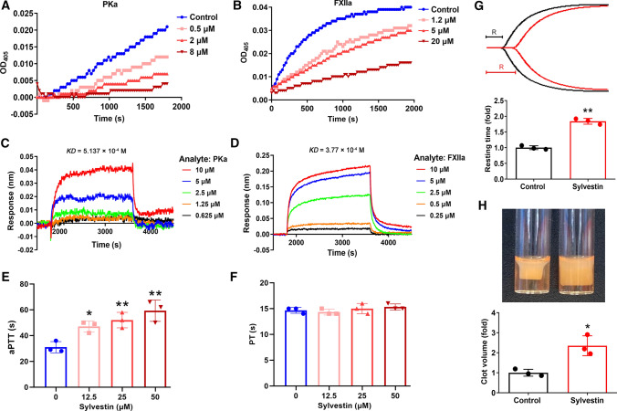
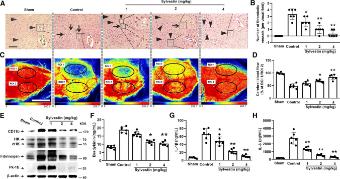
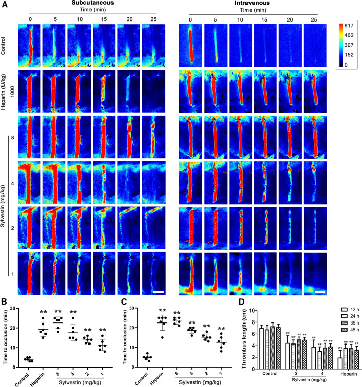
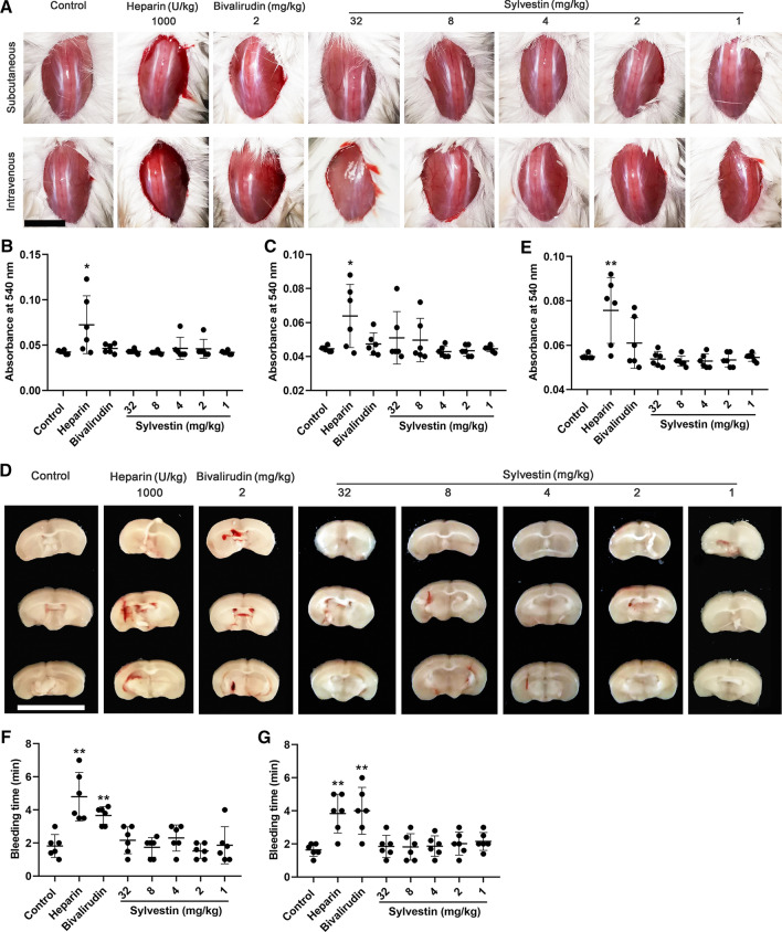
Ischemic stroke is a leading cause of death and disability worldwide. Increasing evidence indicates that ischemic stroke is a thromboinflammatory disease in which the contact-kinin pathway has a central role by activating pro-coagulant and pro-inflammatory processes. The blocking of distinct members of the contact-kinin pathway is a promising strategy to control ischemic stroke. Here, a plasma kallikrein and active FXII (FXIIa) inhibitor (sylvestin, contained 43 amino acids, with a molecular weight of 4790.4 Da) was first identified from forest leeches (Haemadipsa sylvestris). Testing revealed that sylvestin prolonged activated partial thromboplastin time without affecting prothrombin time. Thromboelastography and clot retraction assays further showed that it extended clotting time in whole blood and inhibited clot retraction in platelet-rich plasma. In addition, sylvestin prevented thrombosis in vivo in FeCl3-induced arterial and carrageenan-induced tail thrombosis models. The potential role of sylvestin in ischemic stroke was evaluated by transient and permanent middle cerebral artery occlusion models. Sylvestin administration profoundly protected mice from ischemic stroke by counteracting intracerebral thrombosis and inflammation. Importantly, sylvestin showed no signs of bleeding tendency. The present study identifies sylvestin is a promising contact-kinin pathway inhibitor that can proffer profound protection from ischemic stroke without increased risk of bleeding.

Keywords: Factor XII; Ischemic stroke; Plasma kallikrein; Thromboinflammation.


Conflict of interest statement

The authors declare that they have no competing interest.

Figures

Fig. 1
Fig. 1
Purification and characterization of sylvestin A Homogenate of H. sylvestris was fractionated using a Sephadex G-50 gel filtration column. Absorbance of the eluate was monitored at 280 nm. B Fractions in panel (A) (arrow) were further purified by C4 RP-HPLC using a gradient of acetonitrile in 0.1% trifluoroacetic acid. C Collected fraction from panel (B) was subjected to C18 RP-HPLC using a shallower acetonitrile gradient. D MALDI-TOF analysis of purified sylvestin. E Nucleotide sequence encoding sylvestin precursor and the deduced amino acid sequence. Mature peptide of sylvestin is boxed. F Multiple sequence alignment of sylvestin with other Kunitz-type serine protease inhibitors. Shaded areas in yellow represent the conserved amino acid residues. Amino acids with weak similarity are shaded in green
Fig. 2
Fig. 2
Effects of sylvestin on coagulation factors and blood coagulation Sylvestin was incubated with PKa (A) and FXIIa (B) at indicated molar ratio in presence of specific protease substrate and resulting protease activity was determined at 405 nm. Binding affinity assays of sylvestin and PKa (C) and FXIIa (D) by ForteBio Octet system. Effects of sylvestin on aPTT (E) and PT (F) were determined with human plasma after incubation with sylvestin at indicated concentrations. G Representative TEG tracings and resting time (R) measured by TEG in human whole blood with (red curve) or without (black curve) 50 μM sylvestin. H Representative pictures and quantification of clot retraction in PRP in presence (right tube) or absence (left tube) of 50 μM sylvestin. All results were from three independent experiments. Saline was used as control in A, B, G, and H. Data represent mean ± SD. E, *p < 0.05, **p < 0.01, one-way ANOVA with Dunnett post hoc test compared with control (0 μM sylvestin). G and H, *p < 0.05, **p < 0.01, paired t-test
Fig. 3
Fig. 3
Effects of sylvestin on transient and permanent ischemic stroke C57BL/6 J mice (8 weeks old, male, n = 6) were first subjected to transient MCAO. Sylvestin (1–4 mg/kg) or saline (control) was injected 10 min before (A) or after (B) reperfusion. Representative images of TTC-stained coronal brain sections (top) and quantitative analysis of stained infarct volumes (lower top) in a mouse on day 1 after transient MCAO are shown. Bederson score (upper bottom) and grip test scores (bottom) are also shown. Ischemic infarctions appear white. C Female mice (8 weeks old, n = 6) were subjected to transient MCAO, and application of sylvestin 10 min before reperfusion. On day 1 after transient MCAO, mice were killed. Representative images of TTC-stained of coronal brain sections, quantitative analysis of stained infarct volumes, Bederson score, and grip test score are shown, respectively. D Representative TTC-stained coronal brain sections of 60–62-week-old mice (n = 6) on day 1 after transient MCAO, and infarct volumes, Bederson score, and grip test score are shown. Sylvestin was applied 10 min before reperfusion in this model. E Eight-week-old female mice (n = 6) were first subjected to permanent MCAO. Sylvestin was injected 50 min after MCAO, and mice were killed on day 1 after permanent MCAO and representative images of TTC-stained coronal brain sections (top) and quantitative analysis of stained infarct volumes (bottom) are shown. Long-term functional Bederson score (F), grip test score (G), and survival rate (H) are shown in sylvestin-treated mice and controls (sham and saline-treated (control) groups) until day 7 after permanent MCAO. Scale bars, 1 cm (AE). Data represent mean ± SD. AE, *p < 0.05, **p < 0.01, one-way ANOVA with Dunnett post hoc test compared with control group (saline)
Fig. 4
Fig. 4
Effects of sylvestin on intracerebral thrombosis and inflammation C57BL/6 J mice (8 weeks old, male, n = 6) were first subjected to transient MCAO, followed by application of sylvestin 10 min before reperfusion. Mice were killed 24 h after transient MCAO, and brain specimens were excised, fixed, and cut into 5-μm sections. Representative H&E-stained brain sections (A) and quantitative results of thrombotic vessels (B) are shown. Arrowheads depict patent vessels and arrows indicate thrombotic vessels. C Representative images of cerebral blood flow measured by laser speckle perfusion imaging on day 1 after transient MCAO. Two regions of interest (ROI) in ipsilesional (ROI 1) and contralesional cortex (ROI 2) were used to quantify cerebral blood flow changes, respectively. D Ratio of cerebral blood flow in ROI 1 to ROI 2 is shown. E Immunoblot analysis of PK (Pk-1b), native (HK) and cleaved kininogen (cHK), fibrinogen, and CD11b of ipsilesional cortex from controls and sylvestin-treated mice on day 1 after transient MCAO. β-Actin was a loading control. Plasma concentrations of bradykinin (F), IL-1β (G), and IL-6 (H) in mice described above. Scale bars, 100 μm (A) and 5 mm (C). Data represent mean ± SD. *p < 0.05, **p < 0.01, one-way ANOVA with Dunnett post hoc test compared with control group (saline)
Fig. 5
Fig. 5
Effects of sylvestin on thrombosis in vivo A Representative images of carotid artery blood flow in FeCl3-treated C57BL/6 J mice (n = 6) by laser speckle perfusion imaging. Mice were injected subcutaneously (left) or intravenously (right) with 0.9% saline (control), heparin (1 000 U/kg), or sylvestin (1–8 mg/kg) 10 min before injury. Blood flow was monitored for 25 min. Red: blood flow; blue and black area: background. Color bar on right indicates perfusion unit scale (0–617). Scale bars, 1 mm. Time of complete vessel occlusion was also recorded after subcutaneous (B) or intravenous (C) administration of samples. D Effects of sylvestin and heparin (500 U/kg) on thrombus formation induced by carrageenan in mouse tail (n = 6). Length of thrombus in tails of mice was measured 12, 24, 36, and 48 h after treatment. Data represent mean ± SD. B and C, **p < 0.01, one-way ANOVA with Dunnett post hoc test compared with control (saline). D, **p < 0.01, two-way ANOVA with Dunnett post hoc test compared with control (saline)
Fig. 6
Fig. 6
Effects of sylvestin on bleeding A-C, Mouse skin bleeding model (n = 6) was created using full-thickness skin defects on dorsal skin. Sylvestin and control samples (saline, heparin, and bivalirudin) were applied subcutaneously (A, top, and B) or intravenously (A, bottom, and C). Representative images of skin wounds (A) and quantitative analysis of bleeding (B and C) are shown. B and C, Cerebral bleeding model (n = 6) was used to study the effect of sylvestin on cerebral bleeding. Representative images of coronal brain sections (D) and quantitative analysis of the bleeding (E) are shown. F and G, Tail bleeding assay of control (1 000 U/kg heparin and 2 mg/kg bivalirudin) and sylvestin-treated mice (n = 6, samples were applied subcutaneously 40 min before tail transection (F) or intravenously 20 min before tail transaction (G)). Scale bars, 5 mm (A) and 1 cm (D). Data represent mean ± SD. B, C, E, F, and G, *p < 0.05, **p < 0.01, one-way ANOVA with Dunnett post hoc test compared with control group (saline)

References

    1. Chamorro A, Dirnagl U, Urra X, Planas AM. Neuroprotection in acute stroke: Targeting excitotoxicity, oxidative and nitrosative stress, and inflammation. Lancet Neurol. 2016;15:869–881. doi: 10.1016/S1474-4422(16)00114-9. - DOI - PubMed
    1. Xing C, Arai K, Lo EH, Hommel M. Pathophysiologic cascades in ischemic stroke. Int J Stroke. 2012;7:378–385. doi: 10.1111/j.1747-4949.2012.00839.x. - DOI - PMC - PubMed
    1. Mattson MP, Culmsee C, Yu ZF. Apoptotic and antiapoptotic mechanisms in stroke. Cell Tissue Res. 2000;301:173–187. doi: 10.1007/s004419900154. - DOI - PubMed
    1. Szydlowska K, Tymianski M. Calcium, ischemia and excitotoxicity. Cell Calcium. 2010;47:122–129. doi: 10.1016/j.ceca.2010.01.003. - DOI - PubMed
    1. Spagnoli LG, Mauriello A, Sangiorgi G, Fratoni S, Bonanno E, Schwartz RS, Piepgras DG, Pistolese R, Ippoliti A, Holmes DR., Jr Extracranial thrombotically active carotid plaque as a risk factor for ischemic stroke. JAMA. 2004;292:1845–1852. doi: 10.1001/jama.292.15.1845. - DOI - PubMed