. 2022 Apr 13:11:e76887.
doi: 10.7554/eLife.76887.

Genetic variation of putative myokine signaling is dominated by biological sex and sex hormones

Affiliations

Genetic variation of putative myokine signaling is dominated by biological sex and sex hormones

Leandro M Velez et al. Elife. .

Abstract

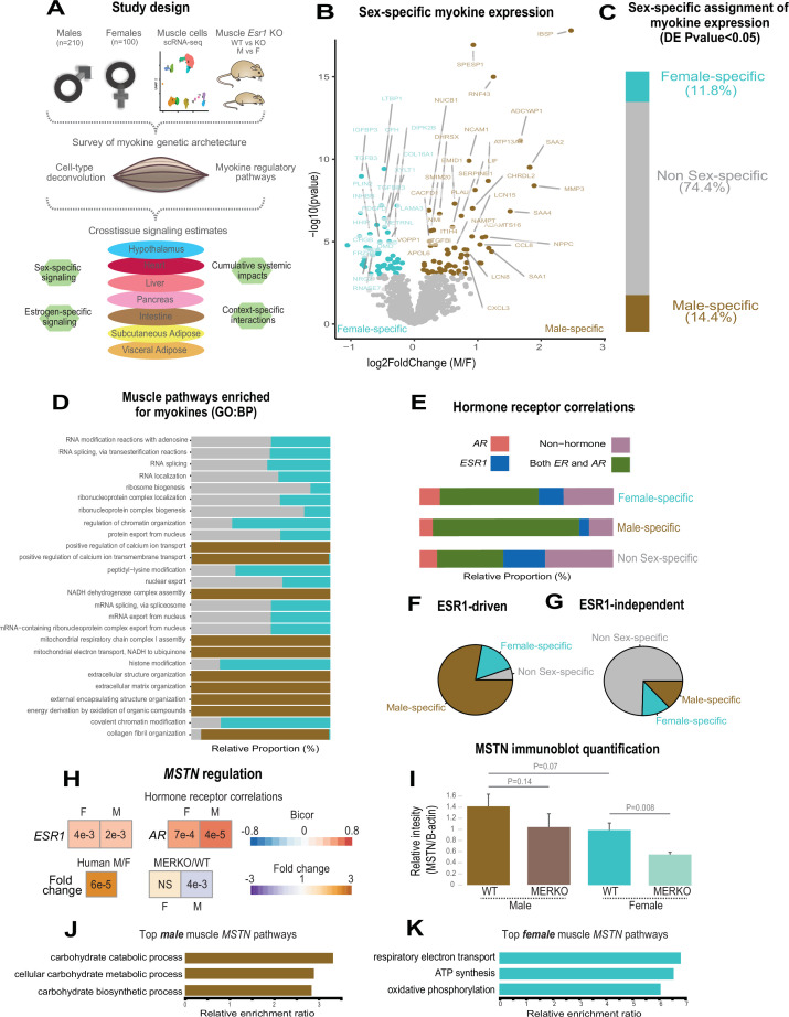
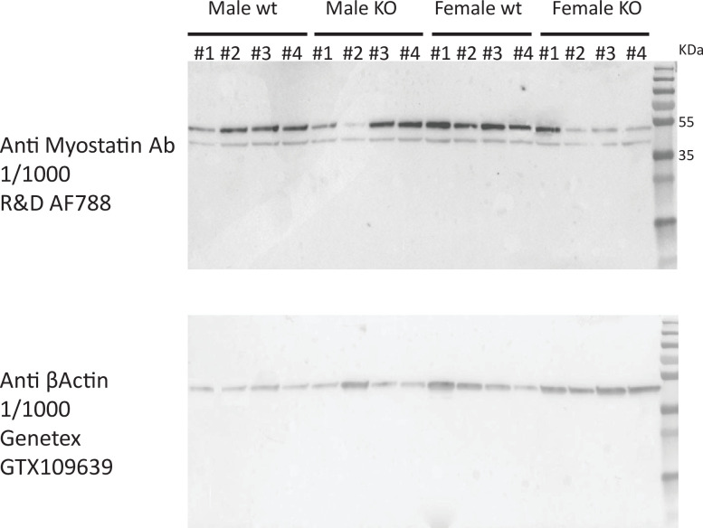
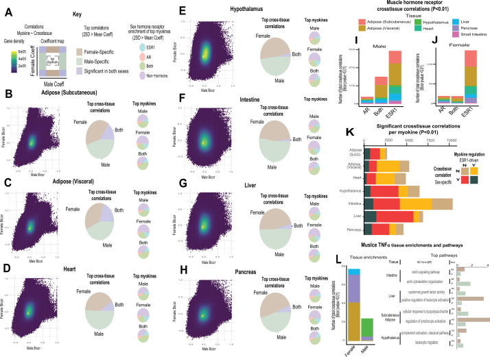
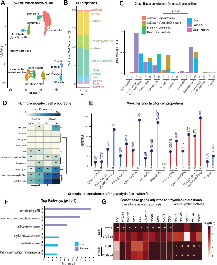
Skeletal muscle plays an integral role in coordinating physiological homeostasis, where signaling to other tissues via myokines allows for coordination of complex processes. Here, we aimed to leverage natural genetic correlation structure of gene expression both within and across tissues to understand how muscle interacts with metabolic tissues. Specifically, we performed a survey of genetic correlations focused on myokine gene regulation, muscle cell composition, cross-tissue signaling, and interactions with genetic sex in humans. While expression levels of a majority of myokines and cell proportions within skeletal muscle showed little relative differences between males and females, nearly all significant cross-tissue enrichments operated in a sex-specific or hormone-dependent fashion; in particular, with estradiol. These sex- and hormone-specific effects were consistent across key metabolic tissues: liver, pancreas, hypothalamus, intestine, heart, visceral, and subcutaneous adipose tissue. To characterize the role of estradiol receptor signaling on myokine expression, we generated male and female mice which lack estrogen receptor α specifically in skeletal muscle (MERKO) and integrated with human data. These analyses highlighted potential mechanisms of sex-dependent myokine signaling conserved between species, such as myostatin enriched for divergent substrate utilization pathways between sexes. Several other putative sex-dependent mechanisms of myokine signaling were uncovered, such as muscle-derived tumor necrosis factor alpha (TNFA) enriched for stronger inflammatory signaling in females compared to males and GPX3 as a male-specific link between glycolytic fiber abundance and hepatic inflammation. Collectively, we provide a population genetics framework for inferring muscle signaling to metabolic tissues in humans. We further highlight sex and estradiol receptor signaling as critical variables when assaying myokine functions and how changes in cell composition are predicted to impact other metabolic organs.

Keywords: biochemistry; chemical biology; computational biology; endocrinology; human; myokine; physiology; systems biology; systems genetics.

Plain language summary

The muscles that are responsible for voluntary movements such as exercise are called skeletal muscles. These muscles secrete proteins called myokines, which play roles in a variety of processes by interacting with other tissues. Essentially, myokines allow skeletal muscles to communicate with organs such as the kidneys, the liver or the brain, which is essential for the body to keep its metabolic balance. Some of the process myokines are involved include inflammation, cancer, the changes brought about by exercise, and even cognition. Despite the clear relevance of myokines to so many physiological outcomes, the way these proteins are regulated and their effects are not well understood. Genetic sex – specified by sex chromosomes in mammals – contributes to critical aspects of physiology. Specifically, many of the metabolic traits impacted by myokines show striking differences arising from hormonal or genetic interactions depending on the genetic sex of the subject being studied. It is therefore important to consider genetic sex when studying the effects of myokines on the body. Velez, Van et al. wanted to gain a better understanding of how skeletal muscles interact with metabolic tissues such as pancreas, liver and brain, taking genetic sex into consideration. To do this they surveyed human datasets for the correlations between the activity of genes that code for myokines, the composition of muscle cells, the signaling between muscles and metabolic tissues and genetic sex. Their results showed that, genetic sex and sex hormones predicted most of the effects of skeletal muscle on other tissues. For example, myokines from muscle were predicted to be more impactful on liver or pancreas, depending on whether individuals were male or female, respectively. The results of Velez, Van et al. illustrate the importance of considering the effects of genetic sex and sexual hormones when studying metabolism. In the future, these results will allow other researchers to design sex-specific experiments to be able to gather more accurate information about the mechanisms of myokine signaling.


Conflict of interest statement

LV, CV, TM, ZZ, CJ, AH, MS No competing interests declared

Figures

Figure 1.
Figure 1.. Sex and hormone effects on myokine regulation.
(A) Overall study design for integration of gene expression from muscle from 310 humans, single-cell RNA-sequencing (RNA-seq), muscle-specific deletion of Esr1 to infer interorgan coregulatory process across major metabolic tissues. (B–C) Differential expression analysis for sex was performed on all genes corresponding to secreted proteins in skeletal muscle (myokines). The specific genes which showed significant changes in each sex are shown as a volcano plot (B) and the relative proportions of myokines corresponding to each category at a least-stringent logistic regression p-value less than 0.05 (C). (D) For each differential expression category based on sex shown in C, myokines were correlated with all other muscle genes for pathway enrichment. Then the top 10 enriched pathways in males, females, or non-sex specific (by overall significance) were visualized together where number of genes corresponding to each category shown as a relative proportion. (E) The same analysis as in D, except instead of myokines being correlated with AR, ESR1, both hormone receptors, or neither, as compared to correlating with all genes. (F–G) Myokines were binned into two categories based on significant differential expression (logistic regression adjusted p-value < 0.05) between muscle-specific WT and MERKO mice (F) or those that showed no change (G), then visualized as relative proportions within each category shown in (C). (H) Midweight bicorrelation (bicor) coefficients (color scheme) and corresponding regression p-values (filled text) are shown for muscle MSTN ~ESR1 or AR in both sexes (top). Below, correlations are shown for differential expression log2FC (color scheme) and corresponding logistic regression p-values (text fill) for MSTN between sexes in humans or WT vs. MERKO mice. (I) Quantification of processed form of myostatin (Figure 1—figure supplement 2, bottom band) relative to β-actin in WT or MERKO muscle in male or female mice. p-Values calculated using a Student’s t-test. (J–K) The top three pathways of genes which significantly (p < 1e-4) correlated with muscle MSTN in males (J) or females (K). For human data, n = 210 males and n = 100 females. For mouse MERKO vs. WT comparisons, n = 3 mice per group per sex. p-Values from midweight bicorrelations were calculated using the Student’s p-value from WGCNA and logistic regression p-values were calculated using DESeq2.
Figure 1—figure supplement 1.
Figure 1—figure supplement 1.. Skeletal muscle sex hormone receptor expression between sexes.
Normalized gene expression levels for androgen receptor (AR) or estrogen receptor (ESR1) (y-axis) in each sex (x-axis). None of the expression levels were significantly different between sexes (Student’s t-test).
Figure 1—figure supplement 2.
Figure 1—figure supplement 2.. Immunoblot for myostatin in EDL muscle from WT and MERKO male and female mice.
Full immunoblots shown for skeletal muscle lysate blotted for myostatin (top) or β-actin (bottom) corresponding to different C57BL/6J male (left) or female (right) mice in either WT (floxed) or KO (floxed-cre) for skeletal muscle Esr1. Band sizes shown to indicate either precursor (top band) or processed/LAP form (bottom band) of myostatin.
Figure 2.
Figure 2.. Sex and hormone effects on myokine regulation.
(A–H) Key illustrating analysis for distribution of midweight bicorrelation coefficients between all myokines in skeletal muscle and global transcriptome measures in each target tissue. Coefficients are plotted between sexes (left), where proportions for 2SD > mean are subdivided into occurrence uniquely in females, males, or shared (middle). The significant (2SD > mean) myokines identified in each category were then binned into hormone receptor correlations for ESR1, AR, both, or neither (right). This analysis was performed on all myokines across subcutaneous adipose tissue (B), visceral adipose (C), heart (D), hypothalamus (E), small intestine (F), liver (G), and pancreas (H). (I–J) Significant cross-tissue correlations between muscle ESR1, AR, or both hormone receptors are colored by tissue and shown for males (I) or females (J). (K) For each tissue (y-axis), the ratio of significant cross-tissue correlations per muscle myokine (x-axis) are shown and colored by categories of either the myokine regulated by ESR1 and/or a significant target tissue regression occurring specifically in one sex. (L) Number of significant cross-tissue correlations with muscle TNFα are shown for each sex and colored by tissue as in I–L (left). The −log10(p-value) of significance in an overrepresentation test (x-axis) are shown for top significant inter-tissue pathways for muscle TNFα in each sex (right).
Figure 3.
Figure 3.. Genetic variation of muscle cell proportions and coregulated cross-tissue processes.
(A) Uniform Manifold Approximation and Projection (UMAP) for skeletal muscle single-cell sequencing used to deconvolute proportions. (B) Mean relative proportions of pseudo-single-cell muscle cell compositions (denoted by color) between sexes. (C) Number of significant cross-tissue correlations (y-axis) corresponding to each skeletal muscle type in each sex (x-axis). Target tissues are distinguished by color, where NS (male platelets) denotes that no significant cross-tissue correlations were observed. (D) Heatmap showing significance of correlations between skeletal muscle hormone receptors and cell proportions, * = p < 0.01. (E) The strongest enriched myokines are plotted for each myokine (y-axis, −log10 p-value of myokine ~ cell composition) are shown for each muscle proportion for each sex (x-axis). Gene symbols for myokines are shown above each line, where red lines indicate positive correlations between myokine and cell type and blue shows inverse relationships. (F) Significant cross-tissue correlated genes in liver (blue) and pancreas (purple) for muscle fast-twitch glycolytic fibers (p < 1e-6) were used for overrepresentation tests where enrichment ratio of significance (x-axis) is shown for each pathway and sex (y-axis). (G) Heatmap showing the regression significance of the top five genes corresponding to inflammation (liver), exocytosis (liver), and protein synthesis (pancreas) for proportions of fast-twitch fiber type (un-adj). Below each correlation between fast-twitch fiber and liver or pancreas gene, the same regressions were performed while adjusting for abundance of select myokines in each sex. * = p < 1e-6.
Figure 3—figure supplement 1.
Figure 3—figure supplement 1.. Comparisons of deconvolution methods.
Cell proportions were estimated from skeletal muscle sequencing across the 310 individuals in GTEx. Here, comparisons of the three most common methods (DCQ, NNLS, and porportionsInAdmixture) were plotted for each pseudo-sc-proportion, where proportionsInAdmixture method captured the largest relative number of cell types.
Figure 3—figure supplement 2.
Figure 3—figure supplement 2.. Cell composition correlations within each sex.
Heatmaps showing regressions for cell proportions in males (left) or females (right), * = regression p-value < 0.01.

References

    1. Battle A, Brown CD, Engelhardt BE, Montgomery SB. Genetic effects on gene expression across human tissues. Nature. 2017;550:204–213. doi: 10.1038/nature24277. - DOI - PMC - PubMed
    1. Chella Krishnan K, Vergnes L, Acín-Pérez R, Stiles L, Shum M, Ma L, Mouisel E, Pan C, Moore TM, Péterfy M, Romanoski CE, Reue K, Björkegren JLM, Laakso M, Liesa M, Lusis AJ. Sex-specific genetic regulation of adipose mitochondria and metabolic syndrome by Ndufv2. Nature Metabolism. 2021;3:1552–1568. doi: 10.1038/s42255-021-00481-w. - DOI - PMC - PubMed
    1. Clegg DJ, Mauvais-Jarvis F. An integrated view of sex differences in metabolic physiology and disease. Molecular Metabolism. 2018;15:1–2. doi: 10.1016/j.molmet.2018.06.011. - DOI - PMC - PubMed
    1. Danziger SA, Gibbs DL, Shmulevich I, McConnell M, Trotter MWB, Schmitz F, Reiss DJ, Ratushny AV. ADAPTS: Automated deconvolution augmentation of profiles for tissue specific cells. PLOS ONE. 2019;14:e0224693. doi: 10.1371/journal.pone.0224693. - DOI - PMC - PubMed
    1. Eckel J. Myokines in metabolic homeostasis and diabetes. Diabetologia. 2019;62:1523–1528. doi: 10.1007/s00125-019-4927-9. - DOI - PubMed

Publication types