. 2022 Apr 13;10(1):52.
doi: 10.1186/s40478-022-01358-z.

Interface astrogliosis in contact sport head impacts and military blast exposure

Affiliations

Interface astrogliosis in contact sport head impacts and military blast exposure

Katharine J Babcock et al. Acta Neuropathol Commun. .

Abstract

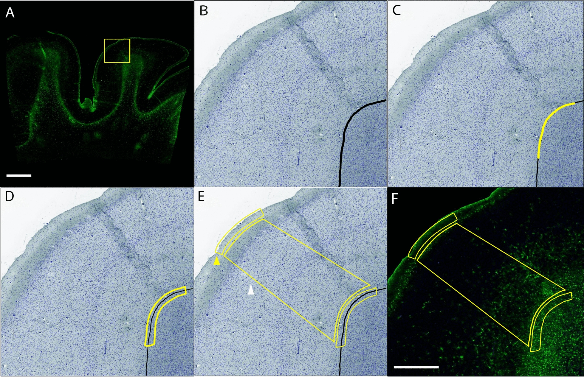
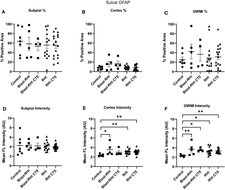
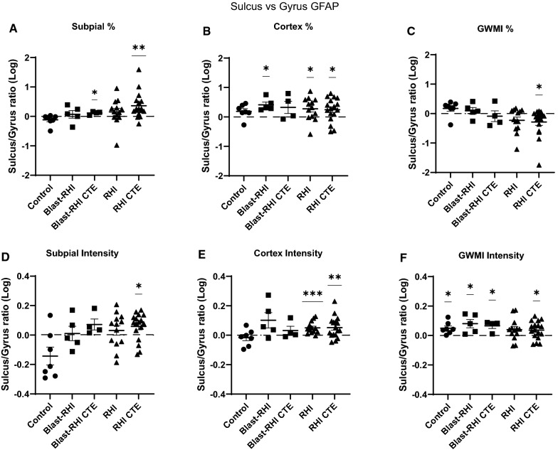
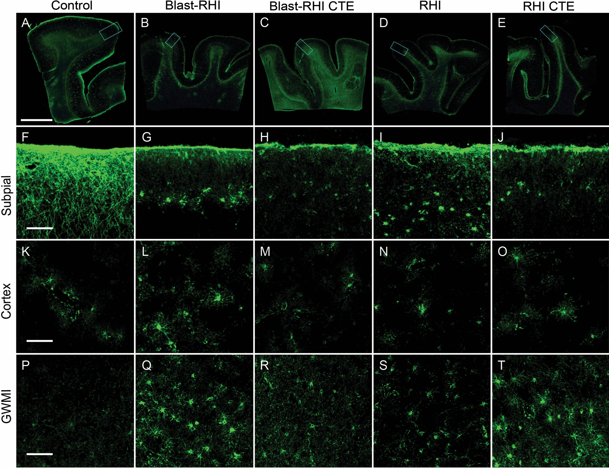
Exposure to military blast and repetitive head impacts (RHI) in contact sports is associated with increased risk of long-term neurobehavioral sequelae and cognitive deficits, and the neurodegenerative disease chronic traumatic encephalopathy (CTE). At present, the exact pathogenic mechanisms of RHI and CTE are unknown, and no targeted therapies are available. Astrocytes have recently emerged as key mediators of the multicellular response to head trauma. Here, we investigated interface astrogliosis in blast and impact neurotrauma, specifically in the context of RHI and early stage CTE. We compared postmortem brain tissue from former military veterans with a history of blast exposure with and without a neuropathological diagnosis of CTE, former American football players with a history of RHI with and without a neuropathological diagnosis of CTE, and control donors without a history of blast, RHI exposure or CTE diagnosis. Using quantitative immunofluorescence, we found that astrogliosis was higher at the grey-white matter interface in the dorsolateral frontal cortex, with mixed effects at the subpial surface and underlying cortex, in both blast and RHI donors with and without CTE, compared to controls. These results indicate that certain astrocytic alterations are associated with both impact and blast neurotrauma, and that different astroglial responses take place in distinct brain regions.

Keywords: Astrogliosis; Blast injury; Chronic traumatic encephalopathy; Mild traumatic brain injury; Repetitive head impacts.


Conflict of interest statement

The authors have no duality or conflicts of interest to declare.

Figures

Fig. 1
Fig. 1
Annotation Regions. The average fluorescent intensity and percent positivity of GFAP were quantified in representative sections of the cortical gyrus (yellow box in A, field of view in BF) and sulcus (not highlighted) using a modified version of the Area Quantification FL analysis algorithm in HALO. Annotations were generated by switching to the brightfield view and using the magnetic pen tool to delineate the grey-white matter border in the dapi and autofluorescence channels (B, black). The pen tool was then used to outline a 2 mm portion of the grey-white border (C, yellow), and a 200 µm GWMI annotation was produced using the marginal partitioning tool (D, yellow). A 2 mm line was then drawn at the cortical surface and the marginal partitioning tool was used to generate a 200 µm subpial annotation (E, yellow arrow head). The brush tool was used to annotate the cortical grey matter spanning between the subpial and GWMI annotations (E, white arrow head). Composite image showing all annotations with the GFAP channel in darkfield view (F). Green = GFAP, blue = DAPI. Scale bars: A = 5 mm, BF = 1 mm
Fig. 2
Fig. 2
Overview of GFAP Comparisons. Summary of the two main types of comparisons carried out in the current study. Comparison 1 (A) directly compares the GFAP immunoreactivity (percent positivity and mean fluorescent intensity) between Controls and each neurotrauma group at both the gyral crest and sulcal depth. Comparison 2 (B) directly compares the GFAP at the sulcus to the GFAP at the gyrus within each cohort using a relative ratio of sulcal:gyral GFAP, where a value over 0 indicates sulcal predominance, and a value under 0 indicates gyral predominance
Fig. 3
Fig. 3
Between Group Comparisons of GFAP at the Gyral Crest. At the crest of the gyrus, GFAP was significantly decreased at the subpial surface in the RHI (A) Blast-RHI CTE (D), and RHI-CTE (A, D). No differences were detected in the underlying grey matter (B, E). At the grey-white matter interface, all neurotrauma groups had significantly higher mean fluorescent GFAP intensity compared to controls (F). No significant differences were detected in percent positivity at the GWMI (C). Error bars represent standard error of the mean. *P < 0.05, **P < 0.01, ***P < 0.001, ****P < 0.0001
Fig. 4
Fig. 4
Between Group Comparisons of GFAP at the Sulcal Depth. At the depth of the sulcus, no significant differences in GFAP were detected at the subpial surface (A, D). Within the underlying cortical grey matter, the Blast-RHI, RHI), and RHI CTE) groups all had significantly higher mean fluorescent GFAP intensity compared to controls (E), with no differences detected in any groups for percent positivity (B). At the grey-white matter junction, all neurotrauma groups had significantly higher GFAP in terms of mean fluorescent intensity compared to controls (F). No differences were detected in percent positivity at the GWMI (C). Error bars represent standard error of the mean. *P < 0.05, **P < 0.01
Fig. 5
Fig. 5
Within Group Comparisons of Sulcal vs Gyral GFAP. Within group comparisons of sulcal versus gyral GFAP were calculated using a relative ratio of sulcal:gyral GFAP. The Blast-RHI CTE group had higher GFAP at the sulcus relative to the gyrus in terms of percent positivity at the subpial surface (A), while the RHI-CTE group had higher GFAP at the sulcus relative to the gyrus at the subpial surface in both percent positivity and mean intensity (A, B). Within the cortex, the Blast-RHI group had significantly higher GFAP in the sulcus in terms of percent positivity (B), while the RHI and RHI CTE groups had significantly greater sulcal predominance in terms of both percent positivity (B) and mean fluorescent intensity (E). At the GWMI (C, F), all groups except RHI had significantly higher GFAP intensity at the sulcus relative to the gyrus (F). The RHI CTE group had significantly less at the sulcus in terms of percent positivity (C). Ratio data were log transformed and analyzed using one-way t-tests. Values significantly greater than zero indicated higher GFAP at the sulcus (sulcal predominance), while values significantly lower than zero indicated higher GFAP at the gyrus (gyral predominance). Error bars represent standard error of the mean. *P < 0.05, **P < 0.01, ***P < 0.001
Fig. 6
Fig. 6
GFAP + Astrocyte Morphology in the Gyral Crest of the Dorsolateral Frontal Cortex. Representative images of the dorsolateral frontal cortex stained with GFAP from Control (A), Blast-RHI (B), Blast-RHI CTE (C), RHI (D), and RHI CTE (E) cases. The blue rectangular boxes in AE indicate the areas analyzed and discussed in F-T. Astrocytes of the glial limitans superficialis demonstrate intralaminar astrocyte processes that extend down from the subpial surface into cortical layers I-II in the control case (F). In all forms of neurotrauma (GJ), there is a dropout of interlaminar astrocytic processes, however astrocytes visible in the subjacent layer display increased GFAP expression and swollen cell bodies indicative of reactive astrocytosis. Throughout the cortex astrocytes appear similar across groups (K-O). At the GWMI, the control case (P) demonstrates fine astrocytic processes and low levels of GFAP expression, while the neurotrauma groups (Q-T) display greater GFAP expression and reactive profiles with thicker astrocyte processes. Green = GFAP. Scale bar, top panel (AE): 5 mm. Scale bar, bottom panels (FT): 50 μm

References

    1. Abbott NJ, Rönnbäck L, Hansson E. Astrocyte-endothelial interactions at the blood-brain barrier. Nat Rev Neurosci. 2006 doi: 10.1038/nrn1824. - DOI - PubMed
    1. Alosco ML, Tripodis Y, Rowland B, Chua AS, Liao H, Martin B, et al. A magnetic resonance spectroscopy investigation in symptomatic former NFL players. Brain Imaging Behav. 2020 doi: 10.1007/s11682-019-00060-4. - DOI - PMC - PubMed
    1. Anderson MA, Burda JE, Ren Y, Ao Y, O’Shea TM, Kawaguchi R, et al. Astrocyte scar formation aids CNS axon regeneration. Nature. 2016;532:195–200. doi: 10.1038/nature17623. - DOI - PMC - PubMed
    1. Argaw AT, Gurfein BT, Zhang Y, Zameer A, John GR. VEGF-mediated disruption of endothelial CLN-5 promotes blood-brain barrier breakdown. Proc Natl Acad Sci U S A. 2009;106:1977–1982. doi: 10.1073/pnas.0808698106. - DOI - PMC - PubMed
    1. Bieniek KF, Cairns NJ, Crary JF, Dickson DW, Folkerth RD, Keene CD, et al. The second NINDS/nibib consensus meeting to define neuropathological criteria for the diagnosis of chronic traumatic encephalopathy. J Neuropathol Exp Neurol. 2021;80:210–219. doi: 10.1007/s00401-015-1515-z. - DOI - PMC - PubMed

Publication types

MeSH terms

LinkOut - more resources