. 2022 Apr 13;12(4):61.
doi: 10.1038/s41408-022-00660-2.

Primary CD33-targeting CAR-NK cells for the treatment of acute myeloid leukemia

Affiliations

Primary CD33-targeting CAR-NK cells for the treatment of acute myeloid leukemia

Nawid Albinger et al. Blood Cancer J. .

Abstract

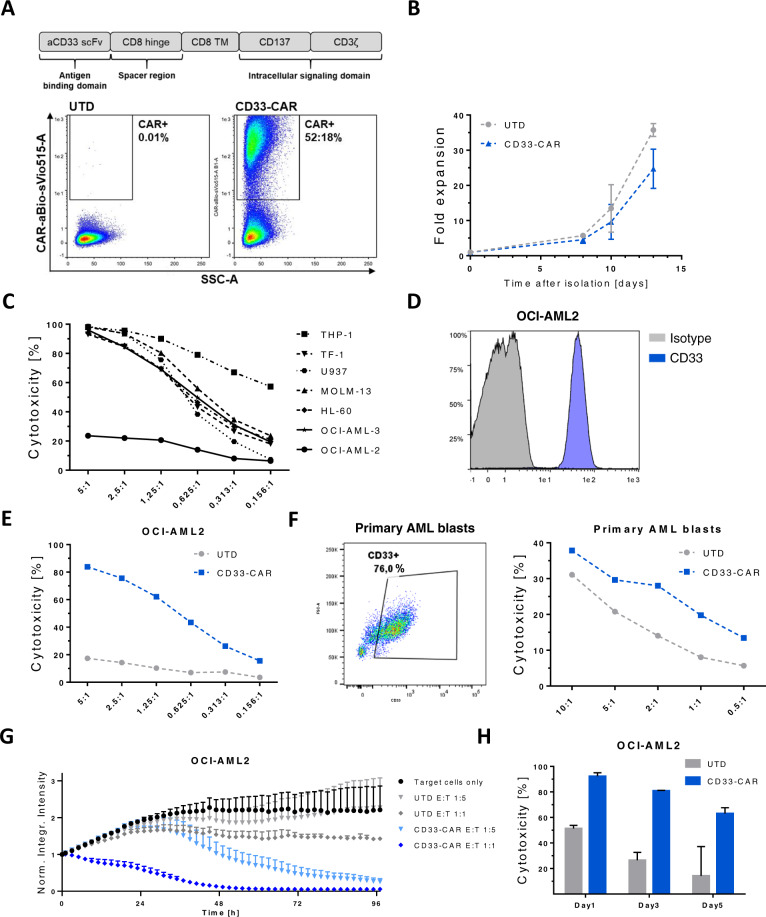
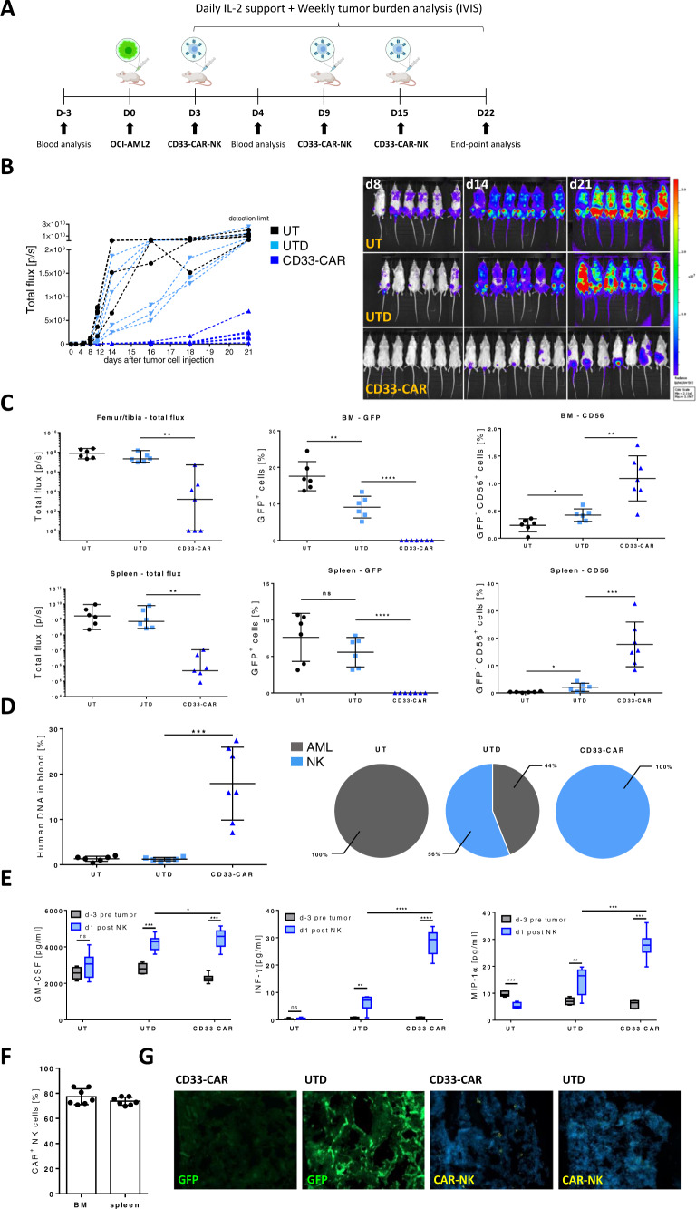
Acute myeloid leukemia (AML) is a malignant disorder derived from neoplastic myeloid progenitor cells characterized by abnormal proliferation and differentiation. Although novel therapeutics have recently been introduced, AML remains a therapeutic challenge with insufficient cure rates. In the last years, immune-directed therapies such as chimeric antigen receptor (CAR)-T cells were introduced, which showed outstanding clinical activity against B-cell malignancies including acute lymphoblastic leukemia (ALL). However, the application of CAR-T cells appears to be challenging due to the enormous molecular heterogeneity of the disease and potential long-term suppression of hematopoiesis. Here we report on the generation of CD33-targeted CAR-modified natural killer (NK) cells by transduction of blood-derived primary NK cells using baboon envelope pseudotyped lentiviral vectors (BaEV-LVs). Transduced cells displayed stable CAR-expression, unimpeded proliferation, and increased cytotoxic activity against CD33-positive OCI-AML2 and primary AML cells in vitro. Furthermore, CD33-CAR-NK cells strongly reduced leukemic burden and prevented bone marrow engraftment of leukemic cells in OCI-AML2 xenograft mouse models without observable side effects.


Conflict of interest statement

RP, MN, MQ, CZ, and NM are employees of Miltenyi Biotec. DS is employee of Lentigen Technology, Inc., a Miltenyi Biotec Company. MWMK is a consultant for Pfizer and Abbvie and receives travel support from Celgene and Daiichi Sankyo. OP and EU have no COIs directly related to this manuscript. OP has received honoraria or travel support from Astellas, Gilead, Jazz, MSD, Neovii Biotech, Novartis, Pfizer, and Therakos. He has received research support from Gilead, Incyte, Jazz, Neovii Biotech, and Takeda and is member of advisory boards to Jazz, Gilead, MSD, Omeros, Priothera, Shionogi, and SOBI. EU has a sponsored research project with Gilead. Remaining authors declare no conflict of interest.

Figures

Fig. 1
Fig. 1. CD33-CAR-NK cells display robust in vitro effector function against CD33-positive AML cells that are partially resistant to natural cytotoxicity.
A Schematic representation and surface expression of the CD33-directed second-generation CAR used in this study. Expression was analyzed by flow cytometry 12 days after transgene transfer into primary NK cells. B Time-lapsed expansion of CAR-transduced (CD33-CAR) and untransduced (UTD)-NK cells in the presence of IL-2 (500 IU/mL) and IL-15 (140 IU/mL) (n = 5). C Expanded NK cells show high cytotoxic activity against various AML cell lines except OCI-AML2. On day 14 of expansion, NK cells were co-incubated with various AML target cells at indicated E:T-ratios. After 24 h, the fraction of viable target cells was quantified by flow cytometry. Data shown are representative of results from two independent experiments. D The AML cell line OCI-AML2 displays high CD33 surface expression. E, F NK cells equipped with a CD33-CAR become highly cytotoxic against OCI-AML2 and CD33-positive primary AML cells. Cells were co-cultivated for 4 h and the viability of target cells was quantitated by flow cytometry. Two representative experiments are shown. G Dynamic monitoring of CAR-NK cell-mediated cytotoxicity. On day 12 after transduction, CAR-NK cells were co-cultured with (GFP+) OCI-AML2 cells and fluorescence emission was measured in the IncuCyte S3 imaging platform over 4 days. Shown is one representative from three separate experiments with a total of 5 donors. H Repetitive tumor-challenge assay revealed superior serial killing capacity of CD33-CAR-NK cells compared to UTD-NK cells. Expanded NK cells at day 12 post transduction were co-cultured with OCI-AML2 cells at an E:T-ratio of 1:1 and re-challenged with AML cells every other day. Shown is one representative experiment with a total of two donors. All graphs show mean of replicated ± SD.
Fig. 2
Fig. 2. A single dose of CD33-CAR-NK cells displays potent anti-tumor efficacy in OCI-AML2 engrafted NSG-SGM3 mice.
A Scheme of the in vivo evaluation of a single treatment with CD33-CAR-NK cells (1 × 107 intravenously) followed by subcutaneous treatment with IL-2 in OCI-AML2 (Luc+) xenograft NSG-SGM3 mice. B Total flux analysis as well as representative BLI images of differently treated OCI-AML2 (Luc+) engrafted NSG-SGM3 mice over time (d7 n = 7; d14 n = 6; d21 n = 5 per group). Mice received a single dose of 1 × 107 NK cells day 3 post AML cell injection. At day 21, 4 out of 5 mice (80%) that were treated with CD33-CAR-NK cells show severely reduced leukemic burden compared to untreated mice (UT) or mice which received untransduced (UTD)-NK cells. C Serum analysis of blood day 3 before AML injection and day 1 post first NK cell application shows significantly increased levels of GM-CSF as well as INF-γ for mice that had received CD33-CAR-NK cells (n = 3). Mean ± SD. D Total flux analysis of femurs/tibiae and spleens, as well as flow cytometry analysis of isolated cells from BMs or spleens at day 7, 14, and 21 post tumor cell injection, revealed the absence of GFP-positive tumor cells in CD33-CAR-NK-treated mice as well as increased NK cell infiltration (day 7/14 n = 1; day 21 n = 2 per group). Values of zero were set to 1 for total flux analysis. Median ± range. Flow cytometry-based CAR expression analysis of BM- (E) or spleen- (F) infiltrating NK cells at day 14 and 21 revealed the presence of mainly CAR-positive cells (day 14 n = 1; day 21 n = 2 per group). Mean ± SD. G Confocal microscopy imaging shows GFP-positive leukemia cells in BM of UTD-NK treated NSG-SGM3 mice at day 21 while absent in mice that received CD33-CAR-NK cells. Images from one representative animal are shown. Statistical analysis was performed by Student’s t test (*P < 0.05, **P < 0.01, ***P < 0.001, ****P < 0.0001).
Fig. 3
Fig. 3. Repetitive administration of CD33-CAR-NK cells displays improved anti-tumor efficacy in OCI-AML2 engrafted NSG-SGM3 mice.
A Scheme of the in vivo evaluation of a repetitive treatment with CD33-CAR-NK cells (1 × 107 intravenously) combined with subcutaneous IL-2 treatment in OCI-AML2 (Luc+) xenograft NSG-SGM3 mice. B Total flux analysis, as well as representative BLI images of differently treated OCI-AML2 (Luc+), engrafted NSG-SGM3 mice over time (n = 7 per group). Mice received a total of three weekly doses of 1 × 107 NK cells. Mice that were treated with CD33-CAR-NK cells show severely reduced leukemic burden compared to untreated mice (UT) or mice which received untransduced (UTD)-NK cells. C Total flux analysis of femurs/tibiae and spleens, as well as flow cytometry analysis of isolated cells from BMs or spleens at day 22 post AML-injection, revealed the absence of GFP-positive leukemic cells in CD33-CAR-NK treated mice as well as increased NK cell infiltration (n = 6–7 per group). Values of zero were set to 1 for total flux analysis. Median ± range. D Chimerism analysis d22 post AML-injection revealed high amounts of DNA from human NK cells without detectable DNA of AML in blood of mice that were treated with CD33-CAR-NK cells (n = 6–7 per group). Mean ± SD. E Serum analysis of blood day 3 before AML injection and day 1 post first NK cell application showed significantly increased pro-inflammatory human cytokines for mice that received CD33-CAR-NK cells (n = 6–7 per group). F Flow cytometry-based CAR-expression analysis of BM- or spleen-infiltrating NK cells in CD33-CAR-NK treated mice revealed the presence of mainly CAR-positive cells (n = 6–7 per group). Mean ± SD. G Confocal microscopy imaging demonstrated GFP-positive leukemia cells in BM of UTD-NK treated NSG-SGM3 mice while absent in mice that received CD33-CAR-NK cells. Additionally, CAR-NK cells could be detected in the BM of CD33-CAR-NK treated mice. One representative image from a total of four are shown. Statistical analysis was performed by Mann–Whitney-test (for total flux analysis) or Student’s t test (for the rest) (*P < 0.05, **P < 0.01, ***P < 0.001, ****P < 0.0001).

References

    1. Döhner H, Weisdorf DJ, Bloomfield CD. Acute myeloid leukemia. N Engl J Med. 2015;373:1136–52. doi: 10.1056/NEJMra1406184. - DOI - PubMed
    1. Döhner H, Estey E, Grimwade D, Amadori S, Appelbaum FR, Büchner T, et al. Diagnosis and management of AML in adults: 2017 ELN recommendations from an international expert panel. Blood. 2017;129:424–47.. doi: 10.1182/blood-2016-08-733196. - DOI - PMC - PubMed
    1. Hahn T, McCarthy, Philip L, Jr, Zhang M-J, Wang D, Arora M, et al. Risk factors for acute graft-versus-host disease after human leukocyte antigen-identical sibling transplants for adults with leukemia. J Clin Oncol. 2008;26:5728–34. doi: 10.1200/JCO.2008.17.6545. - DOI - PMC - PubMed
    1. Kanate AS, Pasquini MC, Hari PN, Hamadani M. Allogeneic hematopoietic cell transplant for acute myeloid leukemia: Current state in 2013 and future directions. World J Stem Cells. 2014;6:69–81. doi: 10.4252/wjsc.v6.i2.69. - DOI - PMC - PubMed
    1. Maude SL, Frey N, Shaw PA, Aplenc R, Barrett DM, Bunin NJ, et al. Chimeric antigen receptor T cells for sustained remissions in leukemia. N Engl J Med. 2014;371:1507–17. doi: 10.1056/NEJMoa1407222. - DOI - PMC - PubMed

Publication types

Substances