. 2022 Aug;81(8):1106-1118.
doi: 10.1136/annrheumdis-2021-221409. Epub 2022 Apr 13.

Deletion of activin A in mesenchymal but not myeloid cells ameliorates disease severity in experimental arthritis

Affiliations

Deletion of activin A in mesenchymal but not myeloid cells ameliorates disease severity in experimental arthritis

Vanessa Waltereit-Kracke et al. Ann Rheum Dis. 2022 Aug.

Abstract

Objective: The aim of this study was to assess the extent and the mechanism by which activin A contributes to progressive joint destruction in experimental arthritis and which activin A-expressing cell type is important for disease progression.

Methods: Levels of activin A in synovial tissues were evaluated by immunohistochemistry, cell-specific expression and secretion by PCR and ELISA, respectively. Osteoclast (OC) formation was assessed by tartrat-resistant acid phosphatase (TRAP) staining and activity by resorption assay. Quantitative assessment of joint inflammation and bone destruction was performed by histological and micro-CT analysis. Immunoblotting was applied for evaluation of signalling pathways.

Results: In this study, we demonstrate that fibroblast-like synoviocytes (FLS) are the main producers of activin A in arthritic joints. Most significantly, we show for the first time that deficiency of activin A in arthritic FLS (ActβAd/d ColVI-Cre) but not in myeloid cells (ActβAd/d LysM-Cre) reduces OC development in vitro, indicating that activin A promotes osteoclastogenesis in a paracrine manner. Mechanistically, activin A enhanced OC formation and activity by promoting the interaction of activated Smad2 with NFATc1, the key transcription factor of osteoclastogenesis. Consistently, ActβAd/d LysM-Cre hTNFtg mice did not show reduced disease severity, whereas deficiency of activin A in ColVI-Cre-expressing cells such as FLS highly diminished joint destruction reflected by less inflammation and less bone destruction.

Conclusions: The results highly suggest that FLS-derived activin A plays a crucial paracrine role in inflammatory joint destruction and may be a promising target for treating inflammatory disorders associated with OC formation and bone destruction like rheumatoid arthritis.

Keywords: arthritis, rheumatoid; fibroblasts; inflammation; rheumatoid Arthritis.


Conflict of interest statement

Competing interests: None declared.

Figures

Figure 1
Figure 1
High levels of activin A under inflammatory conditions. (A) representative images of fluorescence stainings of activin A in synovial tissue samples obtained from patients with OA and RA and corresponding quantification (n=6, unpaired t-test**). (B) Representative images of fluorescence stainings of activin A in hind paws of WT and arthritic hTNFtg mice and corresponding quantification (n=5–6, unpaired t-test**). (C) Representative fluorescence costainings of activin A with CD68, CD90 or MPO in synovial tissue samples obtained from patients with OA and RA and quantification of corresponding activin A-positive cells (n=6, unpaired t-test*,**). (D) Activin A concentrations in serum of WT and arthritic hTNFtg mice. Data represent means±SEM (n=7, unpaired t-test****). (E) PCR analysis of inhibin βA-subunit mRNA in FLS of patients with RA (n=4) and of hTNFtg mice (n=4). (F) Secretion of activin A by WT and (G) hTNFtg FLS on stimulation with IL-1α (20 ng/mL), IL-1β (20 ng/mL), TGF-β1 (20 ng/mL) and IL-17A (20 ng/mL) for 48 hours. (H) Comparison of activin A secretion by WT and hTNFtg FLS. (I) Secretion of activin A by WT and (J) hTNFtg BMDMs on stimulation with IL-1α (20 ng/mL), IL-1β (20 ng/mL), TGF-β1 (20 ng/mL) and IL-17A (20 ng/mL) for 48 hours. (K) Comparison of activin A secretion by WT and hTNFtg BMDMs. All data are means±SEM (n=3, paired t-test, comparison WT/hTNFtg unpaired t-test*). *P≤0.05, **P≤0.01, ****P≤0.0001. Ctrl, control; FLS, fibroblast-like synoviocytes; IL, interleukin; ns, not significant; OA, osteoarthritis; RA, rheumatoid arthritis; WT, wild type.
Figure 2
Figure 2
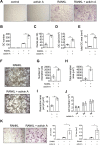
Enhanced RANKL-mediated differentiation and activity of OCs by activin A. (A) Representative images of TRAP staining after 4 days of OC differentiation in the presence of 30 ng/mL macrophage colony-stimulating factor (M-CSF, control) together with activin A (30 ng/mL) or RANKL (50 ng/mL) or RANKL plus activin A (scale bar 100 µm). (B) Corresponding OC numbers, (C) number of nuclei per OC, (D) OC size and (E) total OC area per well (n=4). (F) Representative images of resorption pit formation of WT BMDMs after 6 days of OC differentiation using calcium phosphate as substrate on stimulation with RANKL or RANKL plus activin A (scale bar 500 µm). (G) Number of resorption pits, (H) total resorption area and (I) resorption area per pit after 6 days of OC differentiation (n=3). (J) Cell numbers of BMDMs with or without activin A stimulation after 1, 2 and 3 days (n=3). (K) Representative TRAP staining of human OCs after 15 days of differentiation in the presence of M-CSF (30 ng/mL) and RANKL (50 ng/mL) with and without 100 ng/mL activin A (scale bar 200 µm) and corresponding OC number and OC size. All data are means±SEM (t-test). *P≤ 0.05, **P≤0.01, ***P≤ 0.001. OC, osteoclast; RANKL, receptor activator of nuclear factor κB ligand.
Figure 3
Figure 3
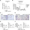
Deficiency of activin A in BMDMs had no impact on OC differentiation in vitro. (A) Secretion of activin A by FLS from ActβAf/f and ActβAd/d LysM-Cre mice after 48 hours. (B) Secretion of activin A by BM cells, BMDMs, pOCs and OCs from ActβAf/f and ActβAd/d LysM-Cre mice after 48 hours. BM cells were not stimulated; BMDMs were stimulated with M-CSF (30 ng/mL) for 3 days; pOCs were stimulated for 3 days with M-CSF (30 ng/mL) followed by stimulation with M-CSF and RANKL (50 ng/mL) for further 2 days. OCs were generated by stimulation of pOCs for a further 2 days with RANKL. All data are means±SEM (n=3–5, Mann-Whitney U test*,**). (C) Representative images of TRAP staining after 5 days of differentiation of BMDMs from ActβAf/f and ActβAd/d LysM-Cre mice (scale bar 100 µm). (D) Corresponding OC number and total OC area per well. All data are means±SEM (n=4, Mann-Whitney U test). (E) Representative images of TRAP staining after 5 days of differentiation of BMDMs from ActβAf/f hTNFtg and ActβAd/d LysM-Cre hTNFtg mice (scale bar 100 µm). (F) Corresponding OC number and total OC area per well. All data are means±SEM (n=4 and 3, respectively; Mann-Whitney U test). *P≤0.05, **P≤0.01. ActβAf/f, ActβAflox/flox; FLS, fibroblast-like synoviocytes; IL, interleukin; OC, osteoclast; RANKL, receptor activator of nuclear factor κB ligand.
Figure 4
Figure 4
Deficiency of activin A in LysM-Cre-expressing cells does not substantially affect disease severity in hTNFtg mice. (A) Paw swelling and (B) grip strength measured in ActβAf/f (n=6), ActβAd/d LysM-Cre (n=6), ActβAf/f hTNFtg (n=6) and ActβAd/d LysM-Cre hTNFtg (n=6) mice over 5–12 weeks. All data are means±SEM (two-way analysis of variance, Bonferroni’s multiple comparison test). (C) Representative images of µCT analysis from the front and back (n=4, each genotype), (D) toluidine blue-stained sections (scale bar 200 µm) and (E) TRAP-stained sections (scale bar 200 µm) from the hind paws of 12-week-old ActβAf/f (n=3), ActβAd/d LysM-Cre (n=3), ActβAf/f hTNFtg (n=6) and ActβAd/d LysM-Cre hTNFtg (n=7) mice. Quantitative histomorphometric assessment of (F) synovial pannus formation, (G) bone erosion and (H) number of OCs in tarsal joints. All data are means±SEM (Mann-Whitney U test). µCT, micro-CT; ActβAf/f, ActβAflox/flox; OC, osteoclast.
Figure 5
Figure 5
Deficiency of activin A in FLS significantly reduces OC formation. (A) Secretion of activin A by FLS from ActβAf/f and ActβAd/d ColVI-Cre mice after 48 hours. (B) Secretion of activin A by BM cells, BMDMs, pOCs and OCs from ActβAf/f and ActβAd/d ColVI-Cre mice after 48 hours. BM cells were not stimulated; BMDMs were stimulated with M-CSF (30 ng/mL) for 3 days; pOCs were stimulated for 3 days with M-CSF (30 ng/mL) followed by stimulation with M-CSF and RANKL (50 ng/mL) for a further 2 days. OCs were generated by stimulation of pOCs for a further 2 days with RANKL. All data are means±SEM (n=3–5, Mann-Whitney U test**). (C) Representative images of TRAP staining after 5 days of differentiation of BMDMs from ActβAf/f and ActβAd/d ColVI-Cre mice (scale bar 100 µm). (D) Corresponding OC number and total OC area per well. All data are means±SEM (n=4, Mann-Whitney U test). (E) Representative images of TRAP staining after 5 days of differentiation of BMDMs from ActβAf/f hTNFtg and ActβAd/d LysM-Cre hTNFtg mice (scale bar 100 µm). (F) Corresponding OC number and total OC area per well. All data are means±SEM (n=4, Mann-Whitney U test). (G) Representative fluorescence images of WT-EGFP BMDMs cocultured with FLS from ActβAf/f hTNFtg and ActβAd/d ColVI-Cre hTNFtg mice for 5 days to induce OC differentiation in presence of 1 µM PGE2 (scale bar 100 µm). (H) Corresponding OC number and total OC area per well. All data are means±SEM (ActβAf/f hTNFtg n=4, ActβAd/d ColVI-Cre hTNFtg n=5, unpaired t-test**). **P≤0.01. FLS, fibroblast-like synoviocytes; OC, osteoclast; RANKL, receptor activator of nuclear factor κB ligand.
Figure 6
Figure 6
Deficiency of activin A in ColVI-Cre-expressing cells ameliorates disease severity in hTNFtg mice. (A) Paw swelling and (B) grip strength measured in ActβAf/f (n=6), ActβAd/d ColVI-Cre (n=6), ActβAf/f hTNFtg (n=6) and ActβAd/d ColVI-Cre hTNFtg (n=6) mice over 5–12 weeks. All data are means±SEM (two-way analysis of variance, Bonferroni’s multiple comparison test). (C) Representative images of µCT analysis from the front and back (n=4, each genotype), (D) Toluidine blue-stained sections (scale bar 200 µm) and (E) TRAP-stained sections (scale bar 200 µm) from the hind paws of 12-week-old ActβAf/f (n=3), ActβAd/d ColVI-Cre (n=3), ActβAf/f hTNFtg (n=6) and ActβAd/d ColVI-Cre hTNFtg (n=6–8) mice. Quantitative histomorphometric assessment of (F) synovial pannus formation, (G) bone erosion and (H) number of OCs in tarsal joints. All data are means±SEM (Mann-Whitney U test). *P≤0.05, **P≤0.01. µCT, micro-CT; OC, osteoclast.
Figure 7
Figure 7
Activin A enhances RANKL-mediated OC formation and expression of OC differentiation markers by activation of Smad2. (A) Representative immunoblots of p-JNK, P-ERK and P-p38 of WT BMDMs stimulated with RANKL (50 ng/mL), activin A (30 ng/mL) or RANKL plus activin A in the presence of M-CSF (30 ng/mL) for 10 and 30 min. Unstimulated BMDMs served as control and glycerinaldehyde-3-phosphate dehydrogenase (GAPDH) as loading control (n=3). (B) Representative immunoblots of IκBα and NF-κB on stimulation of WT BMDMs with RANKL (50 ng/mL), activin A (30 ng/mL) or RANKL plus activin A for 10, 30 and 60 min always in the presence of M-CSF (30 ng/mL). Unstimulated BMDMs were used as control and GAPDH as loading control (n=3). (C) Representative immunoblots of P-Smad2 and Smad2 after stimulation of BMDMs with RANKL (50 ng/mL), activin A (30 ng/mL) or RANKL plus activin A for 10, 30 and 60 min (n=3) and (D) after 3 and 4 days (n=3). (E) Representative immunoblots of coimmunoprecipitation analyses. P-Smad2, coupled to protein G-labelled Dynabeads, was incubated equal amounts of protein lysates of BMDMs stimulated with RANKL, or RANKL plus activin A for 2 days was transferred to the antibody-bead complex. After incubation, pulldown was performed followed by western blot analysis against NFATc1 of the coimmunoprecipitated protein and equal amounts of the total protein lysates (input). Lysis buffer was used as negative control (neg ctrl) (n=3). (F) representative immunoblots of the OC differentiation markers integrin αν, integrin β3, DC-STAMP, NFATc1 and cathepsin K on stimulation of WT BMDMs with activin A (30 ng/mL), RANKL (50 ng/mL) or RANKL plus activin A for 3 and 4 days always in the presence of M-CSF (30 ng/mL). Unstimulated BMDMs served as control. GAPDH was used as loading control (n=3). (G) Representative images of TRAP staining after 4 days of differentiation. (H) Quantification of OC number and area on treatment of BMDMs with an ALK4/5/7 inhibitor together with RANKL (50 ng/mL) or RANKL plus activin A (30 ng/mL) for 4 days. For recovery experiments, ALK4/5/7 inhibitor was removed and BMDMs were stimulated with RANKL or RANKL plus activin A for a further 2 days. The ALK4/5/7 inhibitor was diluted in DMSO; therefore, DMSO-treated BMDMs served as control. All data are means±SEM (n=5–6, Mann-Whitney U test). All experiments were performed in the presence of M-CSF (30 ng/mL). *P≤0.05, **P≤0.01. OC, osteoclast; RANKL, receptor activator of nuclear factor κB ligand.

Similar articles

Cited by

References

    1. Smolen JS, Steiner G. Therapeutic strategies for rheumatoid arthritis. Nat Rev Drug Discov 2003;2:473–88. 10.1038/nrd1109 - DOI - PubMed
    1. Edwards JC, Willoughby DA. Demonstration of bone marrow derived cells in synovial lining by means of giant intracellular granules as genetic markers. Ann Rheum Dis 1982;41:177–82. 10.1136/ard.41.2.177 - DOI - PMC - PubMed
    1. Qu Z, Garcia CH, O'Rourke LM, et al. . Local proliferation of fibroblast-like synoviocytes contributes to synovial hyperplasia. Arthritis & Rheumatism 1994;37:212–20. 10.1002/art.1780370210 - DOI - PubMed
    1. Müller-Ladner U, Kriegsmann J, Franklin BN, et al. . Synovial fibroblasts of patients with rheumatoid arthritis attach to and invade normal human cartilage when engrafted into SCID mice. Am J Pathol 1996;149:1607–15. - PMC - PubMed
    1. Huber LC, Distler O, Tarner I, et al. . Synovial fibroblasts: key players in rheumatoid arthritis. Rheumatology 2006;45:669–75. 10.1093/rheumatology/kel065 - DOI - PubMed

Publication types