. 2022 Apr 13;12(1):6196.
doi: 10.1038/s41598-022-10218-8.

The appropriate and sequential value of standard radiograph, computed tomography and magnetic resonance imaging to characterize a bone tumor

Affiliations

The appropriate and sequential value of standard radiograph, computed tomography and magnetic resonance imaging to characterize a bone tumor

M Gaume et al. Sci Rep. .

Abstract

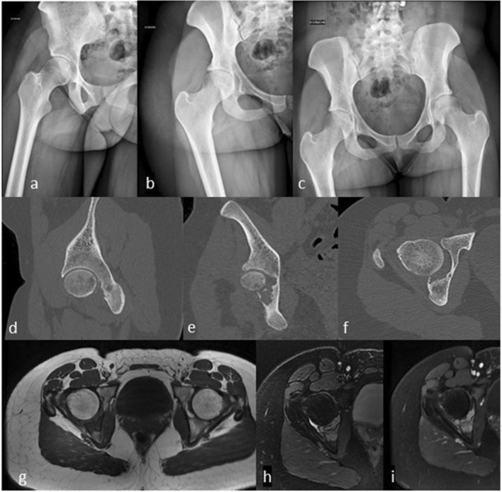
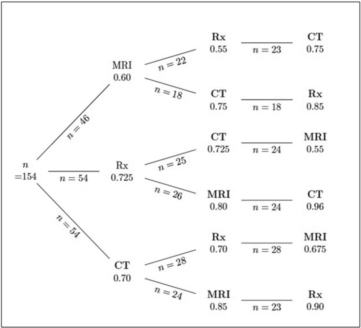
Radiographs (XR), computed tomography (CT) or magnetic resonance imaging (MRI) are regularly analyzed to determine whether a bone lesion is benign or malignant. An online quiz was created providing 15 cases with a clinical summary, MRI, CT, and XR. After each image, participants were asked to rate the probability (0-100%) the bone tumor was malignant. Order and difficulty of the images were randomly determined. Probability statements regarding the diagnosis were actualized along the sequence of exam, to quantify how the degree of belief changed to account for evidence from those exams. 64 physicians participated and provided 154 assessments from 1 (n = 18) to 3 (n = 44) different cases. After the first image, participants favored the correct malignancy status at 70%; 80% after the second and 80% after the third one. Participants were more likely to favor the correct malignancy status when the lesion was malignant and when first confronted with XR or CT, rather than MRI, though the most predictive factor of correct diagnosis was the difficulty of the case. In conclusion, the additional information provided by successive imaging studies was moderate. XR or CT seemed more appropriate than MRI as first imaging study. Bypassing XR should be discouraged.


Conflict of interest statement

The authors declare no competing interests.

Figures

Figure 1
Figure 1
Case of osteosarcoma. A 25 year-old female, no smoker and no drinker, suffered of increasing daily inflammatory right knee pain in the last 6 months. Swelling was noticed few days ago. A tender swelling over the external part of the knee was palpable. Knee mobility was normal. (1a/b) Anteroposterior and lateral radiographs of the knee. (1c/d) Coronal and axial views on computed tomography. (1e/g) T1 sagittal view, T2 fat suppressed axial and T1 sag fat suppressed axial views on magnetic resonance imaging.
Figure 2
Figure 2
Case of Langerhans cell histiocytosis. A 24 years-old female, no smoker and no drinker has been experiencing left hip pain in the last 6 months relieved by Cortisone injection, with recurrence of the pain 4 months later. No general status loss was observed. There was an inflammatory syndrome on blood test. On palpation, pain was localized on the ischium and on the mobilization of the hip without sensitive or motor dysfunction. (1a/c) Standard radiographs of the hip. (1d/f) Sagittal, frontal, and axial views on computed tomography. (1g/i) T1 and T2 axial and T1gado axial views on magnetic resonance imaging.
Figure 3
Figure 3
Probabilities of correct diagnosis along the sequence of exams.

References

    1. Franchi A. Epidemiology and classification of bone tumors. Clin. Cases Miner Bone Metab. 2012;9(2):92–95. - PMC - PubMed
    1. Teo HE, et al. Primary bone tumors of adulthood. Cancer Imaging. 2004;4(2):74–833. doi: 10.1102/1470-7330.2004.0004. - DOI - PMC - PubMed
    1. Peh WC. The role of imaging in the staging of bone tumors. Crit. Rev. Oncol. Hematol. 1999;31:147–167. doi: 10.1016/S1040-8428(99)00007-4. - DOI - PubMed
    1. Aboulafia AJ, Levin AM, Blum J, et al. Prereferral evaluation of patients with suspected bone and soft tissue tumors. Clin. Orthop. Relat. Res. 2002;397:83–88. doi: 10.1097/00003086-200204000-00012. - DOI - PubMed
    1. Schnurr C, Pippan M, Stuetzer H, et al. Treatment delay of bone tumours, compilation of a sociodemographic risk profile: A retrospective study. BMC Cancer. 2008;23(8):22. doi: 10.1186/1471-2407-8-22. - DOI - PMC - PubMed