. 2022 Apr;12(2):307-311.
doi: 10.1177/19418744211039586. Epub 2021 Aug 13.

A 52-Year-Old Man With Progressive Weakness and Incontinence

Affiliations

A 52-Year-Old Man With Progressive Weakness and Incontinence

Ayse Altintas et al. Neurohospitalist. 2022 Apr.

Abstract

Here we report a challenging case of a 52-year-old man presenting with subacute constipation, urinary retention, impotence, absent Achilles reflexes, and hypoesthesia in S2-S5 dermatomes. We review the clinical decision-making as the symptoms evolved and diagnostic testing changed over time. Once the diagnosis is settled, we discuss the sign and symptoms, additional diagnostic tools, treatment options and prognosis.

Keywords: central nervous system infections; cerebrovascular disorders; clinical specialty; general neurology; medical oncology; myelitis; neurooncology; stroke.


Conflict of interest statement

Declaration of Conflicting Interests: The author(s) declared no potential conflicts of interest with respect to the research, authorship, and/or publication of this article.

Figures

Figure 1.
Figure 1.
(A) The FLAIR sequence done in January 2019, showing multiple hyperintense lesions in cerebral white matter, the most prominent lesion being in the left frontal lobe. Frontal lobe lesion showing a patchy enhancement after IV gadolinium injection on postcontrast VIBE sequencing (B). (C-D) diffusion restriction on b1000 diffusion weighted image and ADC sequencing. (E-F) sagittal pre-contrast (E) and post-contrast (F) T1 weighted MRI image of lumbar region showing enhancement of the conus medullaris. (G-H) FLAIR and post-contrast images after treatment (March 2019) with near to complete resolution of the frontal lobe lesion.
Figure 2.
Figure 2.
Microscopic images of the brain biopsy with higher powers of magnification with different staining. A, Complete specimen, red arrow showing the area with foamy macrophages, black arrow showing the area with reactive changes. B, Foamy macrophages with red blood cells. C, Neurofilament protein staining of the infiltrative region on the right and the preserved area on the left. D, Reactive changes with a few intravascular atypical cells (circle). E, CD20 staining with CD20+ intravascular cells (circle). F, Ki-67 staining with ki-67+ positive cells (circle).
Figure 3.
Figure 3.
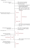
Timeline of diagnostic procedures and treatment until the diagnosis of IVL.

References

    1. Orendácová J, Cízková D, Kafka J, et al. Cauda equina syndrome. Prog Neurobiol. 2001;64(6) 613–637. - PubMed
    1. Eberhardt O, Küker W, Dichgans J, Weller M. HSV-2 Sacral radiculitis (Elsberg syndrome). Neurology. 2004;63(4):758–759. - PubMed
    1. Savoldi F, Kaufmann TJ, Flanagan EP, Toledano M, Weinshenker BG. Elsberg syndrome a rarely recognized cause of cauda equina syndrome and lower thoracic myelitis. Neurol Neuroimmunol Neuroinflamm. 2017;4(4):e355. - PMC - PubMed
    1. Beristain X, Biagio A. The neurological masquerade of intravascular lymphomatosis. Arch Neurol. 2002;59(3):439–443. - PubMed
    1. Ponzoni M, Ferreri AJM. Intravascular lymphoma: a neoplasm of ‘homeless’ lymphocytes? Hematol Oncol. 2006;24(3):105–112. - PubMed