. 2022 Apr 15;149(8):dev200361.
doi: 10.1242/dev.200361. Epub 2022 Apr 22.

Natural killer cells act as an extrinsic barrier for in vivo reprogramming

Affiliations

Natural killer cells act as an extrinsic barrier for in vivo reprogramming

Elena Melendez et al. Development. .

Abstract

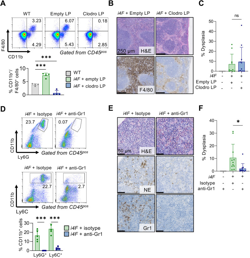
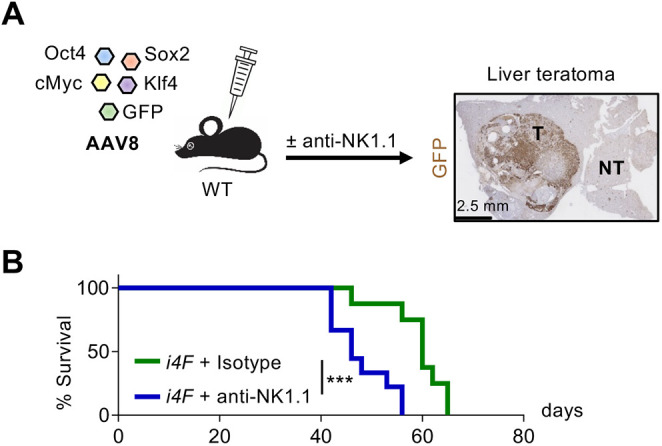
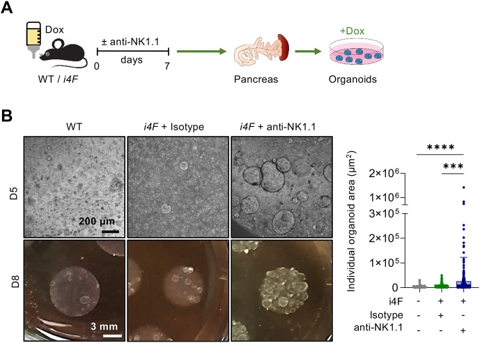
The ectopic expression of the transcription factors OCT4, SOX2, KLF4 and MYC (OSKM) enables reprogramming of differentiated cells into pluripotent embryonic stem cells. Methods based on partial and reversible in vivo reprogramming are a promising strategy for tissue regeneration and rejuvenation. However, little is known about the barriers that impair reprogramming in an in vivo context. We report that natural killer (NK) cells significantly limit reprogramming, both in vitro and in vivo. Cells and tissues in the intermediate states of reprogramming upregulate the expression of NK-activating ligands, such as MULT1 and ICAM1. NK cells recognize and kill partially reprogrammed cells in a degranulation-dependent manner. Importantly, in vivo partial reprogramming is strongly reduced by adoptive transfer of NK cells, whereas it is significantly increased by their depletion. Notably, in the absence of NK cells, the pancreatic organoids derived from OSKM-expressing mice are remarkably large, suggesting that ablating NK surveillance favours the acquisition of progenitor-like properties. We conclude that NK cells pose an important barrier for in vivo reprogramming, and speculate that this concept may apply to other contexts of transient cellular plasticity.

Keywords: Immune system; Mouse; NK receptor ligands; Natural killer cells; Organoids; Plasticity; Pluripotency; Reprogramming.


Conflict of interest statement

Competing interests M.S. is shareholder of Senolytic Therapeutics, Rejuveron Senescence Therapeutics and Life Biosciences.

Figures

Fig. 1.
Fig. 1.
Immune cell populations infiltrate the pancreas during in vivo reprogramming. (A) UMAP plot visualizing immune cell infiltrates in the pancreas. WT (n=2) and i4F (n=3) mice were treated with doxycycline in the drinking water (1 mg/ml) for 1 week to induce partial reprogramming. (B) UMAP plot of immune cell populations. Clusters were annotated using the most-significant markers of each cluster and the FindAllMarkers function of Seurat (v3). The minor clusters correspond to endothelial cells (#), stellate cells (&) and marginal zone B cells ($). (C) Flow cytometry analysis of the main immune populations infiltrating the pancreas after WT (n=9), p53-null (n=5), i4F (n=6) and i4F;p53-null (n=4) mice were treated with doxycycline for 7 days. Cells were gated from DAPI/CD45+ cells. Data are pooled from two independent experiments and represent mean±s.e.m. **P<0.01 (WT versus i4F) and #P<0.05, ##P<0.01 (i4F versus i4F;p53-null) evaluated using the unpaired two-tailed Student's t-test. (D) Previously published RNA-seq data generated in our laboratory (Mosteiro et al., 2016) from the pancreas of i4F and i4F;p53-null (high-reprogramming) mice were used to perform gene set enrichment analysis (GSEA) against a published signature (KEGG entry: mmu04650) of NK cell-mediated cytotoxicity. B, B cells; DC, dendritic cells; Mφ, macrophages; MDSC, myeloid derived suppressor cells; NK, natural killer cells; NT, neutrophils; T, T cells.
Fig. 2.
Fig. 2.
NK cells eliminate partially reprogrammed cells in vitro. (A) i4F MEFs were reprogrammed in vitro with doxycycline (Dox; 1 μg/ml) for 11 days. From days 2 to 6, primed NK cells were added to the medium and cells were then co-cultured for 5 days in co-culture medium. On day 11, iPSC colonies were scored by AP staining. (B,C) Quantification (B) and representative images (C) of NK cells co-cultured with i4F MEFs with or without doxycycline at E:T (NK:MEF) ratios of 0.5:1, 2.3:1 and 4.5:1. Data pooled from two independent experiments. (D) Delta mean fluorescent intensity (ΔMFI) of the total H60, MULT1, ICAM1, RAE1 and CD155 expression in i4F MEFs reprogrammed with doxycycline (1 μg/ml) at different time points (n=3). (E) Co-culture experiment in which primed NK cells were seeded in Transwells (TW) at an NK:MEF ratio of 1:1 to avoid cell–cell contact (n=3). (F) Co-culture experiment using ConA to disrupt the function of lytic granules secreted by NK cells at an NK:MEF ratio of 1:1 (n=4). (G) Co-culture experiment using the blocking antibody anti-NKG2D, which was added to the medium on days 2 and 4 of reprogramming (n=4). All data are mean±s.d.; *P<0.05, **P<0.001, ***P<0.001, ****P<0.0001 evaluated using the unpaired two-tailed Student's t-test (B,E-G) or one-way ANOVA (D). ns, not significant.
Fig. 3.
Fig. 3.
NK cells recruited to the pancreas of i4F mice release lytic granules upon ligand-dependent activation. (A) Delta mean fluorescent intensity (ΔMFI) of the total RAE1, MULT1, ICAM1, CD155 and RAE1 expression in pancreas of WT and i4F mice treated with doxycycline (1 mg/ml) for 7 days (n=4). (B) WT and i4F mice were treated with doxycycline (1 mg/ml) for 7 days and expression levels of NK-activating ligands (Rae1, Mult1, Icam1 and CD155) in pancreatic tissue were assessed by RT-qPCR (n=5). Data in A,B are mean±s.d. *P<0.05, **P<0.01, ****P<0.0001 evaluated using the unpaired two-tailed Student's t-test. (C) Representative H&E (above) and NK1.1 staining (below) of pancreata of WT and i4F mice treated with doxycycline for 7 days (n=5). (D) Subpopulations of infiltrating NK cells in pancreas undergoing reprogramming on days 3 (D3), 5 (D5) and 7 (D7) of doxycycline treatment. Cell populations were gated on CD3/NK1.1+ cells and stained for the indicated activation, degranulation and inhibitory markers. Correlation of NKG2A+ and CD107a+ cells includes all time points. In the box-and-whisker plots, boxes show median and upper and lower quartiles, and whiskers indicate minimum and maximum values; **P<0.01, ***P<0.001, ****P<0.0001 evaluated using one-way ANOVA (data pooled from four independent experiments).
Fig. 4.
Fig. 4.
NK cells are an extrinsic barrier for in vivo reprogramming. (A) WT (n=10) and i4F mice were treated with doxycycline (Dox; 1 mg/ml) for 7 days. i4F mice were treated with either isotype control antibody (1) (n=21) or anti-NK1.1 antibody (2) (n=17) (days −1, 3 and 5), or received adoptive transfer of 3.8×10⁶ NK cells on day 3 of reprogramming (3) (n=10). (B) Representative flow cytometry plots and quantification of NK cells in blood of randomly selected mice on day 5 of reprogramming. NK cells were gated as CD3/NKp46+ cells (n=5 for WT+dox and n=6 for i4F+isotype and i4F+anti-NK1.1 groups). (C) Representative H&E and NANOG staining of pancreatic tissue (upper) and quantifications of the percentage of dysplasia and number of NANOG+ cells (lower). Data pooled from five independent experiments in conditions 1 and 2, and from two independent experiments in condition 3. (D) Representative H&E images of caerulein-induced pancreatitis (100 mg/kg, 7 times/day) treated for 2 days with either isotype control or anti-NK1.1 (n=13) (upper). Quantification of the percentage of acinar-to-ductal metaplasia (ADM) in the pancreata. All data are mean±s.d.; *P<0.05, **P<0.01, ****P<0.0001 evaluated using the unpaired two-tailed Student's t-test. ns, not significant.
Fig. 5.
Fig. 5.
Mφ and Gr1+ cell depletion during in vivo reprogramming. (A) Representative flow cytometry plots and quantification of F4/80 and CD11b double-positive populations in blood 16 h after liposome (LP) were administered intraperitoneally on day 3 of reprogramming. Mice were randomly selected (n=4). Cells were gated from CD45+ cells. (B) Representative H&E and F4/80 immunohistochemistry of the spleen of mice treated with doxycycline and empty or clodronate LP for 7 days. (C) Dysplasia quantification in the pancreas of mice reprogrammed with empty or clodronate LP for 7 days (n=6 for WT, n=10 for empty LP and n=11 for clodronate LP groups). (D) Representative flow cytometry plots and quantification of Ly6C and CD11b, and Ly6G and CD11b populations in blood on day 5 from reprogramming from mice treated with anti-Gr1 or isotype control antibodies. Cells were gated from CD45+ cells. (E) Representative H&E, neutrophil elastase (NE) and Gr1 immunohistochemistry in the spleen of partially reprogrammed mice treated with anti-Gr1 or isotype control antibodies for 7 days. (F) Dysplasia quantification in the pancreas of mice reprogrammed for 7 days with either anti-Gr1 or isotype control (n=8 for WT and n=11 for i4F and anti-Gr1 groups). All data are mean±s.d.; *P<0.05, ***P<0.001 evaluated using the unpaired two-tailed Student's t-test. ns, not significant.
Fig. 6.
Fig. 6.
NK1.1+ cell depletion enables full reprogramming and promotes teratoma formation. (A) WT mice were retro-orbitally injected with scAAV8 SFFV-hCO-O/K/S/M. A scAAV8 vector encoding GFP was additionally added as tracer. Anti-NK1.1 or isotype control antibodies were injected intraperitoneally on days −1, 3 and 5 during the first week, and then once a week until liver teratomas were palpable. (B) Survival curve upon teratoma formation in the liver. Data pooled from two independent experiments (n=9). Data are percentage of alive mice; ***P<0.001 evaluated using log-rank (Mantel-Cox) test. NT, nonteratoma; T, teratoma.
Fig. 7.
Fig. 7.
NK1.1+ cell depletion promotes the survival of pancreatic cells with high plasticity. (A) Mice were treated with doxycycline (Dox) and anti-NK1.1 or isotype control antibodies for 7 days. Pancreata were dissociated to the single cell level and 3×10⁵ cells/well per condition were embedded in Matrigel (n=3) (B). Representative images on day 5 (D5) and day 8 (D8) after seeding (left), and quantification of organoids size at D10 (right) (each dot represents one organoid from three biological replicates; ten images were measured per sample). Data are mean±s.d.; ***P<0.001, ****P<0.0001 evaluated using the unpaired two-tailed Student's t-test.

References

    1. Abad, M., Mosteiro, L., Pantoja, C., Cañamero, M., Rayon, T., Ors, I., Graña, O., Megías, D., Domínguez, O., Martínez, D.et al. (2013). Reprogramming in vivo produces teratomas and iPS cells with totipotency features. Nature 502, 340-345. 10.1038/nature12586 - DOI - PubMed
    1. Ablamunits, V., Henegariu, O., Preston-Hurlburt, P. and Herold, K. C. (2011). NKG2A is a marker for acquisition of regulatory function by human CD8+ T cells activated with anti-CD3 antibody. Eur. J. Immunol. 41, 1832-1842. 10.1002/eji.201041258 - DOI - PMC - PubMed
    1. Alter, G., Malenfant, J. M. and Altfeld, M. (2004). CD107a as a functional marker for the identification of natural killer cell activity. J. Immunol. Methods 294, 15-22. 10.1016/j.jim.2004.08.008 - DOI - PubMed
    1. Arabacı, D. H., Terzioğlu, G., Bayırbaşı, B. and Önder, T. T. (2021). Going up the hill: chromatin-based barriers to epigenetic reprogramming. FEBS J. 288, 4798-4811. 10.1111/febs.15628 - DOI - PubMed
    1. Bankhead, P., Loughrey, M. B., Fernández, J. A., Dombrowski, Y., Mcart, D. G., Dunne, P. D., Mcquaid, S., Gray, R. T., Murray, L. J., Coleman, H. G.et al. (2017). QuPath: open source software for digital pathology image analysis. Sci. Rep. 7, 16878. 10.1038/s41598-017-17204-5 - DOI - PMC - PubMed

Publication types

MeSH terms

Substances