. 2022 Jun 28;13(3):e0043322.
doi: 10.1128/mbio.00433-22. Epub 2022 Apr 14.

The Leishmania donovani Ortholog of the Glycosylphosphatidylinositol Anchor Biosynthesis Cofactor PBN1 Is Essential for Host Infection

Affiliations

The Leishmania donovani Ortholog of the Glycosylphosphatidylinositol Anchor Biosynthesis Cofactor PBN1 Is Essential for Host Infection

Adam Roberts et al. mBio. .

Abstract

Visceral leishmaniasis is a deadly infectious disease caused by Leishmania donovani, a kinetoplastid parasite for which no licensed vaccine is available. To identify potential vaccine candidates, we systematically identified genes encoding putative cell surface and secreted proteins essential for parasite viability and host infection. We identified a protein encoded by LdBPK_061160 which, when ablated, resulted in a remarkable increase in parasite adhesion to tissue culture flasks. Here, we show that this phenotype is caused by the loss of glycosylphosphatidylinositol (GPI)-anchored surface molecules and that LdBPK_061160 encodes a noncatalytic component of the L. donovani GPI-mannosyltransferase I (GPI-MT I) complex. GPI-anchored surface molecules were rescued in the LdBPK_061160 mutant by the ectopic expression of both human genes PIG-X and PIG-M, but neither gene could complement the phenotype alone. From further sequence comparisons, we conclude that LdBPK_061160 is the functional orthologue of yeast PBN1 and mammalian PIG-X, which encode the noncatalytic subunits of their respective GPI-MT I complexes, and we assign LdBPK_061160 as LdPBN1. The LdPBN1 mutants could not establish a visceral infection in mice, a phenotype that was rescued by constitutive expression of LdPBN1. Although mice infected with the null mutant did not develop an infection, exposure to these parasites provided significant protection against subsequent infection with a virulent strain. In summary, we have identified the orthologue of the PBN1/PIG-X noncatalytic subunit of GPI-MT I in trypanosomatids, shown that it is essential for infection in a murine model of visceral leishmaniasis, and demonstrated that the LdPBN1 mutant shows promise for the development of an attenuated live vaccine. IMPORTANCE Visceral leishmaniasis is a deadly infectious disease caused by the parasites Leishmania donovani and Leishmania infantum. It remains a major global health problem, and there is no licensed highly effective vaccine. Molecules that are displayed on the surface of parasites are involved in host-parasite interactions and have important roles in immune evasion, making vaccine development difficult. One major way in which parasite surface molecules are tethered to the surface is via glycophosphatidylinositol (GPI) anchors; however, the enzymes required for all the biosynthetic steps in these parasites are not known. Here, we identified the enzyme required for an essential step in the GPI anchor-biosynthetic pathway in L. donovani, and we show that while parasites lacking this gene are viable in vitro, they are unable to establish infections in mice, a property we show can be exploited to develop a live genetically attenuated parasite vaccine.

Keywords: bioluminescence; enzyme; glycophosphatidylinositol biosynthesis; leishmaniasis; mass spectrometry; parasitology.


Conflict of interest statement

The authors declare no conflict of interest.

Figures

FIG 1
FIG 1
Genetic targeting of LdBPK_061160 caused a cellular adhesion phenotype due to the catastrophic loss of cell surface GPI-anchored molecules. (A) Diagnostic PCRs demonstrating genetic targeting of LdBPK_061160 locus. Schematic of the endogenous locus showing location and orientations of diagnostic primers and PCR products which demonstrate targeted replacement and ectopic overexpression of LdBPK_061160 in the null mutant with genetic rescue from a nonendogenous locus. (B) Procyclic promastigote stage L. donovani parasites lacking LdBPK_061160 exhibit increased adherence to tissue culture flasks (right) compared to the parental control (left). Bar, 20 μm. (C and D) Mutant parasites showed complete loss of cell surface GPI-anchored molecules as measured by flow cytometry, including GP63 (C) and LPG (D), compared to the parental control (Ctrl). In both panels C and D, surface expression was fully rescued by overexpression of LdBPK_061160 (Rescue). (E) Parasites lacking LdBPK_061160 were more brightly stained with convalescent-phase serum from chronically infected mice than the parental control, an effect that was rescued by expression of LdBPK_061160. (F) Images of the parasites analyzed for panel E showing that the epitopes recognized by chronically infected serum were distributed over the entire cell surface of the promastigote-stage L. donovani parasites. A representative image from two independent experiments is shown. Bar, 15 μm.
FIG 2
FIG 2
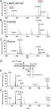
Disruptive targeting of LdBPK_061160 causes an accumulation of the metabolite GlcN-PI demonstrating its requirement for GPI-mannosyltransferase I activity. Negative-ion electrospray mass spectrometry (ES-MS) spectra of lipid extracts from LdBPK_061160−/− (A), parental strain (B), and rescued null (C) showing the selective accumulation of the GlcN-PI precursor ions (red). (D) Schematic showing ES-MS2 product ions of the GlcN-PI precursor ions. The ES-MS2 HCD (high-energy C-trap dissociation) product ion spectra for the [M − H] precursor ion at m/z 984.64 (E) and at m/z 1,012.67 (F) are shown. The difference between the precursors identified at m/z 984.64 (E) and 1,012.67 (F) was due to the length of the alkyl chain attached at position sn-1 containing either C16:0 (E) or C18:0 (F). The product ion assignments for GlcN-PI species are indicated in panel D. The ion at m/z 402.08 representing [GlcN-myo-inositol-1,2-cyclic phosphate] and its dehydration product at m/z 384.07 are characteristic of negative GlcN-PI product ion spectra. The ions at m/z 152.99 and 283.26 are [glycerol-cyclic phosphate] and [CH3(CH2)16COO], respectively. The neutral loss of C18:0/(CH3(CH2)16COO) is represented by ions at m/z 700.37 (E) and 728.40 (F), whereas the neutral loss of C18:0/(CH3(CH2)16COO) and GlcN-inositol is represented by ions at m/z 377.24 (E) and 405.28 (F). The complete lipid profiles observed in negative-ion mode are shown in Fig. S1.
FIG 3
FIG 3
LdBPK_061160 is the L. donovani PBN1 orthologue. (A) Comparison of the protein sequence architecture of LdBPK_061160 with proteins of known flippase activity and the human and yeast PIG-X/PBN1 proteins. Note that LdBPK_061160 shares structural features with the PIG-X/PBN1 proteins and not the flippases. Predicted signal peptides are in blue, and transmembrane regions are in red. (B) Overexpression of the genes encoding Homo sapiens PIG-X and PIG-M in LdBPK_061160 mutants restores presence of GPI-anchored proteins at the cell surface. Histograms show cell surface staining of the GPI-anchored GP63 protein in LdBPK_061160 mutants overexpressing human genes encoding PIG-X, PIG-M, or both using a FITC-conjugated anti-GP63 monoclonal antibody by fluorescence-activated cell sorting (FACS). (C and D) Experimental identification of the PBN1 orthologue in T. cruzi. LdPBN1 mutants that lack surface expression of the GPI-anchored cell surface molecules GP63 and LPG were compared to the parental line and were genetically complemented with the gene encoding T. cruzi PBN1 (TcPBN1) (Contig ADWP02007247; nucleotides 5233.6071) or LdPBN1 as a positive control. Levels of cell surface GP63 (C) and LPG (D) were quantified by staining parasites with monoclonal antibodies and flow cytometry. Results of one representative experiment of three are shown.
FIG 4
FIG 4
L. donovani PBN1 is essential for host infection. (A) Diagram showing examples of how infected animals were regionally gated to quantify infection load using bioluminescence imaging of the liver and the area corresponding to both the spleen and inguinal lymph node. (B) Groups of five mice were inoculated with stationary-phase promastigotes from parental (Ctrl), PBN1−/−, and PBN1−/− parasites transfected with a PBN1 expression rescue plasmid (rescue), and infections were longitudinally monitored by bioluminescence imaging. Quantification of liver (C) and spleen (D) bioluminescent signals from parental control parasites (blue), PBN1-deficient (red) and PBN1-deficient parasites rescued by overexpression of the PBN1 (black). The units of bioluminescence are 1 × 106 photons per second. Data are means and standard deviations (SD) (n = 5). Dotted line represents the average background bioluminescence from five unchallenged mice from a separate experiment measured over 77 days. Quantification of PBN1−/− infection between 4 h to 30 days postinfection in the liver (E). Bioluminescence is reported as 1 × 106 photons per second, and the data are from a single experiment where the group size was 5.
FIG 5
FIG 5
Inoculation with the LdPBN1 mutant is capable of eliciting a robust humoral immune response that offers significant protection against reinfection. Serum harvested from BALB/c mice that had been inoculated with either parental L. donovani parasites (black line) or PBN1−/− parasites (red line) for 14 (A) and 77 (B) days was diluted and used to stain live promastigotes by fluorescence-activated cell sorting, and results were compared to those for control mouse sera (blue line). (C to F) Inoculation with PBN1−/− parasites elicits protection against subsequent L. donovani infection. (C) Mice inoculated with the L. donovani PBN1−/− mutant were rested for 84 days before being infected with the bioluminescent virulent parental L. donovani strain, and the parasitemia was quantified in the livers (C) and spleens (D) of infected animals (red line, n = 3). Two mice were left uninfected to control for the possibility of recrudescence (black line), and four naive mice were infected as a positive infection control (blue line). (E and F) A group of 10 mice were inoculated with PBN1−/− mutant parasites and rested for 8 weeks prior to reinfection with the parental L. donovani strain and parasitemia quantified by bioluminescence in the liver (E) and spleen (F). Significantly different levels of parasitemia were assigned using a two-tailed Student's t test. *, P < 0.05, and **, P < 0.001, compared to a control group of 10 unimmunized mice. Data are from a single experiment, and bioluminescence is reported as 1 × 106 photons per second.

References

    1. WHO. 2021. Leishmaniasis fact sheet. WHO fact sheet.
    1. WHO. 2016. Leishmaniasis in high-burden countries: an epidemiological update based on data reported in 2014. Wkly Epidemiol Rec 91:287–296. - PubMed
    1. Lindoso JAL, Costa JML, Queiroz IT, Goto H. 2012. Review of the current treatments for leishmaniases. Res Rep Trop Med 3:69–77. doi:10.2147/RRTM.S24764. - DOI - PMC - PubMed
    1. Srivastava S, Mishra J, Gupta AK, Singh A, Shankar P, Singh S. 2017. Laboratory confirmed miltefosine resistant cases of visceral leishmaniasis from India. Parasit Vectors 10:49. doi:10.1186/s13071-017-1969-z. - DOI - PMC - PubMed
    1. Croft SL, Sundar S, Fairlamb AH. 2006. Drug resistance in leishmaniasis. Clin Microbiol Rev 19:111–126. doi:10.1128/CMR.19.1.111-126.2006. - DOI - PMC - PubMed

Publication types

Substances