. 2022 Jun 28;13(3):e0030122.
doi: 10.1128/mbio.00301-22. Epub 2022 Apr 14.

Extracellular Vesicles Regulate Biofilm Formation and Yeast-to-Hypha Differentiation in Candida albicans

Affiliations

Extracellular Vesicles Regulate Biofilm Formation and Yeast-to-Hypha Differentiation in Candida albicans

Leandro Honorato et al. mBio. .

Abstract

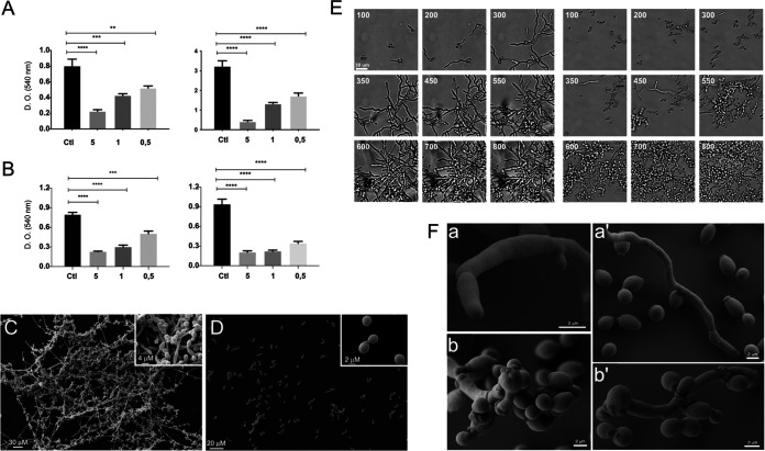
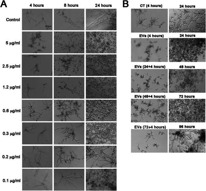
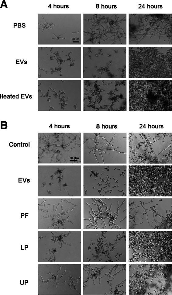
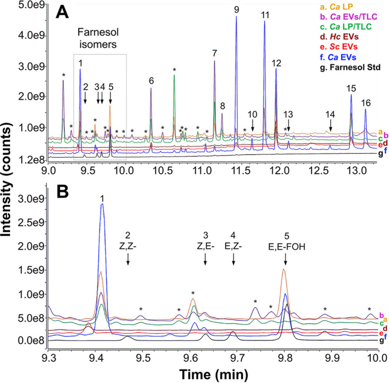
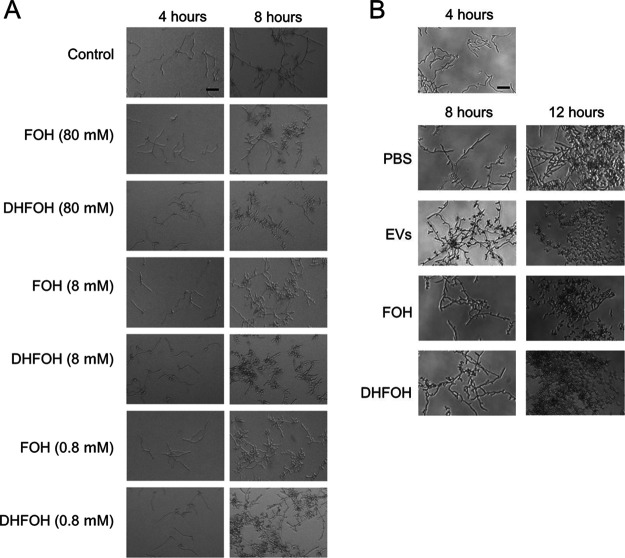
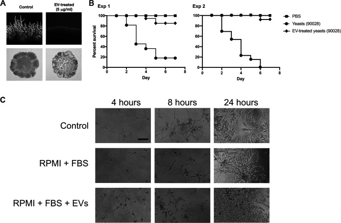
In this study, we investigated the influence of fungal extracellular vesicles (EVs) during biofilm formation and morphogenesis in Candida albicans. Using crystal violet staining and scanning electron microscopy (SEM), we demonstrated that C. albicans EVs inhibited biofilm formation in vitro. By time-lapse microscopy and SEM, we showed that C. albicans EV treatment stopped filamentation and promoted pseudohyphae formation with multiple budding sites. The ability of C. albicans EVs to regulate dimorphism was further compared to EVs isolated from different C. albicans strains, Saccharomyces cerevisiae, and Histoplasma capsulatum. C. albicans EVs from distinct strains inhibited yeast-to-hyphae differentiation with morphological changes occurring in less than 4 h. EVs from S. cerevisiae and H. capsulatum modestly reduced morphogenesis, and the effect was evident after 24 h of incubation. The inhibitory activity of C. albicans EVs on phase transition was promoted by a combination of lipid compounds, which were identified by gas chromatography-tandem mass spectrometry analysis as sesquiterpenes, diterpenes, and fatty acids. Remarkably, C. albicans EVs were also able to reverse filamentation. Finally, C. albicans cells treated with C. albicans EVs for 24 h lost their capacity to penetrate agar and were avirulent when inoculated into Galleria mellonella. Our results indicate that fungal EVs can regulate yeast-to-hypha differentiation, thereby inhibiting biofilm formation and attenuating virulence. IMPORTANCE The ability to undergo morphological changes during adaptation to distinct environments is exploited by Candida albicans and has a direct impact on biofilm formation and virulence. Morphogenesis is controlled by a diversity of stimuli, including osmotic stress, pH, starvation, presence of serum, and microbial components, among others. Apart from external inducers, C. albicans also produces autoregulatory substances. Farnesol and tyrosol are examples of quorum-sensing molecules (QSM) released by C. albicans to regulate yeast-to-hypha conversion. Here, we demonstrate that fungal EVs are messengers impacting biofilm formation, morphogenesis, and virulence in C. albicans. The major players exported in C. albicans EVs included sesquiterpenes, diterpenes, and fatty acids. The understanding of how C. albicans cells communicate to regulate physiology and pathogenesis can lead to novel therapeutic tools to combat candidiasis.

Keywords: Candida albicans; biofilm; extracellular vesicles; lipids; yeast-to-hypha inhibition.


Conflict of interest statement

The authors declare a conflict of interest. Dr. Maurizio Del Poeta, M.D. is a Co-Founder and Chief Scientific Officer (CSO) of MicroRid Technologies Inc.

Figures

FIG 1
FIG 1
EVs from C. albicans inhibit biofilm formation and yeast-to-hyphae differentiation. Both protocols described in Materials and Methods for EV isolation were used with identical results. The results presented here were obtained from C. albicans 90028 EVs (yeast cells) isolated from liquid culture medium. (A) C. albicans (90028) (105 yeasts) were inoculated into RPMI-MOPS in the presence or absence of C. albicans 90028 EVs. Different concentrations of EVs (0.5, 1, and 5 μg/mL, based on sterol content) were added to wells containing C. albicans (90028) yeasts, and cells were incubated to develop biofilm for 24 and 48 h in 96-well microplates. (B) C. albicans (90028) (105 yeasts) were inoculated into RPMI-MOPS and incubated for 90 min. The nonadherent cells were washed out, and fresh medium containing C. albicans 90028 EVs (0.5, 1, and 5 μg/mL, based on sterol content) was added. PBS was used as control. Biofilm formation was quantified by crystal violet method. Group comparisons were submitted to one-way analysis of variance (ANOVA) with Dunnett’s correction (**, P < 0.002; ***, P < 0.001; ****, P < 0.0001). (C and D) For SEM, C. albicans (strain 90028) yeast cells, untreated (C) or treated with C. albicans EVs (5 μg/mL) (D), were inoculated onto coverslips previously coated with poly-l-lysine in RPMI-MOPS and incubated for 24 h. SEM images show the effect of EVs after 24 h of growth. Insets in panels C and D depict higher magnification of each condition. Bar, 30 μm. (E) C. albicans (90028) yeasts were inoculated into M199 medium to induce filamentation in the presence or absence of C. albicans 90028 EVs (5 μg/mL, based on sterol content). Cellular density was monitored, and the pictures represent the minutes of incubation (from 100 to 800 min). Bar, 30 μm. Growth under control conditions (absence of C. albicans 90028 EVs) and in the presence of C. albicans 90028 EVs. The numbers indicate the time in minutes. Bar, 30 μm. (F) C. albicans (90028) yeasts were inoculated into M199 medium to induce filamentation in the presence or absence of EVs (5 μg/mL, based on sterol content). SEM of cells after 24 h showing the hyphae and yeasts under control conditions (a and a′) in contrast with C. albicans 90028 EVs that led to the presence of pseudohyphae with multiple budding sites and yeasts (b and b′).
FIG 2
FIG 2
The inhibitory effect of EVs from C. albicans (strain 90028) on yeast-to-hypha conditions is dose dependent and has a long-term effect. (A) C. albicans (90028) yeasts were inoculated into M199 (pH 7) in the presence or absence of C. albicans 90028 EVs in different concentrations (equivalent to 0.1, 0.2, 0.3, 0.6, 1, 1.2, 2.5, and 5 μg sterol/mL). Bar, 30 μm. (B) The inhibitory effect of a single C. albicans EV treatment was tested after 24, 48, 72, and 96 h. Briefly, overnight-treated yeasts were washed with sterile PBS, and aliquots of (2.5 × 10³ cells/well) cells were transferred to fresh M199 medium. Yeasts were incubated under the same conditions without addition of EVs for 24 h, and the differentiation was accompanied microscopically. This step was repeated until the reestablishment of C. albicans filamentation. Bar, 30 μm. Experiments were performed in biological triplicate with consistent results.
FIG 3
FIG 3
EVs from different C. albicans strains inhibit yeast-to-hyphae transition of strains 90028 and 10231. NTA profile of EVs produced by C. albicans strains SC5314 (A), 90028 (B), and 10231 (C) showed similar properties of EVs. C. albicans yeast cells of strains 90028 (D) or 10231 (E) were inoculated into M199 (pH 7) in the presence or absence of C. albicans EVs (equivalent to 5 μg sterol/mL) from distinct strains of C. albicans (90028, 10231, and SC5314). The effects were visualized after 4, 8, and 24 h. PBS was added alone as a control. Bar, 30 μm. Experiments were performed four times with consistent results.
FIG 4
FIG 4
Thermoresistant molecules carried by C. albicans EVs are associated with yeast-to-hyphae inhibitory effect. (A) C. albicans (90028) yeast cells were inoculated into M199 (pH 7) in the presence or absence of C. albicans 90028 EVs or heat-treated EVs in a concentration equivalent to 5 μg sterol/mL. The effect was visualized after 4, 8, and 24 h. PBS was added alone as a control. Bar, 30 μm. The results shown are representative of three independent experiments. (B) C. albicans (90028) yeasts were inoculated into M199 in the presence or absence of C. albicans EVs, EV-derived protein-rich fraction (PF), or lipid lower (LP) or upper phase (UP) in a concentration equivalent to 5 μg sterol/mL. The effect was visualized after 4, 8, and 24 h. PBS was added alone as a control. Bar, 30 μm.
FIG 5
FIG 5
GC-MS analysis of fungal lipids and EVs. Lipids were extracted from the C. albicans 90028 LP fraction and EVs and from the RP-TLC band comigrating with farnesol in the two samples. Lipids were also extracted from H. capsulatum and S. cerevisiae EVs. The resulting samples were analyzed by GC-MS/MS. (A) GC-MS full-scan chromatogram. (B) The chromatogram region containing the four farnesol isomers is indicated (dashed rectangle) and shown in detail. A standard mixture containing the four farnesol isomers (Z,Z-, Z,E-, E,Z-, and E,E-FOH) was used as reference (black line/trace). The GC-MS full-scan traces were overlaid and labeled (a to g) to facilitate visualization and comparison. Farnesol isomers and peaks of interest are indicated numerically and annotated in Table 1. Asterisks indicate contaminants (e.g., phthalates and other plasticizers) or compounds not identified in the library (low structural match probability). Ca, C. albicans; Hc, H. capsulatum; Sc, S. cerevisiae.
FIG 6
FIG 6
C. albicans EVs, FOH, and DHFOH reversed the yeast-to-hypha differentiation. (A) C. albicans (90028) yeast cells were inoculated into M199 (pH 7) in the presence or absence of distinct concentrations of FOH or DHFOH. (B) C. albicans (90028) yeasts were inoculated into M199 and incubated for 4 h to induce hyphae formation. Then, PBS (control), C. albicans EVs (5 μg/mL), FOH (25 μM), or DHFOH (25 μM) were added to the wells. Cell morphology was visualized after 8 and 12 h. Bars, 30 μm.
FIG 7
FIG 7
The reversing effect on C. albicans morphogenesis caused by db-cAMP is inactivated by preincubation with C. albicans EVs. db-cAMP preincubated with EVs (5 μg/mL) does not reverse the inhibitory effect of C. albicans 90028 EVs on C. albicans phase transition. The addition of db-cAMPc (10 mM) reverses the yeast-to-hyphae inhibition caused by C. albicans 90028 pretreated with C. albicans 90028 EVs. Bar, 30 μm.
FIG 8
FIG 8
C. albicans EVs decrease the capacity of C. albicans to penetrate agar and attenuate virulence in Galleria mellonella. (A) C. albicans (90028) yeasts were treated with PBS (control) or C. albicans 90028 EVs in PBS (5 μg/mL) and then plated onto M199 agar for 7 days. The presence of filamentation (top panels) and agar invasion (bottom panels) was observed only under control conditions. (B) G. mellonella larvae were infected with 105 C. albicans yeast untreated or pretreated with C. albicans 90028 EVs (5 μg/mL for 24 h), and survival was monitored (experiments 1 and 2). PBS indicates uninfected larvae injected with PBS. (C) C. albicans (90028) yeasts were inoculated into RPMI supplemented with FBS (10%) in the presence or absence of C. albicans EVs in a concentration equivalent to 5 μg sterol/mL.

References

    1. Kühbacher A, Burger-Kentischer A, Rupp S. 2017. Interaction of Candida species with the skin. Microorganisms 5:32. doi:10.3390/microorganisms5020032. - DOI - PMC - PubMed
    1. Ranjan A, Dongari-Bagtzoglou A. 2018. Tipping the balance: C. albicans adaptation in polymicrobial environments. JoF 4:112. doi:10.3390/jof4030112. - DOI - PMC - PubMed
    1. Zhang S-L, Wang S-N, Miao C-Y. 2017. Influence of microbiota on intestinal immune system in ulcerative colitis and its intervention. Front Immunol 8:1674. doi:10.3389/fimmu.2017.01674. - DOI - PMC - PubMed
    1. Höfs S, Mogavero S, Hube B. 2016. Interaction of Candida albicans with host cells: virulence factors, host defense, escape strategies, and the microbiota. J Microbiol 54:149–169. doi:10.1007/s12275-016-5514-0. - DOI - PubMed
    1. Basso V, d’Enfert C, Znaidi S, Bachellier-Bassi S. 2018. From genes to networks: the regulatory circuitry controlling Candida albicans morphogenesis. Curr Top Microbiol Immunol 18:61–99. doi:10.1007/82_2018_144. - DOI - PubMed

Publication types