

Stroke Proteomics: From Discovery to Diagnostic and Therapeutic Applications
- PMID: 35420912
- PMCID: PMC9015233
- DOI: 10.1161/CIRCRESAHA.122.320110
Stroke Proteomics: From Discovery to Diagnostic and Therapeutic Applications
Abstract
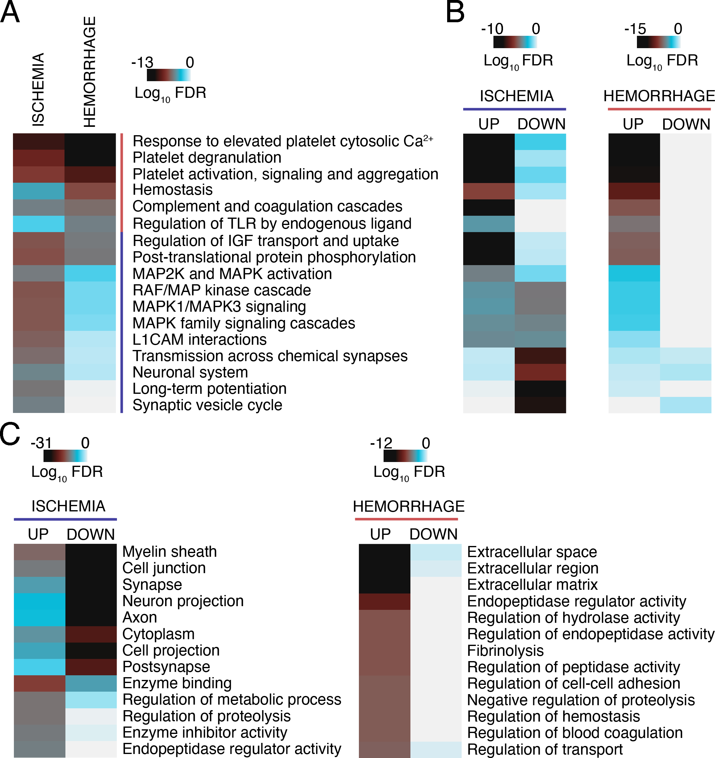
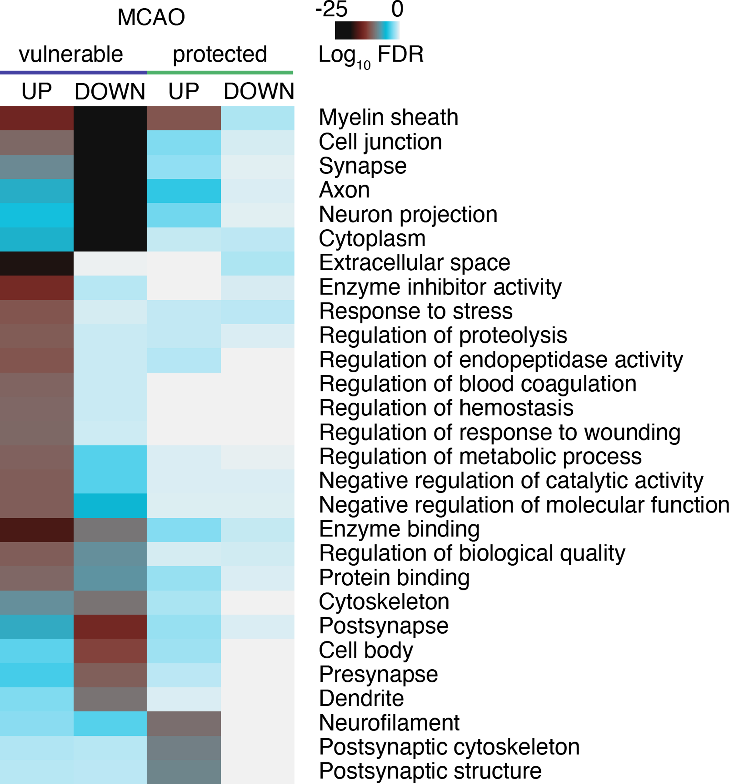
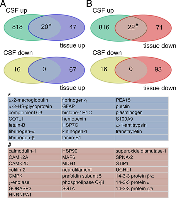
Stroke remains a leading cause of death and disability, with limited therapeutic options and suboptimal tools for diagnosis and prognosis. High throughput technologies such as proteomics generate large volumes of experimental data at once, thus providing an advanced opportunity to improve the status quo by facilitating identification of novel therapeutic targets and molecular biomarkers. Proteomics studies in animals are largely designed to decipher molecular pathways and targets altered in brain tissue after stroke, whereas studies in human patients primarily focus on biomarker discovery in biofluids and, more recently, in thrombi and extracellular vesicles. Here, we offer a comprehensive review of stroke proteomics studies conducted in both animal and human specimen and present our view on limitations, challenges, and future perspectives in the field. In addition, as a unique resource for the scientific community, we provide extensive lists of all proteins identified in proteomic studies as altered by stroke and perform postanalysis of animal data to reveal stroke-related cellular processes and pathways.
Keywords: biomarkers; diagnosis; prognosis; proteomic; stroke; therapy.
Conflict of interest statement
Figures

Similar articles
-
Multilevel omics for the discovery of biomarkers and therapeutic targets for stroke.Nat Rev Neurol. 2020 May;16(5):247-264. doi: 10.1038/s41582-020-0350-6. Epub 2020 Apr 22. Nat Rev Neurol. 2020. PMID: 32322099 Review.
-
Proteomics for Biomarker Identification and Clinical Application in Kidney Disease.Adv Clin Chem. 2018;85:91-113. doi: 10.1016/bs.acc.2018.02.005. Epub 2018 Mar 6. Adv Clin Chem. 2018. PMID: 29655463 Review.
-
Quantitative proteomic analysis of human plasma using tandem mass tags to identify novel biomarkers for herpes zoster.J Proteomics. 2020 Aug 15;225:103879. doi: 10.1016/j.jprot.2020.103879. Epub 2020 Jun 30. J Proteomics. 2020. PMID: 32585426
-
Quantitative clinical proteomic study of autopsied human infarcted brain specimens to elucidate the deregulated pathways in ischemic stroke pathology.J Proteomics. 2013 Oct 8;91:556-68. doi: 10.1016/j.jprot.2013.08.017. Epub 2013 Sep 2. J Proteomics. 2013. PMID: 24007662
-
Emerging Affinity-Based Proteomic Technologies for Large-Scale Plasma Profiling in Cardiovascular Disease.Circulation. 2017 Apr 25;135(17):1651-1664. doi: 10.1161/CIRCULATIONAHA.116.025446. Circulation. 2017. PMID: 28438806 Free PMC article. Review.
Cited by
-
Proteomic analysis of whole blood to investigate the therapeutic effects of nervonic acid on cerebral ischemia-reperfusion injury in rats.Front Cell Dev Biol. 2025 Jan 28;13:1546073. doi: 10.3389/fcell.2025.1546073. eCollection 2025. Front Cell Dev Biol. 2025. PMID: 39936033 Free PMC article.
-
Impact of inflammatory preconditioning on murine microglial proteome response induced by focal ischemic brain injury.Front Immunol. 2024 Apr 9;15:1227355. doi: 10.3389/fimmu.2024.1227355. eCollection 2024. Front Immunol. 2024. PMID: 38655254 Free PMC article.
-
Comprehensive Assessment of Ischemic Stroke in Nonhuman Primates: Neuroimaging, Behavioral, and Serum Proteomic Analysis.ACS Chem Neurosci. 2024 Apr 3;15(7):1548-1559. doi: 10.1021/acschemneuro.3c00826. Epub 2024 Mar 25. ACS Chem Neurosci. 2024. PMID: 38527459 Free PMC article.
-
Investigation of 91 proteins implicated in neurobiological processes identifies multiple candidate plasma biomarkers of stroke outcome.Sci Rep. 2022 Nov 22;12(1):20080. doi: 10.1038/s41598-022-23288-5. Sci Rep. 2022. PMID: 36418382 Free PMC article.
-
Lipidomic Approaches in Common and Rare Cerebrovascular Diseases: The Discovery of Unconventional Lipids as Novel Biomarkers.Int J Mol Sci. 2023 Aug 13;24(16):12744. doi: 10.3390/ijms241612744. Int J Mol Sci. 2023. PMID: 37628924 Free PMC article. Review.
References
-
- Virani SS, Alonso A, Aparicio HJ, Benjamin EJ, Bittencourt MS, Callaway CW, Carson AP, Chamberlain AM, Cheng S, Delling FN, et al. Heart Disease and Stroke Statistics-2021 Update: A Report From the American Heart Association. Circulation. 2021;143:e254–e743. - PubMed
-
- DeGracia DJ. Regulation of mRNA following brain ischemia and reperfusion. Wiley Interdiscip Rev RNA. 2017;8. - PubMed
-
- Focking M, Besselmann M, Trapp T. Proteomics of experimental stroke in mice. Acta Neurobiol Exp (Wars). 2006;66:273–278. - PubMed
Publication types
MeSH terms
Substances
Grants and funding
LinkOut - more resources
Full Text Sources
Medical
