Review
. 2022 Apr 15;130(8):1204-1229.
doi: 10.1161/CIRCRESAHA.121.319949. Epub 2022 Apr 14.

Cerebral Hemorrhage: Pathophysiology, Treatment, and Future Directions

Affiliations
Review

Cerebral Hemorrhage: Pathophysiology, Treatment, and Future Directions

Jessica Magid-Bernstein et al. Circ Res. .

Abstract

Intracerebral hemorrhage (ICH) is a devastating form of stroke with high morbidity and mortality. This review article focuses on the epidemiology, cause, mechanisms of injury, current treatment strategies, and future research directions of ICH. Incidence of hemorrhagic stroke has increased worldwide over the past 40 years, with shifts in the cause over time as hypertension management has improved and anticoagulant use has increased. Preclinical and clinical trials have elucidated the underlying ICH cause and mechanisms of injury from ICH including the complex interaction between edema, inflammation, iron-induced injury, and oxidative stress. Several trials have investigated optimal medical and surgical management of ICH without clear improvement in survival and functional outcomes. Ongoing research into novel approaches for ICH management provide hope for reducing the devastating effect of this disease in the future. Areas of promise in ICH therapy include prognostic biomarkers and primary prevention based on disease pathobiology, ultra-early hemostatic therapy, minimally invasive surgery, and perihematomal protection against inflammatory brain injury.

Keywords: cerebral hemorrhage; epidemiology; hypertension; mortality; stroke.


Conflict of interest statement

No other co-author disclosures

Figures
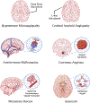
Figure 1.
Figure 1.. Cerebrovascular etiologies of intracerebral hemorrhage. Several vascular pathologies increase the risk for intracerebral hemorrhage (ICH).
Hypertension microangiopathies (top left) present as microhemorrhages primarily localized in the deep brain structures following chronic hypertension due to structural and mechanical age-related changes within arterial walls. Microhemorrhages caused by cerebral amyloid angiopathy (top right) on the other hand, are generally found within lobar regions, and are caused by amyloid deposit which weakens vascular integrity. Arteriovenous malformations (middle left) are abnormal connections between arterial and venous vasculature. Cavernous Angioma disease (middle right) is a neuro-vascular disorder characterized by dilated leaky capillaries following dysfunctional endothelium. Moyamoya (bottom left) is a rare condition causing vascular constriction within the arteries at the Circle of Willis and ensuing abnormal vessel formation compensating for the blockage of normal blood flow to this area. Finally, aneurysms (bottom right) are dilations of blood vessels caused by a weakening in the parent artery angioarchitecture.
Figure 2.
Figure 2.. Mechanisms of Injury After Intracerebral Hemorrhage
Figure 3.
Figure 3.. Open surgery for hematoma evacuation of intracerebral hemorrhage that reached the cortical surface.
After demonstrated stability of the ICH via serial CT scans (ICH volume 81 mL) and etiology screening with an angiogram the patient underwent a craniotomy for clot evacuation. (A) CT showing pre-op ICH, (B) photo of open surgery with a craniotomy (C) photo showing clot evacuation cavity with minimal residual blood products. (D) CT showing 5 days post-op with residual volume of clot measured at <15 mL which predicts a favorable probability of having a good functional outcome.
Figure 4.
Figure 4.. Minimally invasive surgery utilizing the MISTIE protocol of a deep intracerebral hemorrhage from uncontrolled hypertension.
After demonstrated stability of the ICH via serial CT scans (ICH volume 44 mL) and etiology screening with an angiogram the patient underwent real-time clot aspiration in a procedural CT suite. (A) CT showing real-time clot aspiration, (B) photo showing aspiration of clot with gentle suction applied as per the MISTIE protocol, (C) placement of a flexible drain catheter for drainage of residual clot and (D) CT showing residual volume of clot measured at <15 mL which predicts a favorable probability of having a good functional outcome.

References

    1. Virani SS, Alonso A, Benjamin EJ, Bittencourt MS, Callaway CW, Carson AP, Chamberlain AM, Chang AR, Cheng S, Delling FN, et al. Heart disease and stroke statistics-2020 update: A report from the american heart association. Circulation. 2020;141:e139–e596 - PubMed
    1. Feigin VL, Lawes CMM, Bennett DA, Anderson CS. Stroke epidemiology: A review of population-based studies of incidence, prevalence, and case-fatality in the late 20th century. The Lancet Neurology. 2003;2:43–53 - PubMed
    1. van Asch CJJ, Luitse MJA, Rinkel GJE, van der Tweel I, Algra A, Klijn CJM. Incidence, case fatality, and functional outcome of intracerebral haemorrhage over time, according to age, sex, and ethnic origin: A systematic review and meta-analysis. The Lancet Neurology. 2010;9:167–176 - PubMed
    1. Fogelholm R, Murros K, Rissanen A, Avikainen S. Long term survival after primary intracerebral haemorrhage: A retrospective population based study. J Neurol Neurosurg Psychiatry. 2005;76:1534–1538 - PMC - PubMed
    1. Pinho J, CA S, Araujo JM, Amorim JM, Ferreira C. Intracerebral hemorrhage outcome: A comprehensive update. Journal of the Neurologic Sciences. 2019;398:54–66 - PubMed

Publication types