. 2022 May 23;7(10):e149107.
doi: 10.1172/jci.insight.149107.

Estrogen metabolites increase nociceptor hyperactivity in a mouse model of uterine pain

Affiliations

Estrogen metabolites increase nociceptor hyperactivity in a mouse model of uterine pain

Zili Xie et al. JCI Insight. .

Abstract

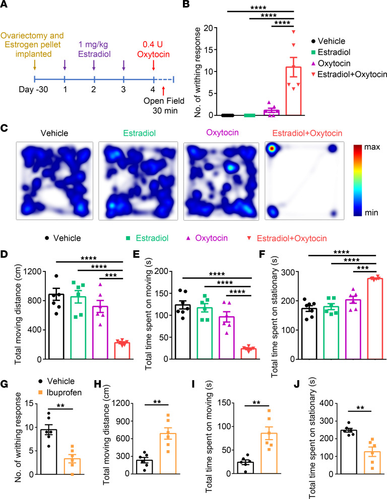
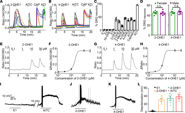
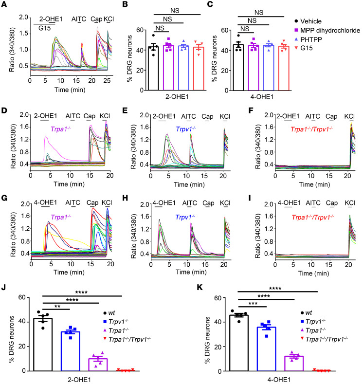
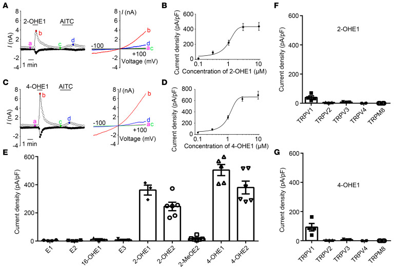
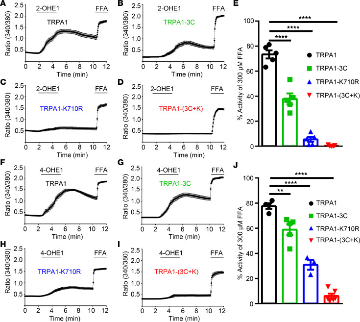
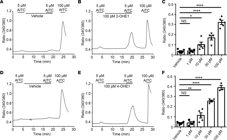
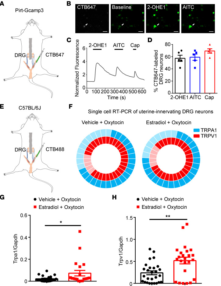
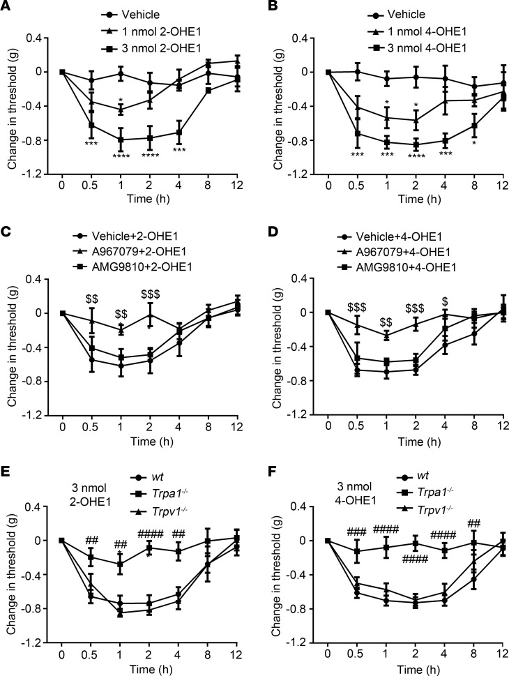
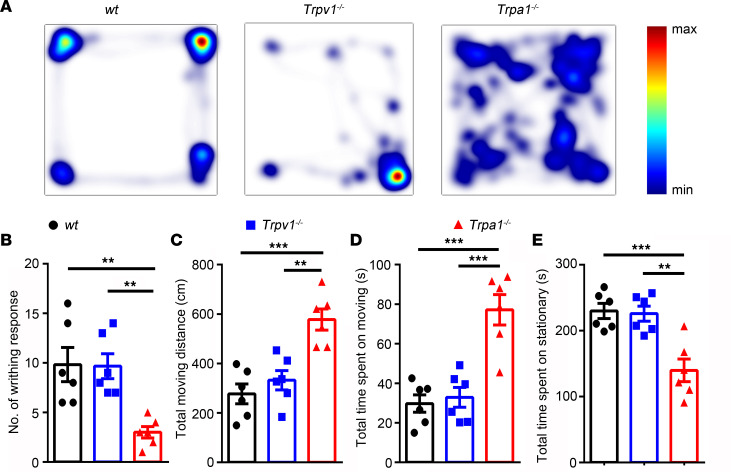
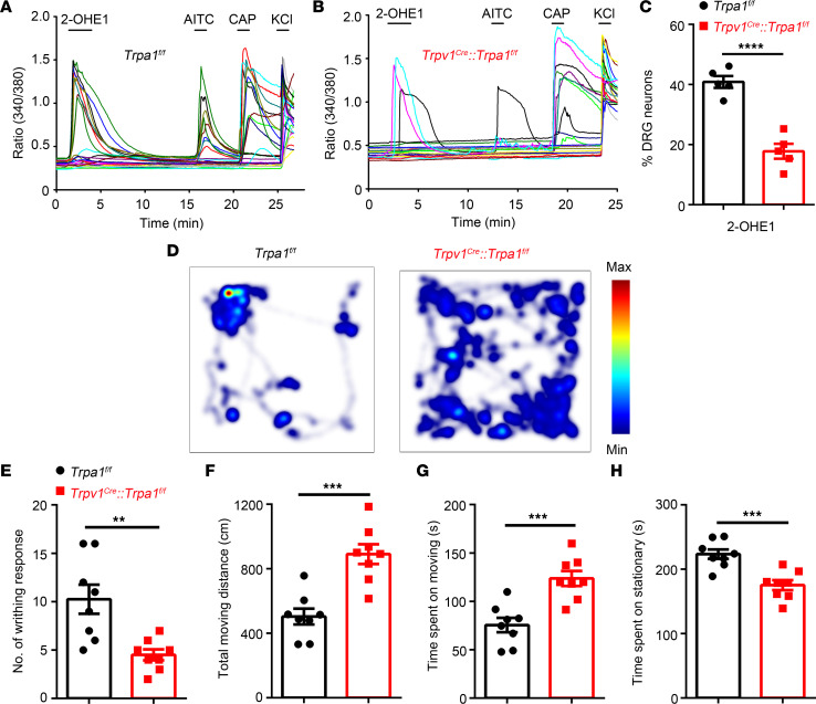
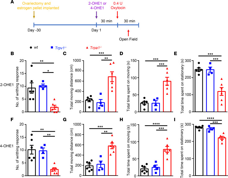
Pain emanating from the female reproductive tract is notoriously difficult to treat, and the prevalence of transient pelvic pain has been placed as high as 70%-80% in women surveyed. Although sex hormones, especially estrogen, are thought to underlie enhanced pain perception in females, the underlying molecular and cellular mechanisms are not completely understood. Here, we showed that the pain-initiating TRPA1 channel was required for pain-related behaviors in a mouse model of estrogen-induced uterine pain in ovariectomized female mice. Surprisingly, 2- and 4-hydroxylated estrogen metabolites (2- and 4-HEMs) in the estrogen hydroxylation pathway, but not estrone, estradiol, or 16-HEMs, directly increased nociceptor hyperactivity through TRPA1 and TRPV1 channels, and picomolar concentrations of 2- and 4-hydroxylation estrone (2- or 4-OHE1) could sensitize TRPA1 channel function. Moreover, both TRPA1 and TRPV1 were expressed in uterine-innervating primary nociceptors, and their expression was increased in the estrogen-induced uterine pain model. Importantly, pretreatment with 2- or 4-OHE1 recapitulated estrogen-induced uterine pain-like behaviors, and intraplantar injections of 2- and 4-OHE1 directly produced a TRPA1-dependent mechanical hypersensitivity. Our findings demonstrated that TRPA1 is critically involved in estrogen-induced uterine pain-like behaviors, which may provide a potential drug target for treating female reproductive tract pain.

Keywords: Ion channels; Neuroscience; Pain.


Figures

Figure 1
Figure 1. Pain-related responses in the mouse model of uterine pain.
(A) Schematic diagram illustrates the protocol for generating a mouse model of uterine pain in ovariectomized female mice implanted with estrogen pellets. (B) Oxytocin-induced writhing response in vehicle group, estradiol group (pretreatment with estradiol benzoate without application of oxytocin), oxytocin group (application of oxytocin without pretreatment with estradiol benzoate) and estradiol + oxytocin group (pretreatment with estradiol benzoate followed by application of oxytocin). n = 6 mice per group, 1-way ANOVA. ****P < 0.0001. (C) Representative images illustrate heatmaps of voluntary movements of mice subjected to different treatments in open-field test in home cages. (DF) Quantification of voluntary movements, including total moving distance (D), total time spent moving (E), and total time spent stationary (F), n = 6 mice per group, 1-way ANOVA. ***P < 0.001, ****P < 0.0001. (GJ) Ibuprofen significantly reduced the number of writhing responses (G) and total time spent stationary (J) and increased the total moving distance (H) and total time spent moving (I); n = 6 mice per group, unpaired Student’s t test. **P < 0.01. All data are expressed as mean ± SEM.
Figure 2
Figure 2. Estrogen metabolites 2- and 4-HEMs directly activate DRG neurons.
(A and B) Representative time-lapse traces of 2-OHE1– (A) and 4-OHE1–induced (B) [Ca2+]i responses in DRG neurons from WT mice. (C) Percentage of DRG neurons responding to estrogen and its metabolites. n = 5 mice (>200 neurons each). (D) Percentage of DRG neurons isolated from female and male mice responding to 2- and 4-OHE1. n = 5 to 6 coverslips per group from 4 mice (>150 neurons each). (EH) Representative [Ca2+]i responses and dose-response curves of [Ca2+]i responses in DRG neurons from WT mice responding to different concentrations of 2-OHE1 (E and F) and 4-OHE1 (G and H). (IK) Representative voltage traces show membrane potential depolarization and action potential firing in DRG neurons isolated from WT mice induced by E1 (I), 2-OHE1 (J), and 4-OHE1 (K). (L) Quantification of E1, 2-OHE1, 4-OHE1, and AITC-induced membrane potential depolarization in DRG neurons isolated from WT mice. n = 4 to 5 from 3 mice.
Figure 3
Figure 3. TRPA1 and TRPV1 are required for 2- and 4-OHE1–induced DRG activation.
(A) Representative traces show that the GPER antagonist G15 had no effect on 2-OHE1–induced [Ca2+]i responses in DRG neurons. (B and C) Summarized data show that estrogen receptor antagonists (at 1 μM) had no effect on 2-OHE1–induced [Ca2+]i responses in DRG neurons. n = 5 coverslips from 3 mice per group (>150 neurons each). One-way ANOVA; NS, not significant. (DI) Representative time-lapse traces of 2-OHE1– (DF) and 4-OHE1–induced (GI) [Ca2+]i responses in the Trpa1−/−, Trpv1−/−, Trpa1−/−Trpv1−/− DRG neurons. (JK) Percentage of DRG neurons responding to 2-OHE1 (J) and 4-OHE1 (K) in DRG neurons isolated from WT, Trpa1−/−, Trpv1−/−, and Trpa1−/−Trpv1−/− mice. n = 5 coverslips from 4 mice per group (>200 neurons each). **P < 0.01, ***P < 0.001, ****P < 0.0001, 1-way ANOVA.
Figure 4
Figure 4. 2- and 4-HEMs activate recombinant TRPA1 and TRPV1 expressed in HEK293T cells.
(A) Representative current traces (left) and I–V curves (right) of TRPA1-dependent currents activated by 2-OHE1 (10 M) and AITC (100 M). (B) Concentration-current density curve of 2-OHE1–activated membrane current in TRPA1-expressing HEK293T cells. The EC50 = 0.97 ± 0.10 μM. n = 5. (C) Representative current traces (left) and I–V curves (right) of TRPA1-dependent currents induced by 10 M 4-OHE1 and 100 M AITC. (D) Concentration-current density curve of 4-OHE1–induced membrane current in TRPA1-expressing HEK293T cells. The EC50 = 1.07 ± 0.09 μM. n = 5. (E) Bar charts illustrate whole-cell current densities produced by estrogen and its metabolites in TRPA1-expressing HEK293T cells. All chemicals were applied at 10 μM. n = 3 to 6. (F and G) Whole-cell current densities produced by 10 μM 2-OHE1 (F) and 10 μM 4-OHE1 (G) in HEK293T cells transfected with TRPV1, TRPV2, TRPV3, TRPV4, and TRPM8 channels. n = 3 to 4 from 3 independent experiments.
Figure 5
Figure 5. Structural basis of 2-OHE1– and 4-OHE1–induced TRPA1 activation.
(AD) Averaged time-lapse traces of 2-OHE1–elicited [Ca2+]i responses in HEK293T cells transfected with TRPA1 (A), TRPA1-3C (B), TRPA1-K710R (C), and TRPA1-(3C+K) (D) constructs; FFA was used as a positive control; (E) Quantification of 2-OHE1–induced [Ca2+]i responses in HEK293T cells transfected with TRPA1 and its mutants. (FI) Averaged time-lapse traces of 4-OHE1–induced [Ca2+]i responses in HEK293T cells transfected with TRPA1 (F), TRPA1-3C (G), TRPA1-K710R (H), and TRPA1-(3C+K) (I) constructs. (J) Quantification of 4-OHE1–induced [Ca2+]i responses in HEK293T cells transfected with TRPA1 and its mutants. **P < 0.01, ****P < 0.0001, 1-way ANOVA, n = 3 to 6 coverslips per group (>100 cells each) from 3 independent experiments.
Figure 6
Figure 6. 2-OHE1 and 4-OHE1 potentiate AITC-induced [Ca2+]i responses in DRG neurons.
(A and B) Preapplication of 100 pM 2-OHE1 (B) but not vehicle (A) for 10 minutes potentiated 5 μM AITC-induced [Ca2+]i responses in DRG neurons isolated from WT mice. (C) Bar charts show the potentiating effect of 2-OHE1 on 5 μM AITC-induced [Ca2+]i responses in a dose-dependent manner. (D and E) Effects of vehicle (D) and 100 pM 4-OHE1 (E) on 5 μM AITC-induced [Ca2+]i responses in DRG neurons isolated from WT mice. (F) Bar charts illustrate the potentiating effect of 4-OHE1 on 5 μM AITC-induced [Ca2+]i responses in a dose-dependent manner. *P < 0.05, **P < 0.01, ****P < 0.0001, 1-way ANOVA, n = 3–6 coverslips from at least 3 mice (>200 neurons each).
Figure 7
Figure 7. TRPA1 and TRPV1 are expressed in uterine-innervated nociceptors.
(A) Schematic representation of intrauterine injections of CTB647 into the PirtGCaMP3 mice. (B) Representative fluorescence images of cultured DRG neurons show that 2-OHE1 and AITC evoked [Ca2+]i responses in a CTB647-labeled neuron. Scale bar: 50 μm. (C) Representative time-lapse trace of 2-OHE1–induced [Ca2+]i response in the CTB647-labeled DRG neuron. (D) Percentage of CTB647-labeled DRG neurons responding to 2-OHE1, AITC, and capsaicin (Cap). n = 5 coverslips from 3 mice. (E) Schematic representation of intrauterine injections of CTB488 into the C57BL/6J mice. (F) Donut plots showing expression and coexpression of TRPA1 and TRPV1 in 32 individual retrogradely traced colon-innervating DRG neurons from control mice (left) and in 29 individual retrogradely traced colon-innervating DRG neurons from the uterine pain group (right). n = 32 cells from 4 mice for control group and n = 29 cells from 4 mice for uterine pain group. (G) Statistical data of single-cell qRT-PCR in CTB647-labeled neurons show the increased expression of TRPA1 in uterine pain model mice compared with control mice. Unpaired t test, *P < 0.05. n = 21 cells from 4 mice for vehicle + oxytocin group and n = 21 cells from 4 mice for estradiol + oxytocin group. (H) Statistical data of single-cell qRT-PCR in CTB647-labeled neurons show the increased expression of TRPV1 in uterine pain model mice compared with control mice. Unpaired t test, **P < 0.01. n = 27 cells from 4 mice for vehicle + oxytocin group and n = 24 cells from 4 mice for estradiol + oxytocin group.
Figure 8
Figure 8. Intraplantar injections of 2-OHE1 and 4-OHE1 produce TRPA1-dependent mechanical allodynia in ovariectomized female mice implanted with estrogen pellets.
(A and B) Intraplantar administration of 2-OHE1 (A) and 4-OHE1 (B) induced mechanical allodynia in ovariectomized WT female mice implanted with estrogen pellets in a dose-dependent manner. *P < 0.05, ***P < 0.001, ****P < 0.0001, 2-way ANOVA, n = 5. (C and D) TRPA1-specific inhibitor A967079 but not TRPV1-specific inhibitor AMG9810 reverted mechanical allodynia induced by 3 nmol 2-OHE1 (C) or 4-OHE1 (D). $P < 0.05, $$P < 0.01, $$$P < 0.001, 2-way ANOVA, n = 5. (E and F) Mechanical allodynia induced by 3 nmol 2-OHE1 (E) and 4-OHE1 (F) was markedly reduced in Trpa1−/− but not Trpv1−/− mice. ##P < 0.01, ###P < 0.001, ####P < 0.0001, 2-way ANOVA with Bonferroni’s post hoc analysis, n = 5.
Figure 9
Figure 9. Genetic ablation of TRPA1 but not TRPV1 significantly inhibits pain-related behaviors in a mouse model of uterine pain induced by estrogen in ovariectomized female mice implanted with estrogen pellets.
(A) Representative images illustrate heatmaps of voluntary movements of WT, Trpv1–/–, and Trpa1–/– mice subjected to 3 days of estrogen treatment followed by oxytocin application. (BE) Statistical data show the number of writhing responses (B), total moving distance (C), total time spent moving (D), and total time spent stationary (E) in WT, Trpa1–/–, and Trpv1–/– mice subjected to 3 days of estrogen treatment followed by oxytocin application. **P < 0.01, ***P < 0.001, n = 6, 1-way ANOVA.
Figure 10
Figure 10. Conditional KO TRPA1 from TRPV1-positive nociceptors significantly inhibits pain-related behaviors in a mouse model of uterine pain.
(A and B) Representative time-lapse trace of 2-OHE1–induced [Ca2+]i response in DRG neurons isolated from Trpa1fl/fl mice (A) and Trpv1Cre Trpa1fl/fl mice (B). (C) Percentage of DRG neurons responding to 2-OHE1 in DRG neurons isolated from Trpa1fl/fl mice and Trpv1Cre Trpa1fl/fl mice. n = 5 coverslips from 3 mice per group. ****P < 0.0001, unpaired t test. (D) Representative images illustrate heatmaps of voluntary movements of Trpa1fl/fl (left) and Trpv1Cre Trpa1fl/fl mice (right) subjected to 3 days of estrogen treatment followed by oxytocin treatment. (EH) Statistical data show the number of writhing responses (E), total moving distance (F), total time spent moving (G), and total time spent stationary (H) in Trpa1fl/fl and Trpv1Cre Trpa1fl/fl mice subjected to 3 days of estrogen treatment followed by oxytocin treatment. n = 8 mice per group. **P < 0.01, ***P < 0.001, unpaired t test.
Figure 11
Figure 11. Acute administration of 3 nmol 2- or 4-OHE1 for 30 minutes followed by oxytocin application recapitulates pain-related behaviors in a mouse model of uterine pain induced by 3 days of estrogen treatment.
(A) Schematic diagram illustrates the protocol for acute i.p. injection of 2-OHE1 and 4-OHE1 to elicit uterine pain in ovariectomized female mice implanted with estrogen pellets. (BE) Quantification of writhing responses (B), total moving distance (C), total time spent moving (D), and total time spent stationary (E) after i.p. injection of 3 nmol 2-OHE1 for 30 minutes followed by oxytocin application in WT, Trpa1−/−, and Trpv1−/− mice. (FI) Quantification of writhing responses (F), total moving distance (G), total time spent moving (H), and total time spent stationary (I) after i.p. injection of 3 nmol 4-OHE1 for 30 minutes followed by oxytocin application in WT, Trpa1−/−, and Trpv1−/− mice. **P < 0.01, ***P < 0.001, ****P < 0.0001, n = 4–6, 1-way ANOVA.

References

    1. Mao J. Challenges of managing chronic pain. BMJ. 2017;356:j741. - PubMed
    1. Fillingim RB, et al. Sex, gender, and pain: a review of recent clinical and experimental findings. J Pain. 2009;10(5):447–485. doi: 10.1016/j.jpain.2008.12.001. - DOI - PMC - PubMed
    1. Dionne RA, et al. Individual responder analyses for pain: does one pain scale fit all? Trends Pharmacol Sci. 2005;26(3):125–130. doi: 10.1016/j.tips.2005.01.009. - DOI - PubMed
    1. Greenspan JD, et al. Studying sex and gender differences in pain and analgesia: a consensus report. Pain. 2007;132(suppl 1):S26–S45. - PMC - PubMed
    1. Miaskowski C. Understanding the genetic determinants of pain and pain management. Semin Oncol Nurs. 2009;25(2 suppl 1):S1–S7. - PubMed

Publication types