. 2022 Apr 14;18(4):e1010152.
doi: 10.1371/journal.pgen.1010152. eCollection 2022 Apr.

Ubiquitin ligase activity inhibits Cdk5 to control axon termination

Affiliations

Ubiquitin ligase activity inhibits Cdk5 to control axon termination

Muriel Desbois et al. PLoS Genet. .

Abstract

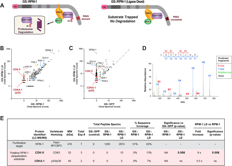
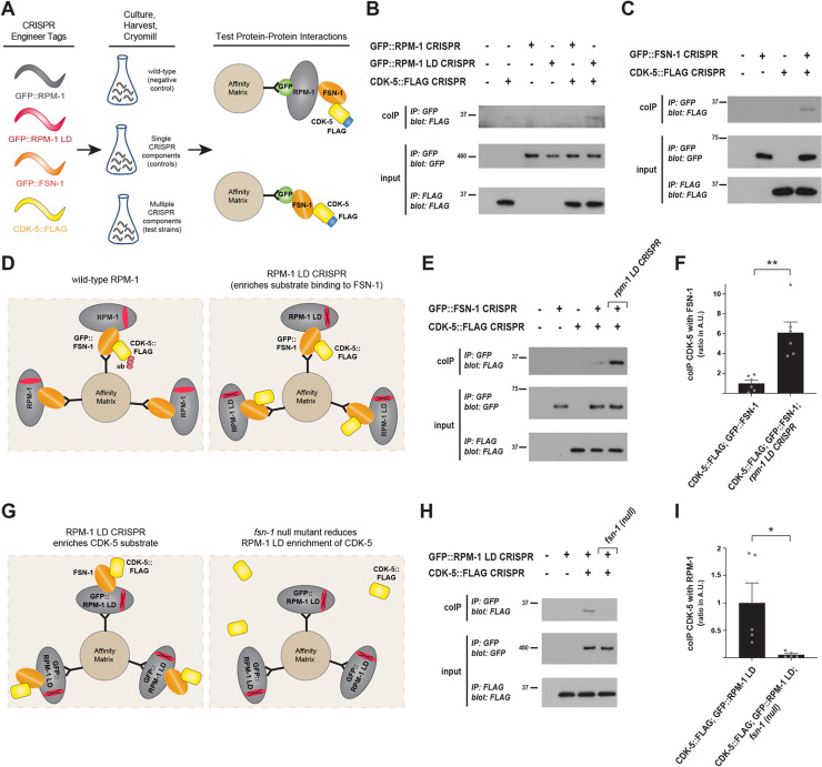
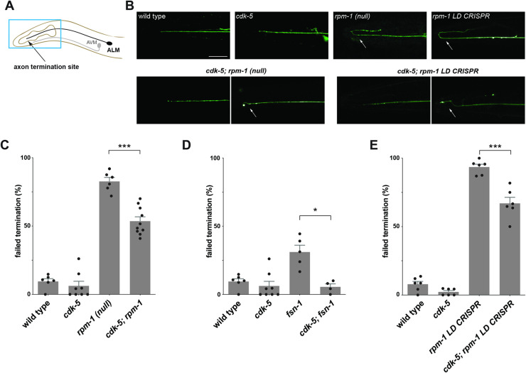
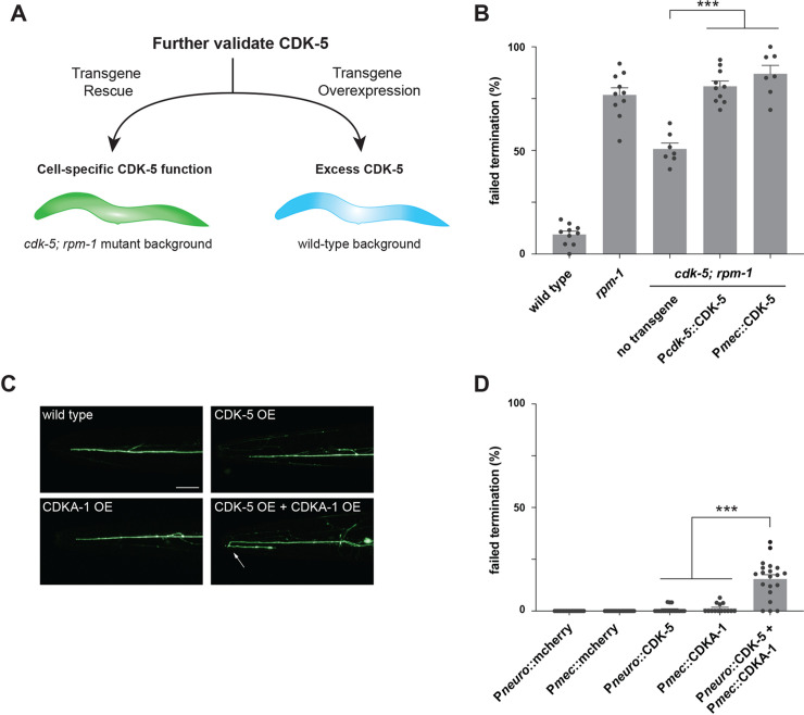
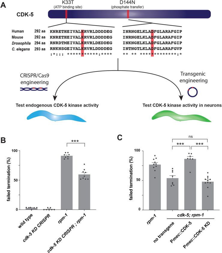
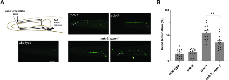
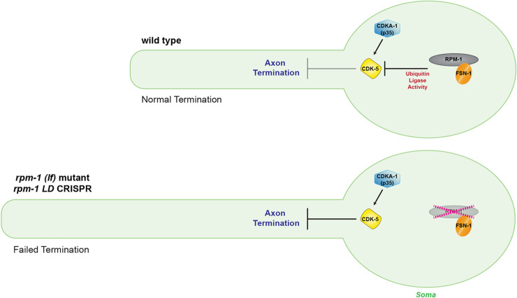
The Cdk5 kinase plays prominent roles in nervous system development, plasticity, behavior and disease. It also has important, non-neuronal functions in cancer, the immune system and insulin secretion. At present, we do not fully understand negative regulatory mechanisms that restrict Cdk5. Here, we use Caenorhabditis elegans to show that CDK-5 is inhibited by the RPM-1/FSN-1 ubiquitin ligase complex. This atypical RING ubiquitin ligase is conserved from C. elegans through mammals. Our finding originated from unbiased, in vivo affinity purification proteomics, which identified CDK-5 as a putative RPM-1 substrate. CRISPR-based, native biochemistry showed that CDK-5 interacts with the RPM-1/FSN-1 ubiquitin ligase complex. A CRISPR engineered RPM-1 substrate 'trap' enriched CDK-5 binding, which was mediated by the FSN-1 substrate recognition module. To test the functional genetic relationship between the RPM-1/FSN-1 ubiquitin ligase complex and CDK-5, we evaluated axon termination in mechanosensory neurons and motor neurons. Our results indicate that RPM-1/FSN-1 ubiquitin ligase activity restricts CDK-5 to control axon termination. Collectively, these proteomic, biochemical and genetic results increase our understanding of mechanisms that restrain Cdk5 in the nervous system.


Conflict of interest statement

The authors have declared that no competing interests exist.

Figures

Fig 1
Fig 1. Proteomics from C. elegans identifies CDK-5 as a putative substrate for the RPM-1 ubiquitin ligase.
A) Schematic of RPM-1 ubiquitin ligase complex and GS tagged constructs used for affinity purification proteomics. GS::RPM-1 LD substrate ‘trap’ does not ubiquitinate substrates which can still bind to the FSN-1 substrate recognition module. As a result, RPM-1/FSN-1 ubiquitination substrates are enriched, but not ubiquitinated and not degraded by the proteasome. Note transgenic GS::RPM-1 LD is generated on an rpm-1 null background to facilitate substrate enrichment and interactions with the endogenous, untagged ubiquitin ligase components FSN-1 and SKR-1(SKP1). B) Scatter plot showing a single affinity purification proteomic experiment comparing proteins identified in GS::RPM-1 LD substrate ‘trap’ versus GS::RPM-1. Highlighted in red are CDK-5 and CDKA-1(p35) which are exclusive to GS::RPM-1 LD. Identified in both samples are known RPM-1 binding proteins (blue), components of the RPM-1 ubiquitin ligase complex (orange) and a previously validated substrate (UNC-51, orange). C) Scatter plot showing the same single proteomic experiment comparing GS::RPM-1 LD versus GS::GFP (negative control). Gray dashed line delineates two-fold enrichment. D) Example LC-MS/MS spectrum for one CDK-5 peptide identified in GS::RPM-1 LD sample. Shown are B ions formed from the amino terminus and Y ions formed from the carboxyl terminus. m/z represents mass to charge ratio with peptide size increasing to the right. E) Summary of results from 7 independent proteomic experiments with RPM-1. Significance determined using Student’s t-test with p-values annotated. ns, not significant (p>0.05); NA, not applicable.
Fig 2
Fig 2. CRISPR-based biochemistry demonstrates substrate-like interactions between CDK-5 and the RPM-1/FSN-1 ubiquitin ligase complex.
A) Schematic of experimental workflow for CRISPR-based, native biochemistry using C. elegans. Multiple rounds of CRISPR/Cas9 engineering was used to affinity tag and point mutate components of the RPM-1/FSN-1 ubiquitin ligase complex and CDK-5. B) CoIP showing increased binding of CDK-5::FLAG to GFP::RPM-1 LD substrate ‘trap’ compared to GFP::RPM-1. C) CoIP showing CDK-5::FLAG binds GFP::FSN-1. D) Schematic showing how rpm-1 LD CRISPR substrate ‘trap’ enriches CDK-5 binding to FSN-1, if CDK-5 is an RPM-1/FSN-1 substrate. E) CoIP showing increased binding between CDK-5::FLAG and GFP::FSN-1 in rpm-1 LD CRISPR animals (far right lane) compared to wildtype background (second lane from right). F) Quantification of CDK-5 coIP with FSN-1 in rpm-1 LD CRISPR animals compared to wildtype background. G) Schematic showing how RPM-1 LD will not bind to CDK-5 in the absence of FSN-1 substrate recognition module (i.e. fsn-1 null mutant). H) CoIP showing reduced binding between CDK-5::FLAG and GFP::RPM-1 LD in fsn-1 (null) mutants (far right lane) compared to wildtype background (second lane from right). I) Quantification of CDK-5 coIP with RPM-1 LD in fsn-1 null mutants compared to wildtype background. For B, C, and E, representative of three or more independent experiments is shown. For H, representative of two independent experiments is shown. For F and I, shown are means from 5–6 replicates from 3 or 2 independent experiments, respectively. Wildtype values were normalized to 1 (Arbitrary Units) to facilitate comparisons. Error bars indicate SEM. Significance assessed using Student’s t-test. ** p<0.01.
Fig 3
Fig 3. RPM-1/FSN-1 ubiquitin ligase complex activity inhibits CDK-5 to regulate axon termination in C. elegans.
A) Schematic shows axon termination sites for anterior mechanosensory neurons, ALM and AVM, of C. elegans. B) Representative images of axon termination in ALM neurons for indicated genotypes visualized using transgenic GFP (muIs32). Shown are severe axon termination defects in rpm-1 (lf) mutants and rpm-1 LD CRISPR animals (arrows). Axon termination defects were not observed in cdk-5 single mutants. In cdk-5; rpm-1 double mutants and cdk-5; rpm-1 LD CRISPR double mutants two examples are shown for wild-type axon termination (left) and mild axon termination defects (right, arrows) which are both observed in these animals. C-E) Quantitation of axon termination defects in ALM neurons for indicated genotypes. Axon termination defects are reduced in C) cdk-5; rpm-1 double mutants, D) cdk-5; fsn-1 double mutants, and E) cdk-5; rpm-1 LD CRISPR double mutants compared to single mutants. For C-E, means (bars) are shown for 4 or more counts (black dots represent individual counts, 20 or more worms/count) for each genotype. Error bars indicate SEM. Significance assessed using Student’s t-test with Bonferroni correction. *** p<0.001, * p<0.05. Scale bars 20 μm.
Fig 4
Fig 4. Transgenic rescue and overexpression studies indicate CDK-5 functions cell-autonomously to regulate axon termination.
A) Schematic of transgenic workflow for evaluating CDK-5 rescue, cell-autonomous function and overexpression. B) Quantitation of axon termination defects in ALM neurons indicates transgenic CDK-5 expressed using native promoter (Pcdk-5) or promoter for mechanosensory neurons (Pmec) rescues reduced axon termination defects in cdk-5; rpm-1 double mutants. C) Representative images of axon termination in ALM neurons of transgenic animals overexpressing indicated constructs. Axon termination is impaired with overexpression of both CDK-5 and CDKA-1(p35). D) Quantitation of ALM axon termination defects caused by transgenic overexpression of indicated constructs. For all experiments, means (bars) are shown from data collected from 3 or more independent transgenic lines (two counts/line, black dots) for each genotype. Error bars indicate SEM. Significance assessed using Student’s t-test with Bonferroni correction. *** p<0.001. Scale bars 20 μm.
Fig 5
Fig 5. CRISPR editing and transgenics demonstrate RPM-1 restricts CDK-5 kinase activity.
A) Protein sequence alignment showing two conserved CDK-5 residues (red) mutated to inactivate kinase activity. Workflow for evaluating CDK-5 kinase activity using CRISPR/Cas9 editing or transgenic engineering. B) CRISPR evaluation of CDK-5 kinase activity. Quantitation indicates axon termination defects in ALM neurons are reduced in rpm-1 mutants when CDK-5 is inactivated by CRISPR (cdk-5 KD CRISPR; rpm-1). C) Transgenic evaluation of CDK-5 kinase activity. Quantitation indicates transgenic expression of CDK-5 KD in mechanosensory neurons fails to rescue reduced axon termination defects in ALM neurons of cdk-5; rpm-1 double mutants. For all experiments, means (bars) are shown from data collected from 3 or more independent transgenic lines (two counts/line, black dots) for each genotype. Error bars indicate SEM. Significance assessed using Student’s t-test with Bonferroni correction. ns, non-significant; *** p<0.001.
Fig 6
Fig 6. CDK-5 colocalizes with RPM-1 LD in neuronal soma.
A) Schematic showing ALM mechanosensory neuron and region imaged for axon termination site (blue) and soma (red). B) CDK-5::mScarlet CRISPR is present in the soma of ALM neurons (bracket) visualized using transgenic GFP expressed in mechanosensory neurons (muIs32). C) CDK-5::mScarlet colocalizes with GFP::RPM-1 LD in ALM soma (bracket). D) Representative images showing CDK-5::mScarlet is not detected at the ALM axon tip visualized using transgenic GFP (arrow). E) GFP::RPM-1 LD accumulates at the ALM axon tip (arrow) where CDK-5::mScarlet is not detected. Scale bars 10 μm.
Fig 7
Fig 7. RPM-1 inhibits CDK-5 to regulate axon termination in SAB motor neurons.
A) Schematic shows axon termination sites for SAB neurons of C. elegans. Representative images of SAB axon termination for indicated genotypes visualized using transgenic GFP (wdIs4). Shown is an example of axon termination defects in rpm-1 (null) mutants (arrow). Axon termination defects were not observed in cdk-5 single mutants. For cdk-5; rpm-1 double mutants, two examples are shown for wild-type (left) and defective axon termination (right, arrow) which are both observed in these animals. B) Quantitation indicates axon termination defects in SAB neurons are reduced in cdk-5; rpm-1 double mutants compared to rpm-1 single mutants. Shown are means (bars) for 12 or more counts (black dots, 20 or more worms/count) for each genotype. Error bars indicate SEM. Significance assessed using Student’s t-test with Bonferroni correction. ** p<0.01. Scale bars 20 μm.
Fig 8
Fig 8. RPM-1/FSN-1 ubiquitin ligase complex inhibits CDK-5 to control axon termination.
Summary showing CDK-5 is restricted by RPM-1/FSN-1 ubiquitin ligase activity in the neuronal soma. In wild-type animals (upper diagram), the RPM-1/FSN-1 ubiquitin ligase complex inhibits CDK-5 to facilitate axon termination. In rpm-1 (lf) mutants and rpm-1 LD CRISPR animals (lower diagram), the activity of the RPM-1/FSN-1 ubiquitin ligase complex is impaired and CDK-5 is not restrained. As a result, excess CDK-5 kinase activity impairs axon termination resulting in excess axon growth. CDKA-1(p35) is a known CDK-5 activator.

References

    1. Su SC, Tsai LH. Cyclin-dependent kinases in brain development and disease. Annu Rev Cell Dev Biol. 2011;27:465–91. doi: 10.1146/annurev-cellbio-092910-154023 . - DOI - PubMed
    1. Cheung ZH, Ip NY. Cdk5: a multifaceted kinase in neurodegenerative diseases. Trends Cell Biol. 2012;22(3):169–75. Epub 2011/12/23. doi: 10.1016/j.tcb.2011.11.003 . - DOI - PubMed
    1. Allnutt AB, Waters AK, Kesari S, Yenugonda VM. Physiological and Pathological Roles of Cdk5: Potential Directions for Therapeutic Targeting in Neurodegenerative Disease. ACS Chem Neurosci. 2020;11(9):1218–30. Epub 2020/04/15. doi: 10.1021/acschemneuro.0c00096 . - DOI - PubMed
    1. Sharma S, Sicinski P. A kinase of many talents: non-neuronal functions of CDK5 in development and disease. Open Biol. 2020;10(1):190287. Epub 2020/01/09. doi: 10.1098/rsob.190287 ; PubMed Central PMCID: PMC7014686. - DOI - PMC - PubMed
    1. Liebl J, Furst R, Vollmar AM, Zahler S. Twice switched at birth: cell cycle-independent roles of the "neuron-specific" cyclin-dependent kinase 5 (Cdk5) in non-neuronal cells. Cell Signal. 2011;23(11):1698–707. Epub 2011/07/12. doi: 10.1016/j.cellsig.2011.06.020 . - DOI - PubMed

Publication types