. 2022 Sep;77(3):723-734.
doi: 10.1016/j.jhep.2022.03.029. Epub 2022 Apr 12.

Mechanotransduction-induced glycolysis epigenetically regulates a CXCL1-dominant angiocrine signaling program in liver sinusoidal endothelial cells in vitro and in vivo

Affiliations

Mechanotransduction-induced glycolysis epigenetically regulates a CXCL1-dominant angiocrine signaling program in liver sinusoidal endothelial cells in vitro and in vivo

Thomas Greuter et al. J Hepatol. 2022 Sep.

Abstract

Background & aims: Liver sinusoidal endothelial cells (LSECs) are ideally situated to sense stiffness and generate angiocrine programs that potentially regulate liver fibrosis and portal hypertension. We explored how specific focal adhesion (FA) proteins parlay LSEC mechanotransduction into stiffness-induced angiocrine signaling in vitro and in vivo.

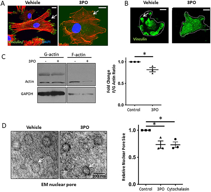
Methods: Primary human and murine LSECs were placed on gels with incremental stiffness (0.2 kPa vs. 32 kPa). Cell response was studied by FA isolation, actin polymerization assay, RNA-sequencing and electron microscopy. Glycolysis was assessed using radioactive tracers. Epigenetic regulation of stiffness-induced genes was analyzed by chromatin-immunoprecipitation (ChIP) analysis of histone activation marks, ChIP sequencing and circularized chromosome conformation capture (4C). Mice with LSEC-selective deletion of glycolytic enzymes (Hk2fl/fl/Cdh5cre-ERT2) or treatment with the glycolysis inhibitor 3PO were studied in portal hypertension (partial ligation of the inferior vena cava, pIVCL) and early liver fibrosis (CCl4) models.

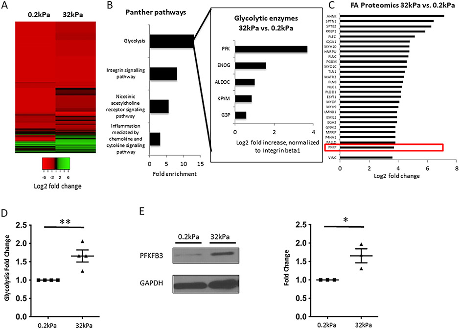
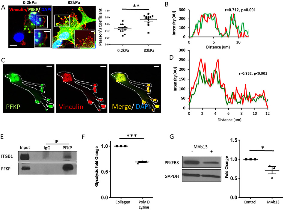
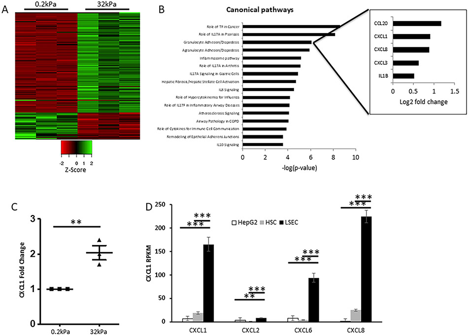
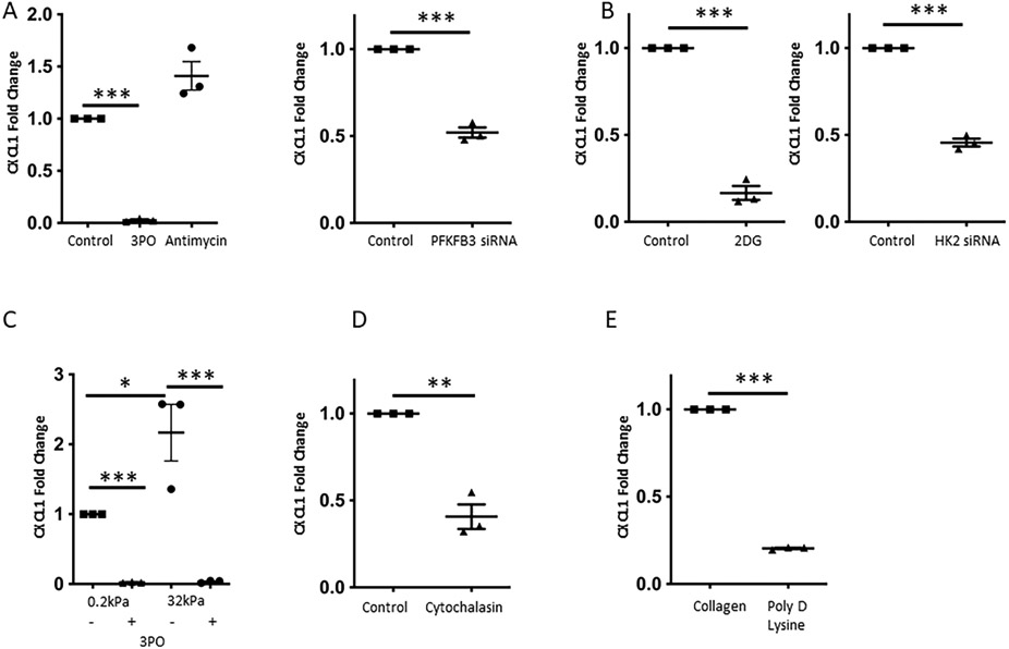
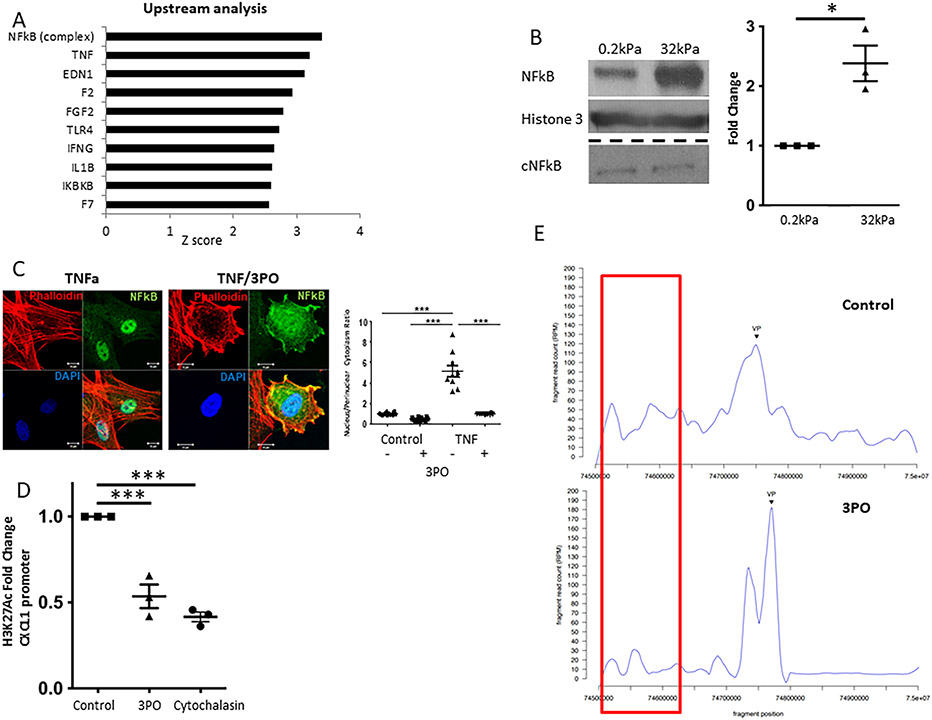
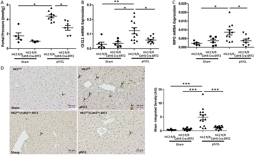
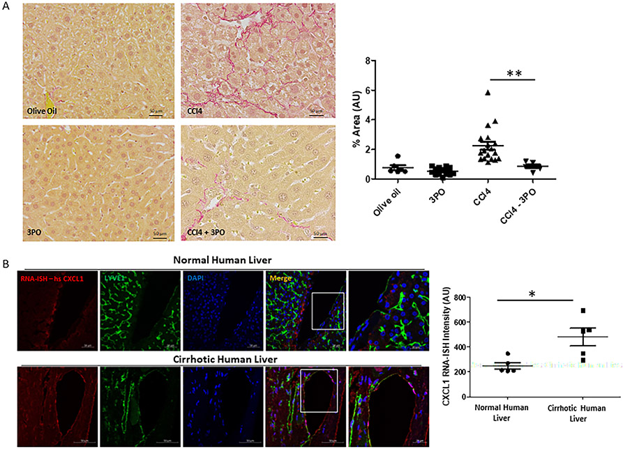
Results: Glycolytic enzymes, particularly phosphofructokinase 1 isoform P (PFKP), are enriched in isolated FAs from LSECs on gels with incremental stiffness. Stiffness resulted in PFKP recruitment to FAs, which paralleled an increase in glycolysis. Glycolysis was associated with expansion of actin dynamics and was attenuated by inhibition of integrin β1. Inhibition of glycolysis attenuated a stiffness-induced CXCL1-dominant angiocrine program. Mechanistically, glycolysis promoted CXCL1 expression through nuclear pore changes and increases in NF-kB translocation. Biochemically, this CXCL1 expression was mediated through spatial re-organization of nuclear chromatin resulting in formation of super-enhancers, histone acetylation and NF-kB interaction with the CXCL1 promoter. Hk2fl/fl/Cdh5cre-ERT2 mice showed attenuated neutrophil infiltration and portal hypertension after pIVCL. 3PO treatment attenuated liver fibrosis in a CCl4 model.

Conclusion: Glycolytic enzymes are involved in stiffness-induced angiocrine signaling in LSECs and represent druggable targets in early liver disease.

Lay summary: Treatment options for liver fibrosis and portal hypertension still represent an unmet need. Herein, we uncovered a novel role for glycolytic enzymes in promoting stiffness-induced angiocrine signaling, which resulted in inflammation, fibrosis and portal hypertension. This work has revealed new targets that could be used in the prevention and treatment of liver fibrosis and portal hypertension.

Keywords: CXCL1; actin polymerization; angiocrine signaling; glycolysis; liver sinusoidal endothelial cells; mechanosensing; nuclear pores; portal hypertension.


Conflict of interest statement

Conflicts of interest The authors declare no conflicts of interest that pertain to this work. Please refer to the accompanying ICMJE disclosure forms for further details.

Figures

Fig. 1.
Fig. 1.. Increased stiffness results in formation of focal adhesions and recruitment of glycolytic enzymes, particularly Phoshofructokinase 1, to focal adhesions.
A. Heatmap for isolated FA proteins from human LSEC plated on hard (32kPa) vs. soft gel (0.2kPa), log2 fold change compared to integrin beta 1 (relative abundance), based on total spectrum counts obtained from mass spectrometry. Green color indicates relative enrichment of FA proteins compared to Integrin beta 1 (main protein within focal adhesions), red color indicates that the FA protein is less abundant than Integrin beta 1. Heatmap reveals considerable differences in the composition of FA on hard vs. soft gels. B. Gene ontology analysis of FA proteins upregulated ≥1.5 fold on hard vs. soft gels reveals glycolysis as the most enriched pathway, which is more enriched than the Integrin signaling pathway. Insert shows glycolytic enzymes contributing to the enrichment of glycolysis and their log2 fold increase on hard vs. soft gels. All values are normalized to Integrin beta 1. PFK is identified as the most upregulated glycolytic enzyme in FA on hard vs. soft gels. C. Mass spectrometry revealed upregulation of typical FA proteins in FA isolates from human LSEC plated on hard vs. soft gels, with upregulation of PFK being comparable to these typical FA proteins. Values represent log2 fold change of total spectrum counts on hard vs. soft gels, normalized to Integrin beta 1. D. Glycolysis was significantly increased in human LSEC plated on hard gels compared to soft gels using radiolabeled glucose experiments (n=4, **p<0.01). E. Protein levels of PFKFB3, a key activator of glycolysis, are significantly upregulated in human LSEC seeded on hard vs. soft gels (western blots on the left, quantification on the right, n=3, *p<0.05), GAPDH was used as a housekeeping cytosolic gene without changes in the whole cell lysate.
Fig. 2.
Fig. 2.. Mechanotransduction activates glycolysis through Integrins.
A. Human LSEC plated on hard gels (32kPa) demonstrated increased co-localization of PFK with the focal adhesion marker, vinculin (yellow in high magnitude inserts) compared to soft gels (0.2kPa). Bar indicates 10μm (insert 5μm). Right panel shows quantification of co-localization of Vinculin and PFK on hard (32kPa) vs soft (0.2kPa) gels with a significant increase of co-localization with incremental stiffness. Co-localization was determined by measuring the Pearson’s coefficient calculated by JACoP plug-in in ImageJ. B. On plastic dish (10MPa), PFK and vinculin show strong and significant co-localization (correlation coefficient r=0.712, p<0.001), respective immunofluorescence can be found in Supplementary Figure 4B). C. Note overlap in PFK and Vinculin (immunofluorescence) in isolated FA of human LSEC plated on plastic dishes. Bar indicates 10μm. D. Quantification of co-localization of PFK and Vinculin in isolated FA (r=0.832, p<0.001). E. Immunoprecipitation studies demonstrated an association of PFK with the focal adhesion protein Integrin beta 1, in human LSEC (after crosslinking). F. Stiffness-induced increase in glycolysis was attenuated by Poly-D-lysine coating promoting non-integrin mediated cell attachment on plastic dishes (10MPa), n=3, ***p<0.001. G. Protein levels of PFKFB3 were decreased with the anti-integrin antibody MAb13 (western blots on the left, quantification on the right, n=3, *p<0.05). AU arbitrary units.
Fig. 3.
Fig. 3.. Mechanotransduction-induced glycolysis promotes actin polymerization, which results in increased nuclear pore size.
A. Inhibition of glycolysis by 3PO (15μM) in human LSEC on plastic (10MPa) attenuated stress fiber (Phalloidin staining, red) and FA formation (Vinculin staining, green). Bar indicates 10μm. B. Immunostaining of FA isolates reveals decreased FA formation with 3PO treatment (15μM). Bar indicates 10μm. C. Quantification of actin polymerization with a decrease in F/G actin ratio upon inhibition of glycolysis with 15 μM 3PO (western blots on the left, quantification on the right, n=3, *p<0.05). D. Inhibition of glycolysis by 15 μM 3PO in human LSEC plated on glass coverslip (represents stiffness above gigapascals) decreased nuclear pore size visualized by electron microscopy on the left (TEM; example of 50 nm nuclear pore is shown en fasse). Quantification of nuclear pore size for cells treated with vehicle, 3PO and cytochalasin shows significant reduction in nuclear pore size with 3PO and cytochalasin (on the right). For each experiment, >30 pores per condition measured, experiments were run in biological triplicates (*p<0.05).
Fig. 4.
Fig. 4.. Neutrophil migration pathway with CXCL1 is one of the top pathways affected by stiffness.
A. Heatmap of RNAseq data with up- and downregulated genes (logFC≥0.8, FDR<0.05) in primary human LSEC plated on hard (32kPa) vs. soft (0.2kPa) gels. Green color indicates upregulation, red color indicates downregulation. Triplicates are shown for both conditions. B. Ingenuity pathway analysis of RNAseq results reveals neutrophil migration (granulocyte diapedesis) as one of the top affected pathways on hard vs. soft gels, ranked by −log p-value. Insert shows contributing genes to this pathway with CXCL1 being among the top upregulated genes on hard vs. soft gel. C. qPCR from human LSEC plated on hard vs. soft gels confirms RNAseq data with significant upregulation of CXCL1 mRNA expression on hard gel (n=3, **p<0.01). D. Comparison of CXCL1, 2, 6 and 8 expression among different human liver cell types based on RPKM levels shows highest expression of CXCL 1, 6 and 8 in LSEC (n=3, **p<0.01, ***p<0.001).
Fig. 5.
Fig. 5.. Inhibition of glycolysis attenuates mechanotransduction-induced CXCL1 production.
A. Increase in CXCL1 mRNA levels is attenuated upon inhibition of glycolysis with 3PO (15 μM), but not after blocking mitochondrial respiration (Antimycin), on plastic dishes (10MPa), on the left (n=3, ***p<0.001). Knockdown of the glycolytic activator PFKFB3 significantly decreased CXCL1 mRNA expression, on the right (n=3, ***p<0.001). B. Blocking glycolysis by 2-DG, an inhibitor of hexokinase 2 (on the left), and knockdown of hexokinase 2 (on the right) resulted in a significant decrease of CXCL1 mRNA levels (n=3, ***p<0.001). C. 3PO (15 μM) decreased CXCL1 mRNA levels on both soft and hard gels, and significantly attenuated the hard-gel induced CXCL1 increase (n=3, *p<0.05, ***p<0.001). D. Disruption of the cytoskeleton by using Cytochalasin resulted in a decrease of CXCL1 mRNA levels (n=3, **p<0.01). E. Poly-D-lysine coating promoting non-Integrin mediated cell attachment significantly decreased CXCL1 mRNA expression compared to collagen I coating, which promotes Integrin mediated cell attachment (n=3, ***p<0.001). All experiments performed with human LSEC.
Fig. 6.
Fig. 6.. Glycolysis promotes CXCL1 expression through NFkB and epigenetic modifications at H3K27 mediated by super-enhancer formation.
A. Upstream regulator analysis of RNAseq data from human LSEC plated on hard (32kPa) vs. soft (0.2kPa) collagen-I coated gels revealed TNF-NFkB as the top pathway involved. B. Nuclear NFkB protein levels are significantly increased on hard compared to soft gels without an increase in cytosolic NFkB (representative western blot on the left, quantification on the right, n=3, *p<0.05. C. TNF-induced nuclear NFkB translocation is significantly attenuated by co-treatment with 3PO (representative immunofluorescence on the left, quantification on the right, n=10, ***p<0.001). D. H3K27 acetylation of the CXCL1 promoter is significantly decreased upon treatment of human LSEC with 3PO and Cytochalasin (n=3, ***p<0.001). E. Circularized chromosome conformation capture (4C) of the CXCL1 promoter (Chr4q13.3) identifies a superenhancer at a genomic site 130-170kb upstream. The interaction between CXCL1 promoter and this genomic site is largely attenuated by inhibition of glycolysis with 3PO (15 μM), n=1. AU arbitrary unit. All experiments performed with human LSEC.
Fig. 7.
Fig. 7.. Endothelial cell-specific inhibition of glycolysis attenuates neutrophil infiltration and development of portal hypertension in vivo.
A. Portal pressure measurement shows attenuated portal hypertension in HK2 fl/fl Cdh5 Cre-ERT2 mice (=endothelial cell specific HK2 knockdown) after pIVCL compared with HK2 fl/fl mice (n=3-9, *p<0.05). B. CXCL1 mRNA expression is significantly increased after pIVCL in HK2 fl/fl, while this increase is attenuated in HK2 fl/fl Cdh5 Cre-ERT2 mice (n=5-11, *p<0.05, **p<0.01). C. MPO mRNA expression is significantly increased after pIVCL in HK2 fl/fl, while this increase is attenuated in HK2 fl/fl Cdh5 Cre-ERT2 mice (n=5-11, *p<0.05). D. Immunohistochemistry for MPO shows attenuated neutrophil infiltration in HK2 fl/fl Cdh5 Cre-ERT2 mice after pIVCL compared with HK2 fl/fl mice (n=7-14, ***p<0.001). Left panel shows representative pictures (arrowheads indicate neutrophils), right panel shows quantification.
Fig 8.
Fig 8.. Inhibition of glycolysis by 3PO attenuates liver fibrosis in a 3-week CCl4 model and human liver samples from patients with cirrhosis indicate involvement of CXCl1.
A. Sirius red staining shows significant attenuation of liver fibrosis in 3PO treated mice after 3-week CCl4 injection compared to non 3PO treated mice (n=3-9, **p<0.01). Right panel shows quantification. B. RNA in situ hybridization of human liver samples from patients with cirrhosis shows significantly increased expression of CXCL1 mRNA in cirrhotic vs non-cirrhotic liver, particularly in LYVE1 positive cells. Right panel shows quantification.
Fig. 9.
Fig. 9.. Summary cartoon.
The glycolytic enzyme PFKP is recruited to FA in response to stiffness. This results in the formation of stress fibers (actin polymerization). Actin tethering increases nuclear pore size enabling translocation of the transcription factor NFkB. Through 3D conformational changes, enhancers and promoter of stiffness-inducible genes (here CXCL1) get in close proximity resulting in histone modification and gene transcription. CXCL1 induces neutrophil migration and finally portal hypertension, early liver fibrosis.

References

    1. Greuter T, Shah VH. Hepatic sinusoids in liver injury, inflammation, and fibrosis: new pathophysiological insights. J Gastroenterol. 2016;51(6):511–9. - PubMed
    1. Dauphinee SM, Karsan A. Lipopolysaccharide signaling in endothelial cells. Lab Invest. 2006;86(1):9–22. - PubMed
    1. Schrage A, Wechsung K, Neumann K, Schumann M, Schulzke JD, Engelhardt B, et al. Enhanced T cell transmigration across the murine liver sinusoidal endothelium is mediated by transcytosis and surface presentation of chemokines. Hepatology. 2008;48(4):1262–72. - PubMed
    1. Kostallari E, Shah VH. Angiocrine signaling in the hepatic sinusoids in health and disease. Am J Physiol Gastrointest Liver Physiol. 2016;311(2):G246–51. - PMC - PubMed
    1. Hilscher MB, Sehrawat T, Arab JP, Zeng Z, Gao J, Liu M, et al. Mechanical Stretch Increases Expression of CXCL1 in Liver Sinusoidal Endothelial Cells to Recruit Neutrophils, Generate Sinusoidal Microthombi, and Promote Portal Hypertension. Gastroenterology. 2019;157(1):193–209.e9. - PMC - PubMed

Publication types