

Myenteric Neurons Do Not Replicate in Small Intestine Under Normal Physiological Conditions in Adult Mouse
- PMID: 35421596
- PMCID: PMC9117811
- DOI: 10.1016/j.jcmgh.2022.04.001
Myenteric Neurons Do Not Replicate in Small Intestine Under Normal Physiological Conditions in Adult Mouse
Abstract
Background & aims: The enteric nervous system (ENS) is the largest part of the peripheral nervous system; moreover, abnormal ENS development and function are associated with multiple human pathologies. Data from several groups suggest that under normal physiological conditions in adult animals, enteric nerve cells do not replicate. A study by Kulkarni et al in 2017 challenged this view and proposed that nearly 70% of enteric neurons in the myenteric ganglia are born in 1 week. The authors of this study suggested that differences in DNA labelling times and DNA denaturation conditions might explain discrepancies with previous reports. Previous studies were carried out using different conditions and labelling techniques in various regions of the gastrointestinal tract; thus, conclusions have remained elusive.
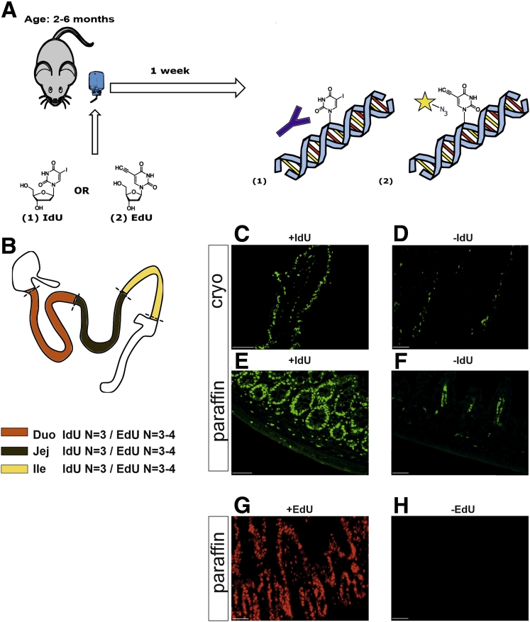
Methods: Here, we have eliminated those variables by analyzing the whole small intestine using the reagents and conditions that Kulkarni et al used. To exclude variables related to immunohistochemistry, we carried out parallel experiments with "click chemistry"-based detection of DNA replication.
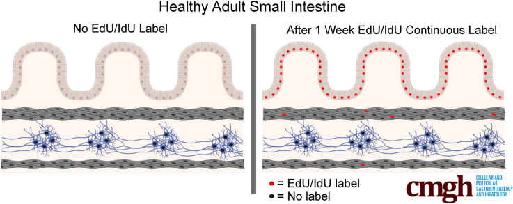
Results: Although proliferation was readily detected in the epithelium, we found no evidence of neuronal replication in the myenteric ganglia.
Conclusions: We conclude that within 1 week under normal physiological conditions, myenteric neurons in the small intestine do not replicate.
Keywords: DNA Labelling; ENS; EdU; IdU; Proliferation.
Copyright © 2022 The Authors. Published by Elsevier Inc. All rights reserved.
Figures

Comment in
-
Enteric Neurons Get Our Undivided Attention.Cell Mol Gastroenterol Hepatol. 2022;14(1):239-240. doi: 10.1016/j.jcmgh.2022.04.007. Epub 2022 May 3. Cell Mol Gastroenterol Hepatol. 2022. PMID: 35523354 Free PMC article. No abstract available.
-
Detecting Adult Enteric Neurogenesis in the Context of Adult ENS Homeostasis.Cell Mol Gastroenterol Hepatol. 2022;14(4):967. doi: 10.1016/j.jcmgh.2022.07.003. Epub 2022 Aug 4. Cell Mol Gastroenterol Hepatol. 2022. PMID: 35932848 Free PMC article. No abstract available.
References
-
- Rademakers G., Vaes N., Schonkeren S., Koch A., Sharkey K.A., Melotte V. The role of enteric neurons in the development and progression of colorectal cancer. Biochim Biophys Acta Rev Cancer. 2017;1868:420–434. - PubMed
-
- Pham T.D., Gershon M.D., Rothman T.P. Time of origin of neurons in the murine enteric nervous system: Sequence in relation to phenotype. J Comp Neurol. 1991;314:789–798. - PubMed
Publication types
MeSH terms
LinkOut - more resources
Full Text Sources
Other Literature Sources
