. 2022 Apr 15;20(1):158.
doi: 10.1186/s12916-022-02359-4.

Deep immunophenotyping reveals endometriosis is marked by dysregulation of the mononuclear phagocytic system in endometrium and peripheral blood

Affiliations

Deep immunophenotyping reveals endometriosis is marked by dysregulation of the mononuclear phagocytic system in endometrium and peripheral blood

Júlia Vallvé-Juanico et al. BMC Med. .

Abstract

Background: Endometriosis is a chronic, estrogen-dependent disorder where inflammation contributes to disease-associated symptoms of pelvic pain and infertility. Immune dysfunction includes insufficient immune lesion clearance, a pro-inflammatory endometrial environment, and systemic inflammation. Comprehensive understanding of endometriosis immune pathophysiology in different hormonal milieu and disease severity has been hampered by limited direct characterization of immune populations in endometrium, blood, and lesions. Simultaneous deep phenotyping at single-cell resolution of complex tissues has transformed our understanding of the immune system and its role in many diseases. Herein, we report mass cytometry and high dimensional analyses to study immune cell phenotypes, abundance, activation states, and functions in endometrium and blood of women with and without endometriosis in different cycle phases and disease stages.

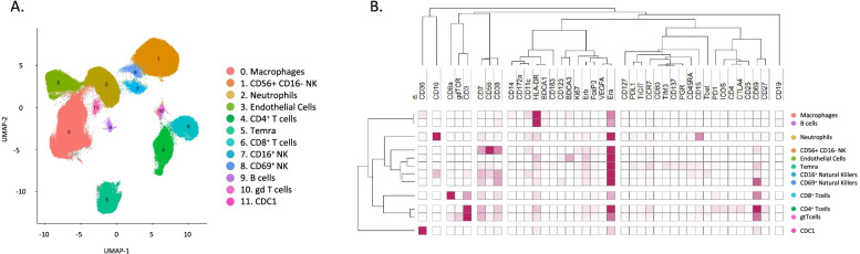
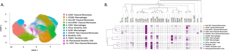
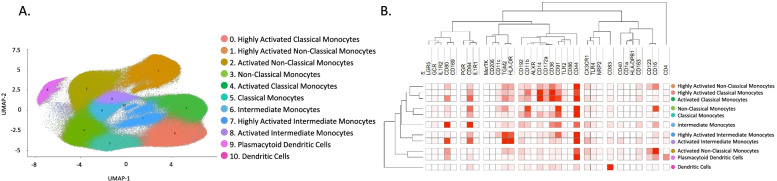
Methods: A case-control study was designed. Endometrial biopsies and blood (n = 60 total) were obtained from women with (n = 20, n = 17, respectively) and without (n = 14, n = 9) endometriosis in the proliferative and secretory cycle phases of the menstrual cycle. Two mass cytometry panels were designed: one broad panel and one specific for mononuclear phagocytic cells (MPC), and all samples were multiplexed to characterize both endometrium and blood immune composition at unprecedented resolution. We combined supervised and unsupervised analyses to finely define the immune cell subsets with an emphasis on MPC. Then, association between cell types, protein expression, disease status, and cycle phase were performed.

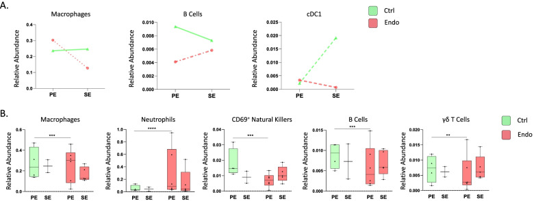
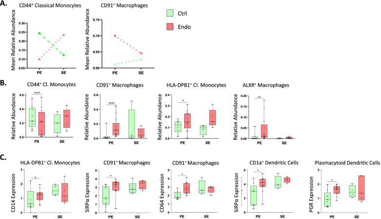
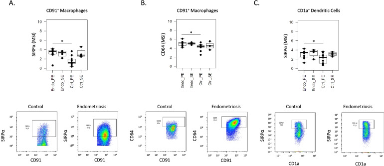
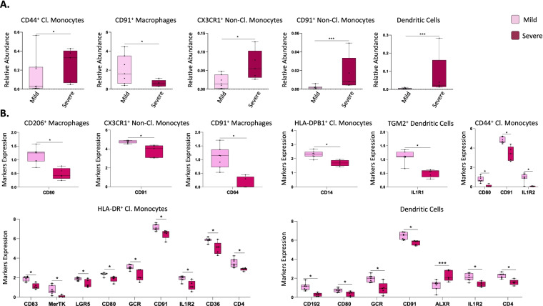
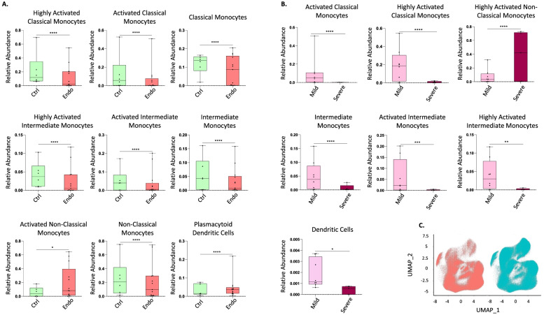
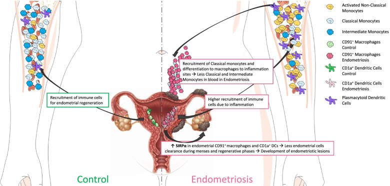
Results: The broad panel highlighted a significant modification of MPC in endometriosis; thus, they were studied in detail with an MPC-focused panel. Endometrial CD91+ macrophages overexpressed SIRPα (phagocytosis inhibitor) and CD64 (associated with inflammation) in endometriosis, and they were more abundant in mild versus severe disease. In blood, classical and intermediate monocytes were less abundant in endometriosis, whereas plasmacytoid dendritic cells and non-classical monocytes were more abundant. Non-classical monocytes were higher in severe versus mild disease.

Conclusions: A greater inflammatory phenotype and decreased phagocytic capacity of endometrial macrophages in endometriosis are consistent with defective clearance of endometrial cells shed during menses and in tissue homeostasis, with implications in endometriosis pathogenesis and pathophysiology. Different proportions of monocytes and plasmacytoid dendritic cells in blood from endometriosis suggest systemically aberrant functionality of the myeloid system opening new venues for the study of biomarkers and therapies for endometriosis.

Keywords: Biomarker; CyTOF; Endometriosis; Endometrium; Innate immune; Macrophages; Monocytes; Mononuclear phagocytes; SIRPα.


Conflict of interest statement

The authors declare that they have no competing interests. LCG: consultant for Myovant Sciences, ForEndo Pharmaceuticals, NextGen Jane, and Celmatix, Inc.

Figures

Fig. 1
Fig. 1
Identified populations from the unsupervised clustering analysis in endometrium in the broad panel. A UMAP showing the 12 identified clusters. B Heatmap with the marker’s level expression in each of the populations identified from the clustering. n = 17 (4 controls PE, 2 controls SE, 6 endometriosis PE, and 5 endometriosis SE). PE: proliferative, SE: secretory
Fig. 2
Fig. 2
Broad panel association analysis in endometrium. A Populations with significantly different relative abundance between proliferative and secretory phases in control and endometriosis patients and showing contrary fluctuation between both groups (median relative abundance is shown). p-values: dotted and dashed lines, p ≤ 0.05; dashed lines, p ≤ 0.0005; dotted lines, p ≤ 0.00005; straight lines: no significant. B Significant differences in abundance between cases and control immune populations. All analyses were performed in both phases of the menstrual cycle (PE: proliferative, SE: secretory). p-values: *, p ≤ 0.05; **, p ≤ 0.005; ***, p ≤ 0.0005; **** p ≤ 0.00005. n = 17 (4 controls PE, 2 controls SE, 6 endometriosis PE, and 5 endometriosis SE). PE: proliferative, SE: secretory
Fig. 3.
Fig. 3.
Identified populations from the unsupervised clustering analysis in endometrium in the focused panel. A UMAP showing the 12 identified clusters. B Heatmap with the marker’s relative level expression in each of the populations identified from the clustering. n = 13 controls (9 PE and 4 SE) and n = 18 endometriosis (13 PE (8 mild and 5 severe stages) and 5 SE (all mild stage of disease)). PE: proliferative, SE: secretory
Fig. 4
Fig. 4
Focused panel association analysis of endometrium in cases and controls and different cycle phases. A Populations with significantly different relative abundance between proliferative and secretory phases in control and endometriosis patients and showing contrary fluctuation between both groups (median relative abundance is shown). p-values: dashed lines, p ≤ 0.0005; dotted lines, p ≤ 0.00005. B Significant differences in abundance of immune populations between control and endometriosis patients. p-values: *, p ≤ 0.05; **, p ≤ 0.005; ***, p ≤ 0.0005; **** p ≤ 0.00005. C Significant differences in marker expression in immune populations between control and endometriosis patients (p ≤ 0.1). n = 13 controls (9 PE and 4 SE) and n = 18 endometriosis (13 PE (8 mild and 5 severe stages) and 5 SE (all mild stage of disease)). Green: controls; red: cases. All analyses were performed in both phases of the menstrual cycle. PE: proliferative, SE: secretory
Fig. 5
Fig. 5
Differentially expressed SIRPα and CD64 in CD91+ macrophages and SIRPα in CD1a+ dendritic cells from endometrium of women with versus without endometriosis. The figure shows the box plots from the mean signal intensity (MSI) obtained by manual gating using FlowJo® (p-value ≤ 0.05) and the dot plots show the number of cells and intensity of each marker. A SIRPα expression in CD91+ macrophages. B CD64 expression in CD91+ macrophages. C SIRPα expression in CD1a+ dendritic cells. n = 9 controls PE and n = 13 endometriosis PE. PE: proliferative
Fig. 6
Fig. 6
Focused panel association analysis between stages of endometriosis in endometrium. A Differences in abundance of populations between samples from patients with mild and severe disease. p-values: *, p ≤ 0.05; **, p ≤ 0.005; ***, p ≤ 0.0005; **** p ≤ 0.00005. B Significant differences in marker expression in specific populations. Both analyses were performed in the proliferative phase of the menstrual cycle. p-values: *, p ≤ 0.1; ***, p ≤ 0.005. n = 13 endometriosis PE (8 mild and 5 severe stages). PE: proliferative
Fig. 7
Fig. 7
Identified populations from the unsupervised clustering analysis in blood (PBMCs) using the focused panel. A UMAP showing the 11 identified clusters. B Heatmap with the marker level expression in each of the populations identified from the clustering. n = 6 controls and n = 13 endometriosis PE (8 mild stage and 5 severe stage). PE: proliferative
Fig. 8
Fig. 8
Focused panel association analysis results in blood. A The figure shows the significant differences in abundance of immune populations in the proliferative phase of the menstrual cycle in cases and controls. B Differences in abundance of populations between stages of endometriosis in blood. Analysis performed in the proliferative phase of the menstrual cycle. C UMAP from controls (red) and endometriosis (blue) samples, including both proliferative and secretory phases and both stages of the disease. Arrows show the cluster of highly activated non-classical monocytes. Y axis: Relative abundance of cells per cluster. The total cells per cluster represents all the cells from each specific population from all conditions analyzed (controls, endometriosis, proliferative and secretory phase). Ctrl: controls, Endo: endometriosis. Analysis performed in the proliferative phase of the menstrual cycle. p-values: *, p ≤ 0.05; **, p ≤ 0.005; **** p ≤ 0.00005. n = 6 controls and n = 13 endometriosis PE (8 mild stage and 5 severe stage). PE: proliferative
Fig. 9
Fig. 9
Proposed model of involvement of specific immune populations in the proliferative phase of the menstrual cycle in endometriosis patients compared to controls. The model shows that, in controls, there is a recruitment of immune cells during regenerative-proliferative phase of the menstrual cycle for endometrial regeneration and proliferation. In addition, there is less recruitment of classical and intermediate monocytes to the endometrium in controls, as the inflammation is not as enhanced in this tissue as it is in disease. The right panel shows the model in endometriosis. It shows that there is higher recruitment of immune populations (specifically classical and intermediate monocytes, which will differentiate to macrophages) to sites of inflammation, such as the endometriotic lesions and endometrium, decreasing like this the proportion of these monocytes in circulation of women with endometriosis. It also shows that there is a higher proportion of plasmacytoid dendritic cells and non-classical monocytes in circulation in women with endometriosis. Finally, we hypothesize that the increase of SIRPα in endometrial macrophages and dendritic cells might decrease the phagocytic capacity of these cells, by allowing endometrial cells to scape clearance during menses and regenerative phases of the cycle, which, in turn, would allow their migration to the peritoneal cavity, implant, and develop the endometriotic lesions

References

    1. Zondervan KT, Becker CM, Koga K, Missmer SA, Taylor RN, Viganò P. Endometriosis. Nat Rev Dis Primers. 2018;4:9. doi: 10.1038/s41572-018-0008-5. - DOI - PubMed
    1. Wang Y, Kristen Nicholes I-MS. The origin and pathogenesis of endometriosis. Physiol Behav. 2020;24:71–95. - PMC - PubMed
    1. Riccio L d GC, Santulli P, Marcellin L, Abrão MS, Batteux F, Chapron C. Immunology of endometriosis. Best Pract Res Clin Obstet Gynaecol. 2018;50:39–49. doi: 10.1016/j.bpobgyn.2018.01.010. - DOI - PubMed
    1. Vallvé-Juanico J, Houshdaran S, Giudice LC. The endometrial immune environment of women with endometriosis. Hum Reprod Update. 2019;25(5):564–591. doi: 10.1093/humupd/dmz018. - DOI - PMC - PubMed
    1. Capobianco A, Rovere-Querini P. Endometriosis, a disease of the macrophage. Front Immunol. 2013;28(4):9. - PMC - PubMed

Publication types