. 2022 Mar 29:13:809804.
doi: 10.3389/fmicb.2022.809804. eCollection 2022.

Photobiont Diversity in Lichen Symbioses From Extreme Environments

Affiliations

Photobiont Diversity in Lichen Symbioses From Extreme Environments

Roberto De Carolis et al. Front Microbiol. .

Abstract

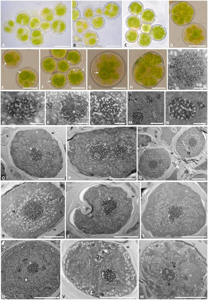
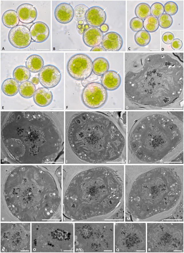
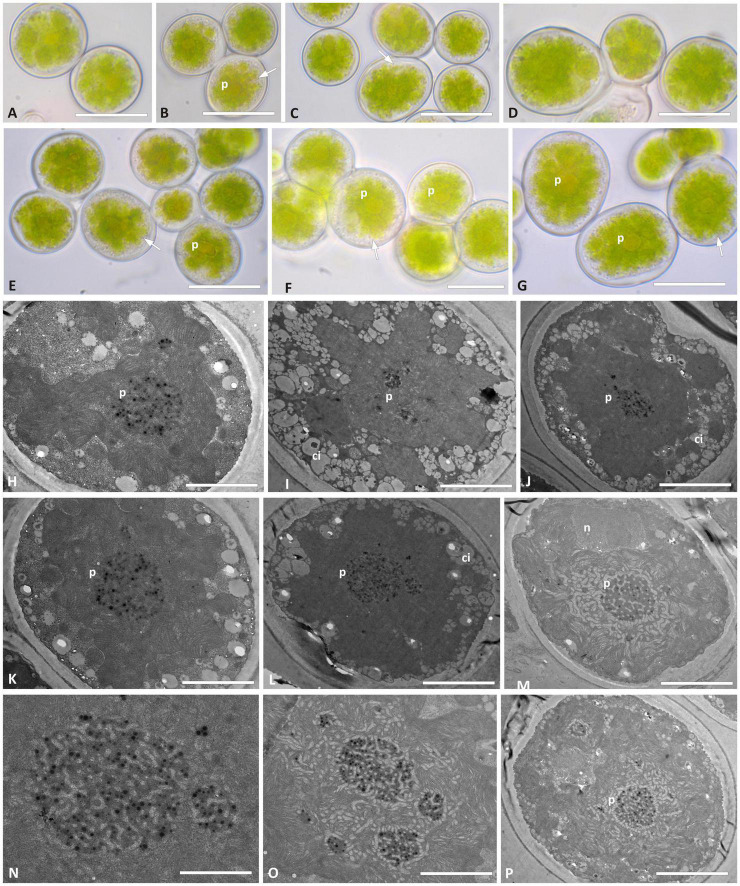
Fungal-algal relationships-both across evolutionary and ecological scales-are finely modulated by the presence of the symbionts in the environments and by the degree of selectivity and specificity that either symbiont develop reciprocally. In lichens, the green algal genus Trebouxia Puymaly is one of the most frequently recovered chlorobionts. Trebouxia species-level lineages have been recognized on the basis of their morphological and phylogenetic diversity, while their ecological preferences and distribution are still only partially unknown. We selected two cosmopolitan species complexes of lichen-forming fungi as reference models, i.e., Rhizoplaca melanophthalma and Tephromela atra, to investigate the diversity of their associated Trebouxia spp. in montane habitats across their distributional range worldwide. The greatest diversity of Trebouxia species-level lineages was recovered in the altitudinal range 1,000-2,500 m a.s.l. A total of 10 distinct Trebouxia species-level lineages were found to associate with either mycobiont, for which new photobionts are reported. One previously unrecognized Trebouxia species-level lineage was identified and is here provisionally named Trebouxia "A52." Analyses of cell morphology and ultrastructure were performed on axenically isolated strains to fully characterize the new Trebouxia "A52" and three other previously recognized lineages, i.e., Trebouxia "A02," T. vagua "A04," and T. vagua "A10," which were successfully isolated in culture during this study. The species-level diversity of Trebouxia associating with the two lichen-forming fungi in extreme habitats helps elucidate the evolutionary pathways that this lichen photobiont genus traversed to occupy varied climatic and vegetative regimes.

Keywords: Rhizoplaca; Tephromela; Trebouxia; chloroplast morphology; culture; phylogeny.


Conflict of interest statement

The authors declare that the research was conducted in the absence of any commercial or financial relationships that could be construed as a potential conflict of interest.

Figures

FIGURE 1
FIGURE 1
(A) Schematic ML and Bayesian phylogenetic hypothesis based on the ITS locus of Trebouxia genus including the newly generated sequences. The definition of the major clades follows the multi-locus phylogeny of Muggia et al. (2020). (B) Relative abundances of Trebouxia species-level lineages recovered in this study.
FIGURE 2
FIGURE 2
Phylogenetic hypothesis based on the ITS locus of Trebouxia Clade “A”: the 50% majority rule consensus tree of the Bayesian analysis is presented; ML bootstrap values higher than 70% are reported with bold branches; Bayesian PP values > 0.8 are reported above branches. DNA extraction numbers of the new Trebouxia sequences coming from the original lichen thalli are in italics, while those obtained from the cultured strains are in bold. Correspondence between the original lichen thallus and the axenically isolated Trebouxia strains is indicated by an asterisk and a number in parenthesis (*1–64; as in Supplementary Table 2). Sequences coming from either lichen species are color coded: green for Rhizoplaca melanophthalma and black for Tephromela atra.
FIGURE 3
FIGURE 3
Correlation of relative abundance of Trebouxia species-level lineages (OTUs) recovered in thalli of Rhizoplaca melanophthalma and Tephromela atra (A) with the geographic areas according to the continents (Table 1), (B) with the altitudinal ranges (m a.s.l.) at which the lichen samples were collected (Table 1). (C) Percentage of intrathalline co-occurrence of two or more Trebouxia species-level lineages in both R. melanophthalma and T. atra in relation to the altitudinal range.
FIGURE 4
FIGURE 4
Morphology and pyrenoid ultrastructure of Trebouxia “A52” isolated in axenic culture and in the corresponding original thalli. DNA extraction numbers (Table 1 and Supplementary Table 2) identify the samples as follows: (A–C,U) culture L2918(*3); (D–F,W) culture L2906(*2); (G,H,M,V) culture L2903(*2); (I,Q,R) from thallus L2388(*2); (J,K,S,T) from thallus L2385(*1); (L,O,P) from thallus L2389(*3); (N) culture L2912(*1). (A–G) Cultured algal cells observed by light microscopy: arrows indicate the lobes of the chloroplast (central green body) and the nucleus. (H) Asexual autospore cells. (I–W) TEM microphotographs of algal cells: (I–N) detail of pyrenoid ultrastructure of gigantea type; (O–S) algal cells from thallus; (T–W) axenically cultured algae. The letters indicate cytoplasmic inclusion (ci), mycobiont hyphae (h), nucleus (n), pyrenoid (p), starch grain (sg), and cell wall (w); multiple pyrenoid bodies are visible in panels (M,P,Q,U,W). Scale bars: (A–C) 20 μm; (D–F,U) 10 μm; (G,H,O–T,V,W) 5 μm; (M) 2 μm; (I–L,N) 1 μm.
FIGURE 5
FIGURE 5
Morphology and pyrenoid ultrastructure of Trebouxia “A02” cells isolated in axenic culture. DNA extraction numbers (Table 1 and Supplementary Table 2) identify the samples as follows: (A–C,E) L3000; (D,H–K) L3015; (F,M–Q) L3202; (G) L3169. (A–H) Algal cells observed by light microscopy; arrows indicate the lobes of the massive central chloroplast. (I–Q) TEM microphotographs: (I,J,L) multiple pyrenoid bodies are visible; (P,Q) detail of pyrenoid ultrastructure of gigantea type. The letters indicate cytoplasmic inclusion (ci), pyrenoid (p), and cell wall (w). Scale bars: (A–H,J) 10 μm; (I,K,M,N) 5 μm; (L) 2 μm; (P,Q) 1 μm.
FIGURE 6
FIGURE 6
Morphology and pyrenoid ultrastructure of the Trebouxia vagua “A04” axenically cultured strain L2943 (Supplementary Table 2). (A–F) Algal cells observed by light microscopy; arrows indicate the lobes of the chloroplast (central green body) and the nucleus. (G–R) TEM microphotographs of algal cells: (N–R) detail of pyrenoid ultrastructure. The letters indicate cytoplasmic inclusion (Ci), nucleus (n), and pyrenoid (p). Scale bars: (A–F) 10 μm; (G–M) 5 μm; (N–R) 2 μm.
FIGURE 7
FIGURE 7
Morphology and pyrenoid ultrastructure of the Trebouxia vagua “A10” axenically cultured strain L2957. (A–G) Algal cells observed by light microscopy; arrows indicate the lobes of the chloroplast (central green body). (H–P) TEM microphotographs; (N,O) detail of pyrenoid ultrastructure gigantea type. The letters indicate cytoplasmic inclusion (ci), nucleus (n), and pyrenoid (p). Scale bar: (E,G) 20 μm; (A–D,F) 15 μm; (H–J,M,P) 5 μm; (K,L) 2 μm; (N,O) 1 μm.

References

    1. Ahmadjian V. (1967). A guide to the algae occurring as lichen symbionts: isolation, culture, cultural physiology, and identification. Phycologia 6 127–160. 10.2216/i0031-8884-6-2-127.1 - DOI
    1. Altschul S. F., Gish W., Miller W., Myers E. W., Lipman D. J. (1990). Basic local alignment search tool. J. Mol. Biol. 215 403–410. 10.1016/S0022-2836(05)80360-2 - DOI - PubMed
    1. Beck A., Kasalicky T., Rambold G. (2002). Myco-photobiontal selection in a Mediterranean cryptogam community with Fulgensia fulgida. New Phytol. 153 317–326. 10.1046/j.0028-646x.2001.00315.x - DOI
    1. Bischoff H. W., Bold H. C. (1963). Phycological Studies IV. Some Soil Algae from Enchanted Rock and Related Algal Species. Univeristy of Texas Publication 6318. Austin, TX: University of Texas, 1–95.
    1. Blaha J., Baloch E., Grube M. (2006). High photobiont diversity associated with the euryoecious lichen-forming ascomycete Lecanora rupicola. Biol. J. Linn. Soc. 88 283–293.