

Phycobiliproteins: Structural aspects, functional characteristics, and biotechnological perspectives
- PMID: 35422968
- PMCID: PMC8983314
- DOI: 10.1016/j.csbj.2022.02.016
Phycobiliproteins: Structural aspects, functional characteristics, and biotechnological perspectives
Abstract
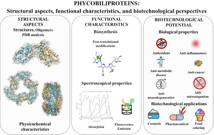
Phycobiliproteins (PBPs) are fluorescent proteins of various colors, including fuchsia, purple-blue and cyan, that allow the capture of light energy in auxiliary photosynthetic complexes called phycobilisomes (PBS). PBPs have several highly preserved structural and physicochemical characteristics. In the PBS context, PBPs function is capture luminous energy in the 450-650 nm range and delivers it to photosystems allowing photosynthesis take place. Besides the energy harvesting function, PBPs also have shown to have multiple biological activities, including antioxidant, antibacterial and antitumours, making them an interesting focus for different biotechnological applications in areas like biomedicine, bioenergy and scientific research. Nowadays, the main sources of PBPs are cyanobacteria and micro and macro algae from the phylum Rhodophyta. Due to the diverse biological activities of PBPs, they have attracted the attention of different industries, such as food, biomedical and cosmetics. This is why a large number of patents related to the production, extraction, purification of PBPs and their application as cosmetics, biopharmaceuticals or diagnostic applications have been generated, looking less ecological impact in the natural prairies of macroalgae and less culture time or higher productivity in cyanobacteria to satisfy the markets and applications that require high amounts of these molecules. In this review, we summarize the main structural characteristics of PBPs, their biosynthesys and biotechnological applications. We also address current trends and future perspectives of the PBPs market.
Keywords: Bioactive molecules; Biotechnology; PBPs; Protein structure.
© 2022 Published by Elsevier B.V. on behalf of Research Network of Computational and Structural Biotechnology.
Conflict of interest statement
The authors declare that they have no known competing financial interests or personal relationships that could have appeared to influence the work reported in this paper.
Figures

References
-
- Kirk JT. O. Light and Photosynthesis in Aquatic Ecosystems 3rd Edition. (Cambridge, 2011).
-
- Sven Beer, Mats Bjork, J. B. Photosynthesis in the Marine Environment. 2014).
-
- Fischer W.W., Hemp J., Johnson J.E. Evolution of oxygenic photosynthesis. Annu Rev Earth Planet Sci. 2016;44(1):647–683.
-
- Tanaka A, Tanaka R. The biochemistry, physiology, and evolution of the chlorophyll cycle. Advances in Botanical Research 90, (Elsevier Ltd., 2019).
-
- Morançais M., Mouget J.-L., Dumay J. Microalgae in Health and Disease Prevention. Elsevier; 2018. Proteins and pigments; pp. 145–175. - DOI
Publication types
LinkOut - more resources
Full Text Sources