

Maxillary Odontogenic Keratocyst
- PMID: 35422993
- PMCID: PMC9004408
- DOI: 10.1093/jscr/rjac078
Maxillary Odontogenic Keratocyst
Abstract
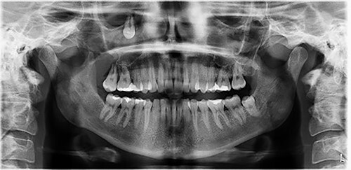
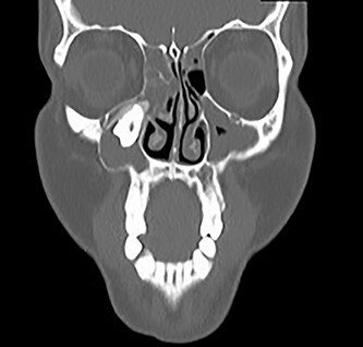
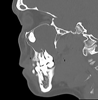
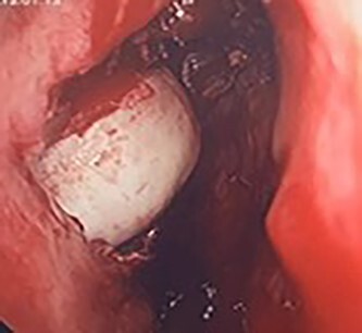
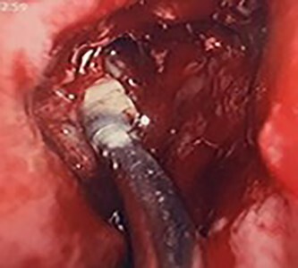
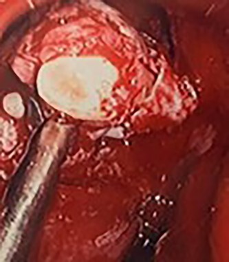
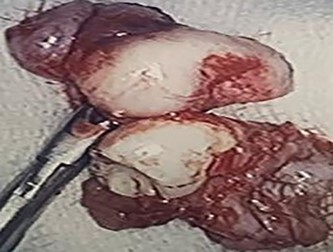
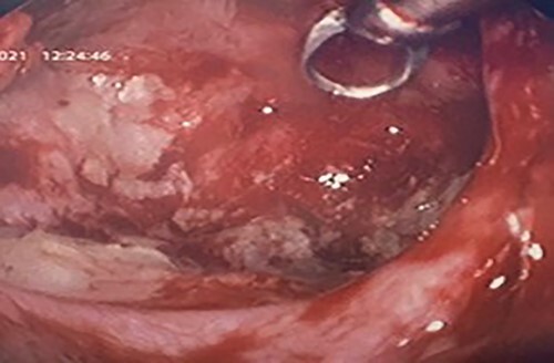
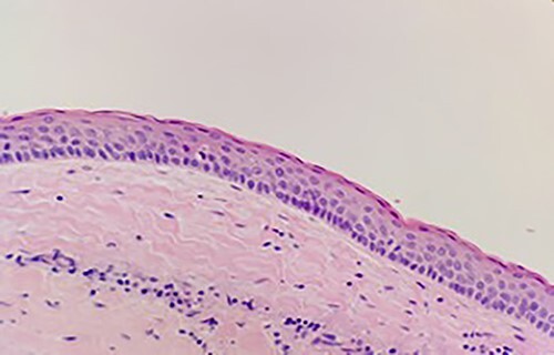
The Odontogenic Keratocyst (OKC) is one of the most aggressive odontogenic cysts. OKCs of the maxilla are particularly rare with less than 1% of cases reported in the literature. A 29-year-old female patient presented with pain and loose upper molars. Imaging confirmed an ectopic tooth at the osteomeatal complex and a maxillary OKC. These were endoscopically surgically removed and two teeth were encountered at the maxillary antrum. Histopathology confirmed the diagnosis of OKC of the maxilla. Surveillance with CT imaging and clinical assessment at 6 months shows no evidence of recurrence.
Keywords: Carnoy’s solution; Odontogenic Keratocyst; endoscopic sinus surgery; maxillary sinus.
Published by Oxford University Press and JSCR Publishing Ltd. © The Author(s) 2022.
Figures
References
-
- Karthiga KS, Sivapatha Sundharam B, Manikandan R. Nevoid basal cell carcinoma syndrome. Indian J Dent Res 2006;17:50–3. - PubMed
-
- Kokila MJ, Laxmidevi BL. Odontogenic keratocyst of maxilla involving the sinus-OKC to be a cyst or a tumour. J Dent Sci Res 2010;1:83–90.
-
- Agaram NP, Collins BM, Barnes L, Lomago D, Aldeeb D, Swalsky P, et al. Molecular analysis to demonstrate that odontogenic keratocysts are neoplastic. Arch Pathol Lab Med 2004;128:313–7. - PubMed
-
- Silva GC, Silva EC, Santiago Gomez R, Vieira TC. Odontogenic keratocyst in the maxillary sinus: report of two cases. Oral Oncol 2006;42:231–4.
Publication types
LinkOut - more resources
Full Text Sources
