. 2021 Apr 26;11(25):15400-15409.
doi: 10.1039/d1ra02198b. eCollection 2021 Apr 21.

Inhibitory properties of saponin from Eleocharis dulcis peel against α-glucosidase

Affiliations

Inhibitory properties of saponin from Eleocharis dulcis peel against α-glucosidase

Yipeng Gu et al. RSC Adv. .

Abstract

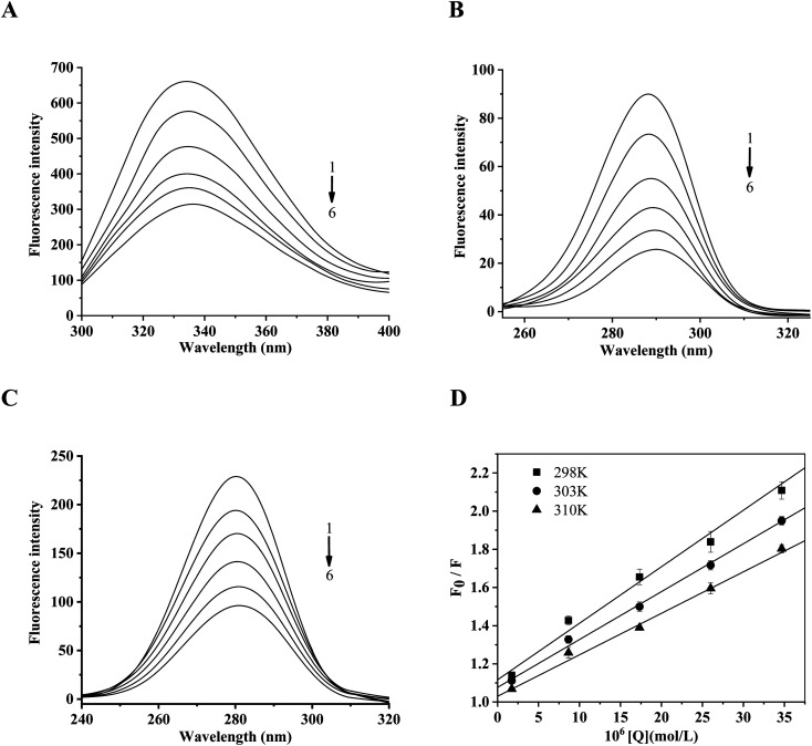
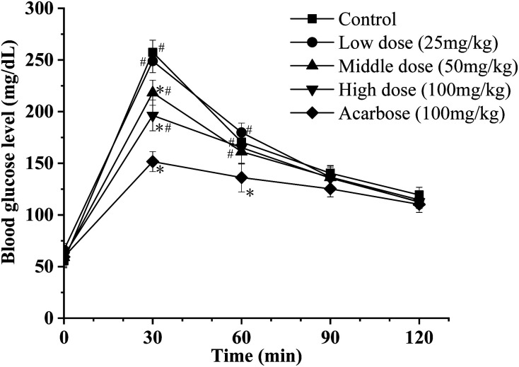
The inhibitory properties towards α-glucosidase in vitro and elevation of postprandial glycemia in mice by the saponin constituent from Eleocharis dulcis peel were evaluated for the first time. Three saponins were isolated by silica gel and HPLC, identified as stigmasterol glucoside, campesterol glucoside and daucosterol by NMR spectroscopy. Daucosterol presented the highest content and showed the strongest α-glucosidase inhibitory activity with competitive inhibition. Static fluorescence quenching of α-glucosidase was caused by the formation of the daucosterol-α-glucosidase complex, which was mainly derived from hydrogen bonds and van der Waals forces. Daucosterol formed 7 hydrogen bonds with 4 residues of the active site and produced hydrophobic interactions with 3 residues located at the exterior part of the binding pocket. The maltose-loading test results showed that daucosterol inhibited elevation of postprandial glycemia in ddY mice. This suggests that daucosterol from Eleocharis dulcis peel can potentially be used as a food supplement for anti-hyperglycemia.


Conflict of interest statement

The authors state that there is no conflict of interest.

Figures

Fig. 1
Fig. 1. Flowchart for the isolation of saponins from Eleocharis dulcis peel.
Fig. 2
Fig. 2. The chemical structures of the three compounds isolated from Eleocharis dulcis peel. The structures from 1 to 3 are stigmasterol glucoside, campesterol glucoside and daucosterol, respectively.
Fig. 3
Fig. 3. The inhibition kinetics of α-glucosidase by daucosterol (pH 7.4, T = 310 K). Values are means ± SD, n = 3.
Fig. 4
Fig. 4. The fluorescence quenching of α-glucosidase by daucosterol. (A–C) The fluorescence emission spectra (A) and the synchronous fluorescence spectra (B and C) of α-glucosidase in the presence of daucosterol at different concentrations (pH 7.4, T = 298 K). (A) λex = 280 nm; (B) Δλ = 15 nm; (C) Δλ = 60 nm. The concentrations of daucosterol from 1 to 6 are 0.00, 1.00, 5.00, 10.00, 15.00 and 20.00 mg L−1, respectively. (D) The linear-fitting graph of Stern–Volmer for the fluorescence quenching of α-glucosidase by daucosterol at different temperatures (pH 7.4, T = 298 K, 303 K, 310 K). λex = 280 nm, λem = 334 nm. Values are means ± SD, n = 3. The concentrations of daucosterol are 0.00, 0.17, 0.87, 1.73, 2.60 and 3.47 × 10⁻⁵ mol L−1, respectively.
Fig. 5
Fig. 5. Cluster analysis of conformations from the AutoDock docking runs of daucosterol with α-glucosidase.
Fig. 6
Fig. 6. Molecular docking analysis of α-glucosidase and daucosterol (A and B): 3D-diagram (A) and 2D-diagram (B) of molecular docking of daucosterol and α-glucosidase. The black dashed lines represent hydrogen-bonding interactions; the green solid line represents hydrophobic interactions.
Fig. 7
Fig. 7. The effect of daucosterol on the blood glucose level after maltose loading in mice. Values are means ± SEM, n = 6. *p < 0.05, compared with control group; #p < 0.05, compared with acarbose group.

References

    1. Odaci D. Telefoncu A. Timur S. Maltose biosensing based on co-immobilization of α-glucosidase and pyranose oxidase. Bioelectrochemistry. 2010;79:108–113. doi: 10.1016/j.bioelechem.2009.12.010. - DOI - PubMed
    1. Stefano E. D. Oliviero T. Udenigwe C. C. Functional significance and structure-activity relationship of food-derived α-glucosidase inhibitors. Curr. Opin. Food Sci. 2018;20:7–12. doi: 10.1016/j.cofs.2018.02.008. - DOI
    1. Santos C. M. Freitas M. Fernandes E. A comprehensive review on xanthone derivatives as α-glucosidase inhibitors. Eur. J. Med. Chem. 2018;157:1460–1479. doi: 10.1016/j.ejmech.2018.07.073. - DOI - PubMed
    1. Akaza M. Akaza I. Kanouchi T. Sasano T. Sumi Y. Yokota T. Nerve conduction study of the association between glycemic variability and diabetes neuropathy. Diabetol. Metab. Syndr. 2018;10:69. doi: 10.1186/s13098-018-0371-0. - DOI - PMC - PubMed
    1. Wang Q. Rehman M. Peng D. Liu L. Antioxidant capacity and α-glucosidase inhibitory activity of leaf extracts from ten ramie cultivars. Ind. Crops Prod. 2018;122:430–437. doi: 10.1016/j.indcrop.2018.06.020. - DOI