. 2022 Mar 8;12(13):7742-7756.
doi: 10.1039/d1ra08061j.

Magnetic resonance imaging of tumor-associated-macrophages (TAMs) with a nanoparticle contrast agent

Affiliations

Magnetic resonance imaging of tumor-associated-macrophages (TAMs) with a nanoparticle contrast agent

Junhan Zhou et al. RSC Adv. .

Abstract

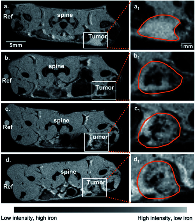
In the tumor micro-environment, tumor associated macrophages (TAMs) represent a predominant component of the total tumor mass, and TAMs play a complex and diverse role in cancer pathogenesis with potential for either tumor suppressive, or tumor promoting biology. Thus, understanding macrophage localization and function are essential for cancer diagnosis and treatment. Typically, tissue biopsy is used to evaluate the density and polarization of TAMs, but provides a limited "snapshot" in time of a dynamic and potentially heterogeneous tumor immune microenvironment. Imaging has the potential for three-dimensional mapping; however, there is a paucity of macrophage-targeted contrast agents to specifically detect TAM subtypes. We have previously found that sulfated-dextran coated iron oxide nanoparticles (SDIO) can target macrophage scavenger receptor A (SR-A, also known as CD204). Since CD204 (SR-A) is considered a biomarker for the M2 macrophage polarization, these SDIO might provide M2-specific imaging probes for MRI. In this work, we investigate whether SDIO can label M2-polarized cells in vitro. We evaluate the effect of degree of sulfation on uptake by primary cultured bone marrow derived macrophages (BMDM) and found that a higher degree of sulfation led to higher uptake, but there were no differences across the subtypes. Further analysis of the BMDM showed similar SR-A expression across stimulation conditions, suggesting that this classic model for macrophage subtypes may not be ideal for definitive M2 subtype marker expression, especially SR-A. We further examine the localization of SDIO in TAMs in vivo, in the mammary fat pad mouse model of breast cancer. We demonstrate that uptake by TAMs expressing SR-A scales with degree of sulfation, consistent with the in vitro studies. The TAMs demonstrate M2-like function and secrete Arg-1 but not iNOS. Uptake by these M2-like TAMs is validated by immunohistochemistry. SDIO show promise as a valuable addition to the toolkit of imaging probes targeted to different biomarkers for TAMs.


Conflict of interest statement

There are no conflicts to declare.

Figures

Scheme 1
Scheme 1. Synthetic route of SDIO/DIO.
Fig. 1
Fig. 1. BMDMs cell uptake study. (a) T2 values of BMDMs lysates. 10 : 1 SDIO and 1 : 1 SDIO have significant higher uptake compared to untargeted DIO and untreated control. (b) Iron concentration of BMDMs lysates. 10 : 1 SDIO has significant higher uptake compared to 1 : 1 SDIO, untargeted DIO and untreated control. The differences were considered significant with P values * < 0.05, ** < 0.01, and *** < 0.001 as shown. P values are corrected for multiple comparisons within a given outcome with the Bonferroni Holm–correction for 42 tests.
Fig. 2
Fig. 2. Expression of SR-A receptor in different polarization states of BMDMs. (a) Representative immunoblot of SR-A expression in polarized macrophages after 24 h of stimulation. GAPDH was used as a housekeeping gene. (b) Quantification of SR-A in macrophages polarized as in (a). Quantification is an average of three blots. Relative SR-A expression on stimulated BMDMs. The raw blot is shown in Fig. S3 in ESI. The differences were considered significant with P values * < 0.05, ** < 0.01, and *** < 0.001 as shown. P-values were corrected for 3 tests via Bonferroni–Holm.
Fig. 3
Fig. 3. Representative reconstituted MR images of BMDMs lysates (a) and mean T2 values (b). Samples containing reconstituted cell lysate samples from different macrophages subtype incubated with 1 : 1 SDIO (top row), DIO (2nd row), media only (3rd row), and a reference of pure water (bottom row). (n = 3) All three subtypes of macrophage incubated with 1 : 1 SDIO showed the highest signal suppression characteristic of T2 agents (darker images), while DIO incubated cells and untreated cells were similar to pure water. Scale bar = 4 mm. The average T2 values (n = 3) were reported in panel b with standard deviation as the error bar. The differences were considered significant with P values * < 0.05, ** < 0.01, and *** < 0.001 as shown. P values are corrected for multiple comparisons within a given outcome with the post hoc Tukey HSD correction.
Fig. 4
Fig. 4. Relative TNF-α (a), IL-6 (b) and MCP-1 (c) secretion by bone marrow derived macrophages (BMDMs) treated with dextran sulfate, DIO, 1:1 SDIO, 10:1 SDIO and media. BMDMs incubated with 1 : 1 SDIO show more production of TNF-α, IL-6 and MCP-1 in M1 stimulated cells. 10 : 1 SDIO incubated M2 stimulated BMDMs showed significantly more production of MCP-1. The differences were considered significant with P values * < 0.05, ** < 0.01, and *** < 0.001 as shown. P values are corrected for multiple comparisons within a given outcome with the Bonferroni Holm–correction for 46 tests.
Fig. 5
Fig. 5. Representative MR images in axial plane of breast cancer models. MR images for a representative adult Balb/c mouse with 10 : 1 SDIO 15 mg Fe per kg I.V. injection (a) pre-injection scan, (b) 4 h-post injection scan, (c) 24 h-post injection scan, and (d) 48 h-post injection scan. The left tumor is indicated by a white box and the zoomed in images are shown in (a1–d1). Tumor margins are circled in orange.
Fig. 6
Fig. 6. Representative zoomed in MR images and parametric T2* maps for 25 mm3 tumor (a and c) and 100 mm3 tumor models (b and d) injected with either 15 mg Fe kg−1 10 : 1 SDIO, 15 mg Fe kg−1 1 : 1 SDIO, 30 mg Fe kg−1 1 : 1 SDIO, or 30 mg Fe kg−1 DIO at Pre-, 4 h-post, 24 h-post, or 48 h-post injection time point (tumor margins are outlined in orange). SDIO-injected group in both sizes of tumors have stronger negative contrast and lower T2 * after injection compared to untargeted DIO-injected control. Scale bar = 2 mm.
Fig. 7
Fig. 7. T2* of 25 mm3 tumor and 100 mm3 tumor at pre-, 4 h-post, 24 h-post, or 48 h-post injection imaging with SDIO/DIO injection. 10 : 1 SDIO with 15 mg Fe kg−1 dose (dark blue) have significantly higher accumulation within tumors compared to untargeted DIO-injected controls (orange and red) at all MR imaging time points. The 1 : 1 SDIO with 30 mg Fe kg−1 dose (medium blue) injected group has significant shorter T2 than untargeted DIO injection (orange) at 24 h-post and 48 h-post injection imaging, indicating higher uptake. The differences were considered significant with P values * < 0.05, ** < 0.01, and *** < 0.001 as shown. The *'s below the boxplot showed the P values when comparing to pre-injection. P-values were corrected for 33 tests via Bonferroni–Holm.
Fig. 8
Fig. 8. Representative histology and immunohistochemistry images of mouse breast cancer model. (a) Bright-field H&E, CD204, and Prussian Blue staining for iron after 48 hours post-injection MR imaging. Black arrowhead indicate iron containing cells. Scale bar = 1 mm. (b) Representative immunohistochemistry for Arginase-1, iNOS and F4/80 staining images after 48 h post-injection MR imaging. The cell nuclei were counterstained with Hoechst 33342. Scale bar = 500 μm. TAMs do not express iNOS, an M1 marker, but do express Arg-1, an M2 marker.
Fig. 9
Fig. 9. Representative zoomed in Prussian blue stained image (a) and immunohistochemistry for Arginase-1, CD204 and F4/80 staining images (b). (a) Prussian blue staining image for 10 : 1 SDIO injected animals after 48 h post-injection MRI, which clearly shows the morphology of the tumor cells (arrows) and macrophages (arrowheads) within the tumor microenvironment. Iron is found in macrophages, but not in tumor cells. (b) F4/80, CD204, and Arg-1 co-stained images after 48 h-post injection MRI. The cell nuclei were counterstained with Hoechst 33342. The tumor associated macrophages are CD204 and Arg-1 double positive. Scale bar = 20 μm.

References

    1. Murray P. J. Allen J. E. Biswas S. K. Fisher E. A. Gilroy D. W. Goerdt S. Gordon S. Hamilton J. A. Ivashkiv L. B. Lawrence T. et al., Macrophage Activation and Polarization: Nomenclature and Experimental Guidelines. Immunity. 2014;41(1):14–20. doi: 10.1016/j.immuni.2014.06.008. doi: 10.1016/j.immuni.2014.06.008. - DOI - DOI - PMC - PubMed
    1. Sica A. Invernizzi P. Mantovani A. Macrophage Plasticity and Polarization in Liver Homeostasis and Pathology. Hepatology. 2014:2035–2043. doi: 10.1002/hep.26754. - DOI - PubMed
    1. Stöger J. L. Gijbels M. J. J. Velden S. V. D. Manca M. Loos C. M. V. D. Biessen E. A. L. Daemen M. J. A. P. Lutgens E. Winther M. P. J. D. Distribution of Macrophage Polarization Markers in Human Atherosclerosis. Atherosclerosis. 2012;225(2):461–468. doi: 10.1016/j.atherosclerosis.2012.09.013. doi: 10.1016/j.atherosclerosis.2012.09.013. - DOI - DOI - PubMed
    1. Corporation H. P. Understanding the Mysterious M2 Macrophage through Activation Markers and Effector Mechanisms. Mediat. Inflamm. 2015;2015:16–18. - PMC - PubMed
    1. Lewis M. Merched A. J. Tumor-Associated Macrophages, Inflammation and Pathogenesis of Hepatocellular Carcinoma. J. Mol. Genet. Med. 2014;8(3):132. doi: 10.4172/1747-0862.1000132. - DOI