Clinical Trial
. 2022 May 1;234(5):748-758.
doi: 10.1097/XCS.0000000000000132.

Impact of Intraoperative Molecular Imaging after Fluorescent-Guided Pulmonary Metastasectomy for Sarcoma

Affiliations
Clinical Trial

Impact of Intraoperative Molecular Imaging after Fluorescent-Guided Pulmonary Metastasectomy for Sarcoma

Feredun Azari et al. J Am Coll Surg. .

Abstract

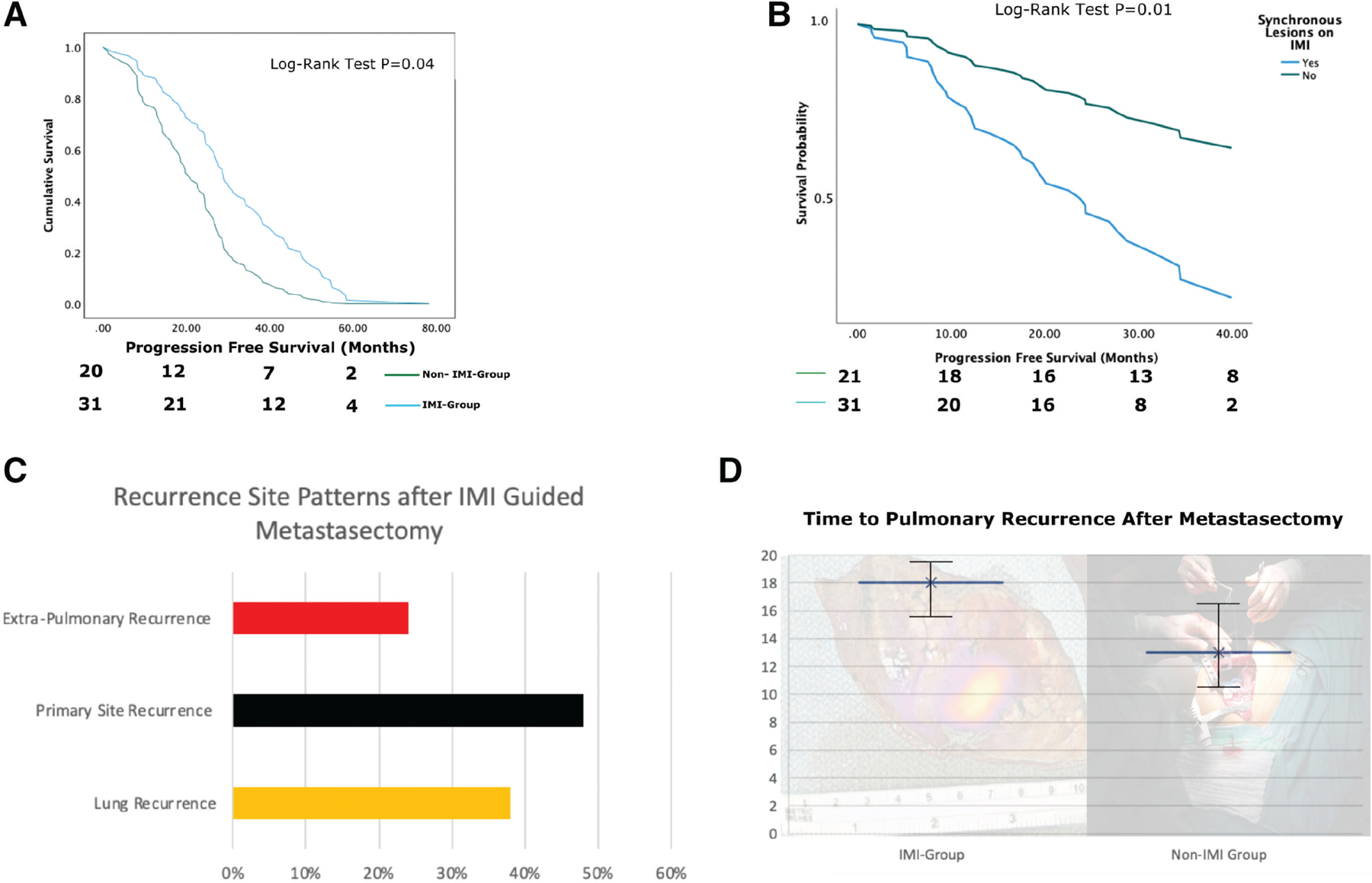
Background: Intraoperative molecular imaging (IMI) has been shown to improve lesion detection during pulmonary sarcomatous metastasectomy. Our goal in this study was to evaluate whether data garnered from IMI-guided resection of pulmonary sarcoma metastasis translate to improved patient outcomes.

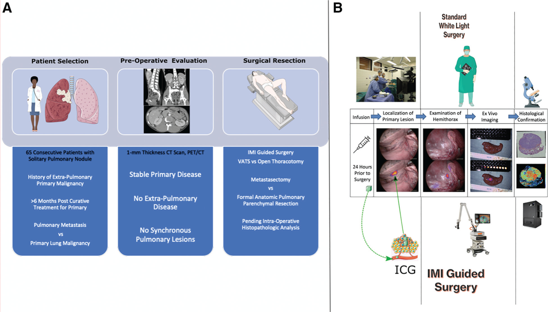
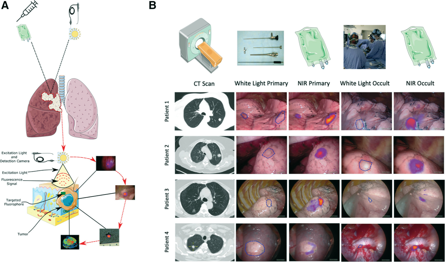
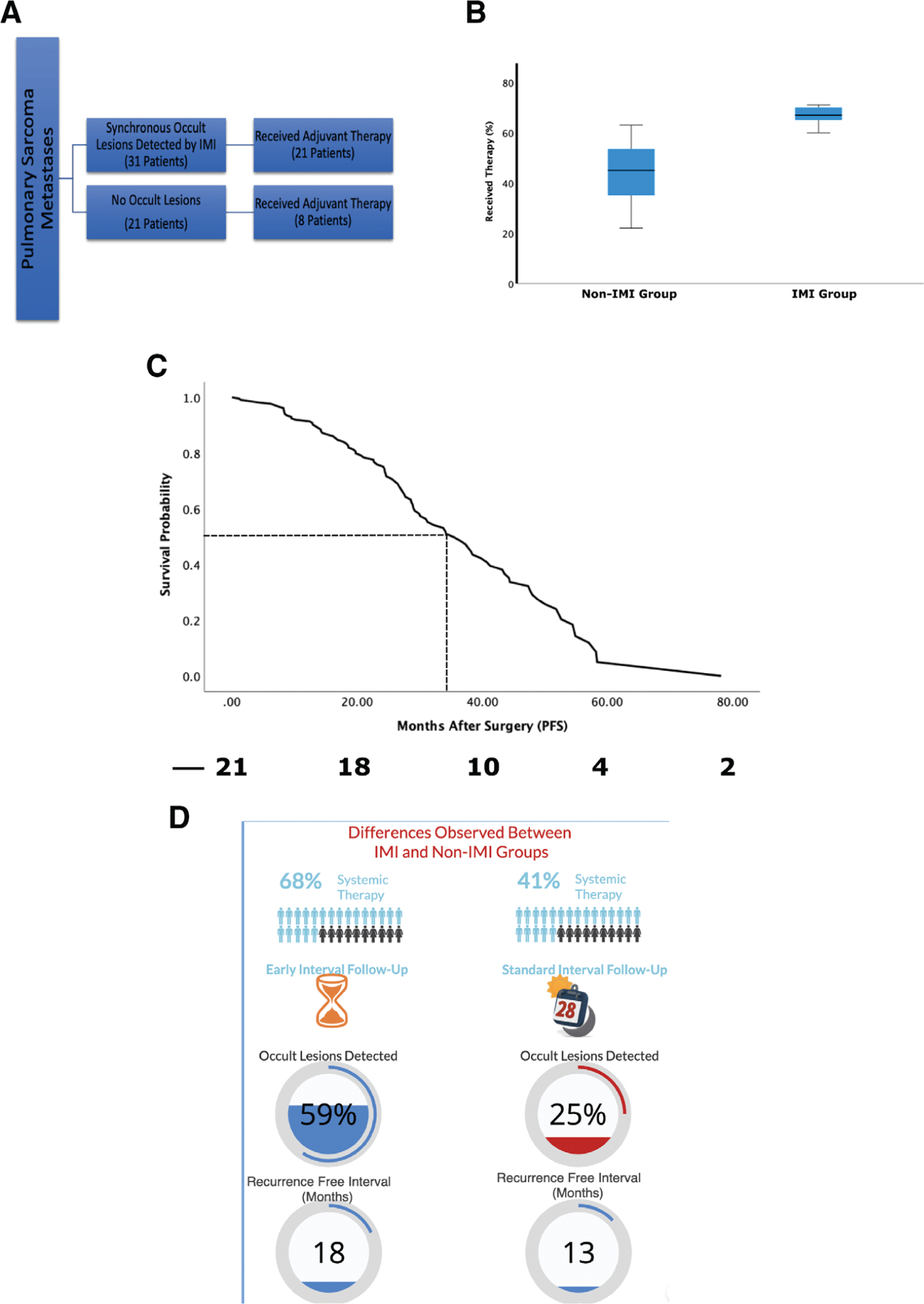
Study design: Fifty-two of 65 consecutive patients with a previous history of sarcomas found to have pulmonary nodules during screening were enrolled in a nonrandomized clinical trial. Patients underwent TumorGlow the day before surgery. Data on patient demographics, tumor biologic characteristics, preoperative assessment, and survival were included in the study analysis and compared with institutional historical data of patients who underwent metastasectomy without IMI. p values < 0.05 were considered significant.

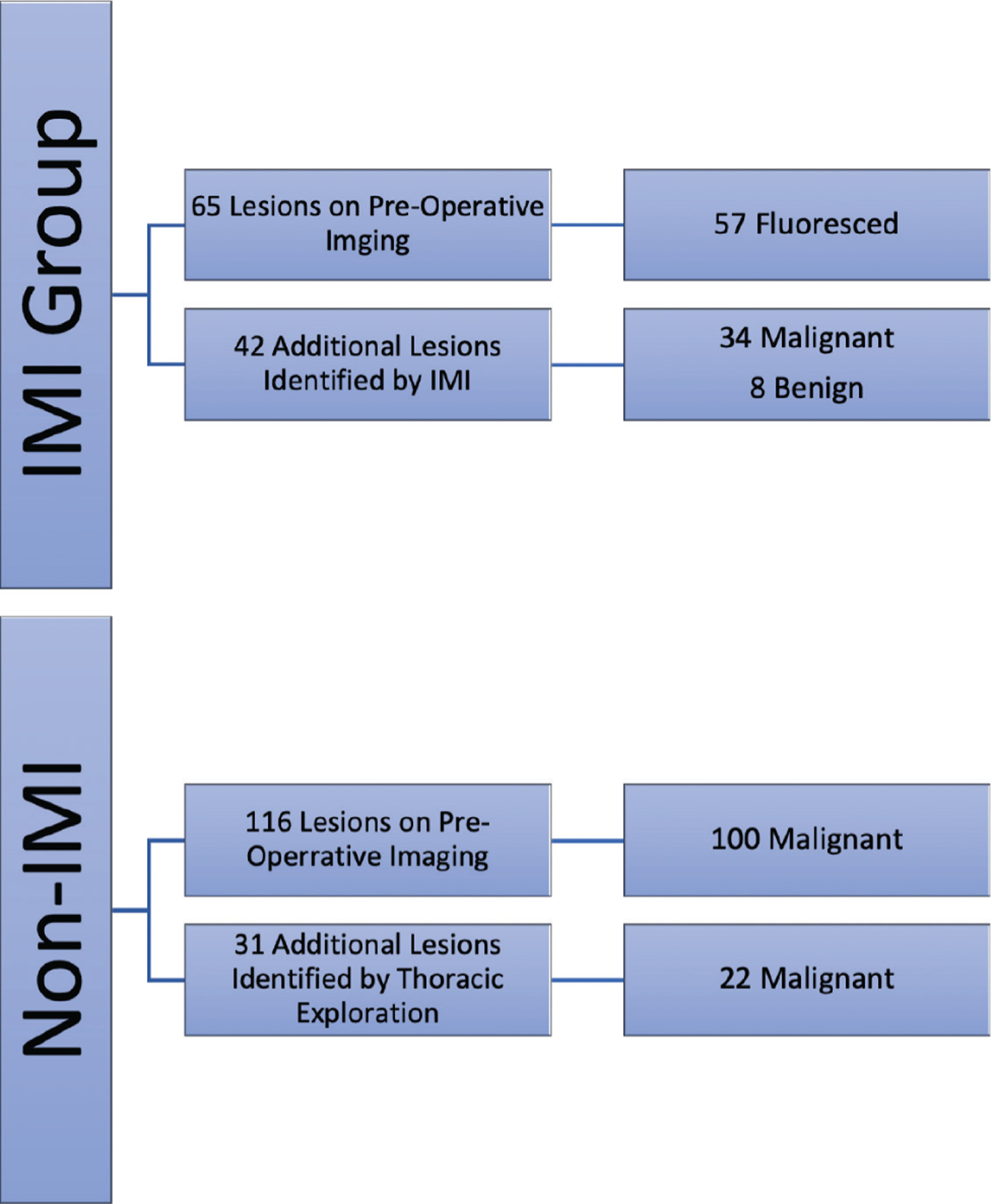
Results: IMI detected 42 additional lesions in 31 patients (59%) compared with the non-IMI cohort where 25% percent of patients had additional lesions detected using tactile and visual feedback only (p < 0.05). Median progression-free survival (PFS) for patients with IMI-guided pulmonary sarcoma metastasectomy was 36 months vs 28.6 months in the historical cohort (p < 0.05). IMI-guided pulmonary sarcoma metastasectomy had recurrence in the lung with a median time of 18 months compared with non-IMI group at 13 months (p < 0.05). Patients with synchronous lesions in the IMI group underwent systemic therapy at a statistically higher rate and tended to undergo routine screening at shorter interval.

Conclusions: IMI identifies a subset of sarcoma patients during pulmonary metastasectomy who have aggressive disease and informs the medical oncologist to pursue more aggressive systemic therapy. In this setting, IMI can serve both as a diagnostic and prognostic tool without conferring additional risk to the patient.


Figures

Figure 1.
Figure 1.
(A) Flowchart depicting the patient selection process, which involves evaluation of patients with pulmonary nodules with a history of primary nonpulmonary sarcoma who were in remission after treatment for primary disease. Differential for the patients included pulmonary metastasis vs primary lung malignancy. All the patients were evaluated with high-fidelity 1-mm thickness cut CT scan and positron emission tomography (PET)/CT for synchronous lesions or extra-pulmonary disease. Appropriate patients were then scheduled for intraoperative molecular imaging (IMI)–guided resection of the nodule (metastasectomy if nonpulmonary lesion or anatomic resection if primary lung disease). (B) Pictorial description of differences between standard white light surgery (top column) vs IMI-guided resection (bottom column) for patients enrolled in the study. ICG, indocyanine green; VATS, video-assisted thoracoscopic surgery.
Figure 2.
Figure 2.
(A) Anatomic distribution of the sarcoma metastases in the thoracic cavity, which shows nonspecific and nonstatistically significant distribution of lesions in all 5 lobes with right middle lobe having the lowest frequency. (B) Distribution of sarcoma subtype by sex in the study group. (C) Anatomic distribution primary sarcomas in the cohort with more than half of the primary lesions arising from the extremities correlating with sarcoma subtypes.
Figure 3.
Figure 3.
Summary of metastases identified in both sarcoma and non-sarcoma pulmonary metastases using gold-standard techniques and intraoperative molecular imaging (IMI). IMI identified 34 malignant sarcomatous pulmonary metastases that would otherwise be missed.
Figure 4.
Figure 4.
Representative intraoperative findings of intraoperative molecular imaging (IMI)–guided metastasectomy of sarcomatous pulmonary lesions. (A) Technological pictorial overview of IMI-guided pulmonary sarcoma metastasectomy. (B) Sample pictorial representation of various IMI guided metastasectomy including preoperative 1-mm cut thickness high-fidelity CT scan images, traditional white light view of the lesions showing identification of known primary metastatic lesions during video-assisted thoracoscopic surgery or open thoracic resection as well as near infrared (NIR) merged views depicting real-time intraoperative visualization primary and occult lesions. As seen in Patients #3 and #4, white light view did not provide clear visual identification of occult lesions, particularly near critical structures which was easily identified using IMI.
Figure 5.
Figure 5.
(A) Overall survival curves for the intraoperative molecular imaging (IMI) and non–IMI-based pulmonary metastasectomy groups. (B) Survival curves displaying worse overall survival for patients who had occult lesions detected with IMI vs those who had no occult lesions. (C) Initial recurrence patterns in patients with pulmonary sarcoma metastases after index resection of metastatic lesion. (D) Time to pulmonary recurrence after pulmonary metastasectomy for IMI and non-IMI groups.
Figure 6.
Figure 6.
(A) Summary of postoperative systemic treatment trends for pulmonary sarcoma metastases based on occult lesion detection with intraoperative molecular imaging (IMI). (B) Box-whisker plot analyzing the rate of postoperative systemic treatment rates in those who had occult lesions detected by IMI showing statistically significant higher rate of postoperative systemic therapy compared with those in the non-IMI group. (C) Survival curve for patients with occult lesions with IMI who underwent postoperative systemic therapy. (D) Summary of differences observed between the IMI- and non–IMI-based pulmonary metastasectomy with IMI patients noted to undergo adjuvant systemic therapy at a higher rate, have a trend for shorter postoperative surveillance, having higher occult lesion detected (59% of patients in IMI group vs 25% in non-IMI group; p < 0.05), and having a higher recurrence-free interval from operation (18 months vs 13 months). PFS, progression-free survival.

Comment in

References

    1. Digesu CS, Wiesel O, Vaporciyan AA, et al. Management of sarcoma metastases to the lung. Surg Oncol Clin N Am 2016;25:721–733. - PMC - PubMed
    1. Marcove RC, Miké V, Hajek JV, et al. Osteogenic sarcoma under the age of twenty-one. A review of one hundred and forty-five operative cases. J Bone Joint Surg Am 1970;52:411–423. - PubMed
    1. Saltzman DA, Snyder CL, Ferrell KL, et al. Aggressive metastasectomy for pulmonic sarcomatous metastases: A follow-up study. Am J Surg 1993;166:543–547. - PubMed
    1. Predina JD, Puc MM, Bergey MR, et al. Improved survival after pulmonary metastasectomy for soft tissue sarcoma. J Thorac Oncol 2011;6:913–919. - PubMed
    1. Cerfolio RJ, Bryant AS, McCarty TP, et al. A prospective study to determine the incidence of non-imaged malignant pulmonary nodules in patients who undergo metastasectomy by thoracotomy with lung palpation. Ann Thorac Surg 2011;91:1696–700; discussion 1700. - PubMed

Publication types