. 2023 Jan 18;118(17):3331-3345.
doi: 10.1093/cvr/cvac052.

Neutrophil inhibition improves acute inflammation in a murine model of viral myocarditis

Affiliations

Neutrophil inhibition improves acute inflammation in a murine model of viral myocarditis

Paolo Carai et al. Cardiovasc Res. .

Abstract

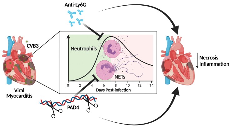
Aims: Viral myocarditis (VM) is an inflammatory pathology of the myocardium triggered by a viral infection that may cause sudden death or heart failure (HF), especially in the younger population. Current treatments only stabilize and improve cardiac function without resolving the underlying inflammatory cause. The factors that induce VM to progress to HF are still uncertain, but neutrophils have been increasingly associated with the negative evolution of cardiac pathologies. The present study investigates the contribution of neutrophils to VM disease progression in different ways.

Methods and results: In a coxsackievirus B3- (CVB3) induced mouse model of VM, neutrophils and neutrophil extracellular traps (NETs) were prominent in the acute phase of VM as revealed by enzyme-linked immunosorbent assay analysis and immunostaining. Anti-Ly6G-mediated neutrophil blockade starting at model induction decreased cardiac necrosis and leucocyte infiltration, preventing monocyte and Ly6CHigh pro-inflammatory macrophage recruitment. Furthermore, genetic peptidylarginine deiminase 4-dependent NET blockade reduced cardiac damage and leucocyte recruitment, significantly decreasing cardiac monocyte and macrophage presence. Depleting neutrophils with anti-Ly6G antibodies at 7 days post-infection, after the acute phase, did not decrease cardiac inflammation.

Conclusion: Collectively, these results indicate that the repression of neutrophils and the related NET response in the acute phase of VM improves the pathological phenotype by reducing cardiac inflammation.

Keywords: Coxsackievirus B3; Inflammation; Neutrophil extracellular traps; Neutrophils; Viral myocarditis.


Conflict of interest statement

Conflict of interest: S.H. receives personal fees for scientific advice to Astra-Zeneca, Cellprothera, CSL Behring, and Merck; unrestricted research grant from Pfizer. K.M. has received consulting fees for scientific advice to PEEL Therapeutics. All the remaining authors, P.C., L.F.G., S.V.B., V.S., D.D.G., N.G., and E.A.V.J., declare no competing interests.

Figures

Graphical Abstract
Graphical Abstract
In CVB3-induced viral myocarditis, neutrophil or NET blockade before 7 days post-infection reduces cardiac necrosis and inflammation. CVB3, coxsackievirus B3; NET, neutrophil extracellular traps.
Figure 1
Figure 1
Neutrophil recruitment and NET formation peaks during the acute phase in CVB3-induced VM. (A) Circulating neutrophil levels measured by flow cytometry analysis in sham controls and VM at 4, 7, and 14 dpi (n ≥ 3). (B) Circulating NETs during VM were measured as MPO-DNA complexes in the plasma of sham controls and VM at 4, 7, and 14 dpi by ELISA detection, n = 4. (CE) Quantification and representative images of cardiac neutrophils (MPO+, C) and NETs (MPO+ H3Cit+, D) in sham controls and VM at 4, 7, and 14 dpi. Murine cardiac sections were immune-stained for MPO, H3Cit, and counterstained with DAPI, n = 5. Scale bars = 100 µm. All values are expressed as mean ± SEM. Significance is assessed by one-way ANOVA, followed by Tukey’s test: *P ≤ 0.05; **P ≤ 0.01; ***P ≤ 0.001.
Figure 2
Figure 2
Early neutrophil depletion reduced cardiac inflammation in acute VM. (A) Experimental layout of anti-Ly6G-induced neutrophil depletion at 7 dpi. Representative images and quantification of cardiac necrosis (B), assessed by haematoxylin and eosin staining (n ≥ 8), and cardiac inflammation (C, CD45+ leucocytes, n ≥ 4) in control and anti-Ly6G Ab-treated mice under sham or VM (7 dpi) conditions. Scale bars = 500 µm. Relative quantification of cardiac monocytes (Ly6GF4/80, Ly6C+, D and E) and macrophages (Ly6G F4/80+ Ly6C+, D and G) and pro-inflammatory Ly6CHigh subpopulation (F and H, respectively) by flow cytometry analysis of the cardiac immune fraction of control and anti-Ly6G Ab-treated mice under sham or VM (7 dpi) conditions (n ≥ 5). (IK) Quantification of plasmatic concentrations of CXCL1 (I), CCL2 (J), and CXCL10 (K), assessed by multiplex ELISA assay in control and anti-Ly6G Ab-treated mice under sham or VM (7 dpi) conditions (n ≥ 5). All values are expressed as mean ± SEM. Significance is assessed by an unpaired Student t-test for (A), Kruskal–Wallis test, followed by Tukey’s test for (IK), and by two-way ANOVA, followed by Tukey’s test for the other panels: *P ≤ 0.05; **P ≤ 0.01; ***P ≤ 0.001.
Figure 3
Figure 3
Neutrophil depletion after the acute phase does not improve VM-induced cardiac necrosis. (A) Experimental layout of anti-Ly6G-induced neutrophil depletion at 14 dpi. Representative images and quantification of cardiac necrosis (B), assessed by haematoxylin and eosin staining (n = 13), and cardiac inflammation (C, CD45+ leucocytes, n = 5) in control and anti-Ly6G Ab-treated mice under sham or VM (14 dpi) conditions. Scale bars = 500 µm. Relative quantification of cardiac monocytes (Ly6G F4/80 Ly6C+, D and E) and macrophages (Ly6G F4/80+ Ly6C+, D and G) and pro-inflammatory Ly6CHigh subpopulation (F and H, respectively) by flow cytometry analysis of the cardiac immune fraction of control and anti-Ly6G Ab-treated mice under sham or VM (14 dpi) conditions (n = 6). All values are expressed as mean ± SEM. Significance is assessed by an unpaired Student t-test for (B), and by two-way ANOVA, followed by Tukey’s test for the other panels: *P ≤ 0.05; **P ≤ 0.01.
Figure 4
Figure 4
NET inhibition decreases monocyte recruitment during early acute VM. (A) Experimental layout of genetic NET inhibition at 7 dpi. Representative images and quantification of cardiac necrosis (B), assessed by haematoxylin and eosin staining (n ≥ 7), and cardiac inflammation (C, CD45+ leucocytes, n ≥ 7) in PAD4 WT mice and Ne-KO littermates under sham and VM (7 dpi) conditions. Scale bars = 500 µm. Relative quantification of cardiac monocytes (Ly6G F4/80 Ly6C+, D and E) and macrophages (Ly6G F4/80+ Ly6C+, D and G) and pro-inflammatory Ly6CHigh subpopulation (F and H, respectively) by flow cytometry analysis of the cardiac immune fraction of PAD4 WT mice and Ne-KO littermates under sham and VM (7 dpi) conditions (n ≥ 3). (IK) Quantification of plasmatic concentrations of CXCL1 (I), CCL2 (J), and CXCL10 (K), assessed by multiplex ELISA assay in PAD4 WT mice and Ne-KO littermates under sham or VM (7 dpi) conditions (n ≥ 6). All values are expressed as mean ± SEM. Significance is assessed by a Mann–Whitney U test for (B), Kruskal–Wallis test, followed by Tukey’s test for (I), and by two-way ANOVA, followed by Tukey’s test for the other panels: *P ≤ 0.05; ***P ≤ 0.001.
Figure 5
Figure 5
Lack of PAD4 reduces cardiac inflammation during late acute VM. (A) Experimental layout of genetic NET inhibition at 14 dpi. Representative images and quantification of cardiac necrosis (B), assessed by haematoxylin and eosin staining (n ≥ 14), and cardiac inflammation (C, CD45+ leucocytes, n ≥ 5) in PAD4 WT mice and Ne-KO littermates under sham and VM (14 dpi) conditions. Scale bars = 500 µm. Relative quantification of cardiac monocytes (Ly6G F4/80 Ly6C+, D and E) and macrophages (Ly6G F4/80+ Ly6C+, D and G) and pro-inflammatory Ly6CHigh subpopulation (F and H, respectively) by flow cytometry analysis of the cardiac immune fraction of PAD4 WT mice and Ne-KO littermates under sham and VM (14 dpi) conditions (n ≥ 4). All values are expressed as mean ± SEM. Significance is assessed by a Mann–Whitney U test for (A), and by two-way ANOVA, followed by Tukey’s test for the other panels: *P ≤ 0.05; ***P ≤ 0.001.

References

    1. Fabre A, Sheppard MN. Sudden adult death syndrome and other non-ischaemic causes of sudden cardiac death. Heart 2006;92:316–320. - PMC - PubMed
    1. Weintraub RG, Semsarian C, Macdonald P. Dilated cardiomyopathy. Lancet 2017;390:400–414. - PubMed
    1. Tschöpe C, Ammirati E, Bozkurt B, Caforio ALP, Cooper LT, Felix SB, Hare JM, Heidecker B, Heymans S, Hübner N, Kelle S, Klingel K, Maatz H, Parwani AS, Spillmann F, Starling RC, Tsutsui H, Seferovic P, van Linthout S. Myocarditis and inflammatory cardiomyopathy: current evidence and future directions. Nat Rev Cardiol 2021;18:169–193. - PMC - PubMed
    1. Corsten MF, Schroen B, Heymans S. Inflammation in viral myocarditis: friend or foe? Trends Mol Med 2012;18:426–437. - PubMed
    1. Ponikowski P, Voors AA, Anker SD, Bueno H, Cleland JGF, Coats AJS, Falk V, González-Juanatey JR, Harjola V-P, Jankowska EA, Jessup M, Linde C, Nihoyannopoulos P, Parissis JT, Pieske B, Riley JP, Rosano GMC, Ruilope LM, Ruschitzka F, Rutten FH, van der Meer P. 2016 ESC Guidelines for the diagnosis and treatment of acute and chronic heart failure. E Heart J 2016;37:2129–2200. - PubMed

Publication types