Review
. 2022 Apr 15;14(1):105.
doi: 10.1007/s40820-022-00847-z.

Anti-Parkinsonian Therapy: Strategies for Crossing the Blood-Brain Barrier and Nano-Biological Effects of Nanomaterials

Affiliations
Review

Anti-Parkinsonian Therapy: Strategies for Crossing the Blood-Brain Barrier and Nano-Biological Effects of Nanomaterials

Guowang Cheng et al. Nanomicro Lett. .

Abstract

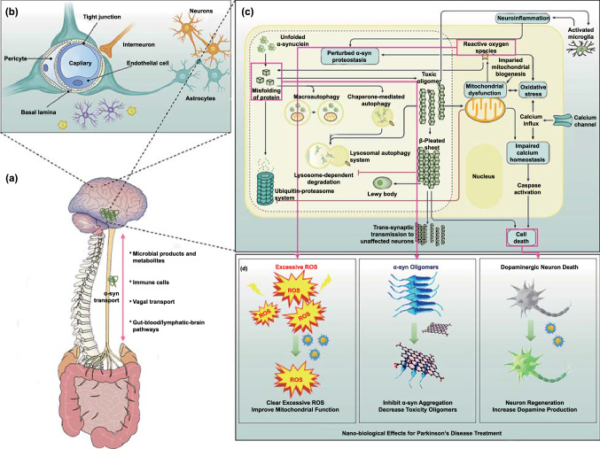
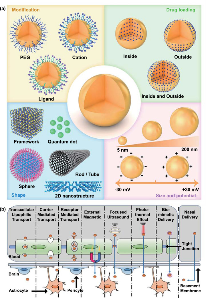
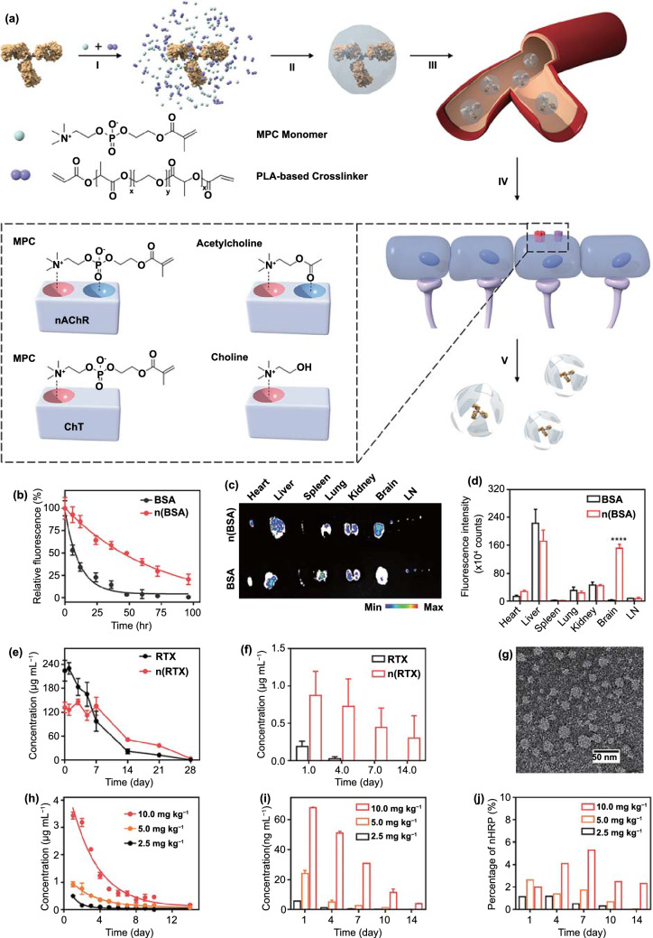
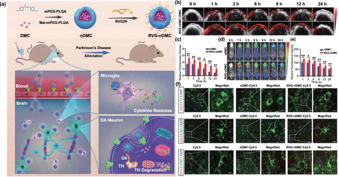
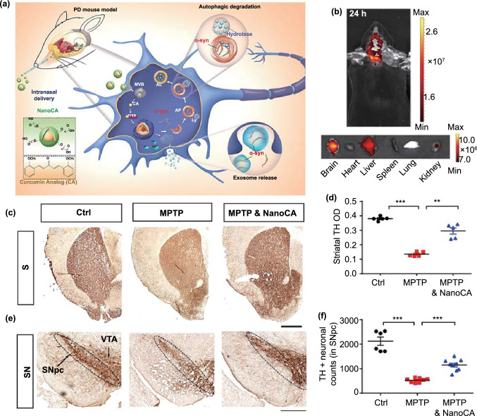
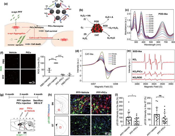
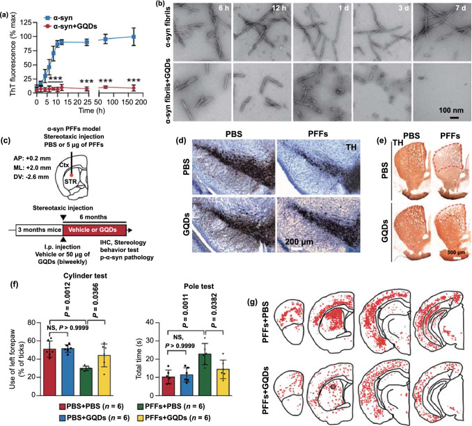
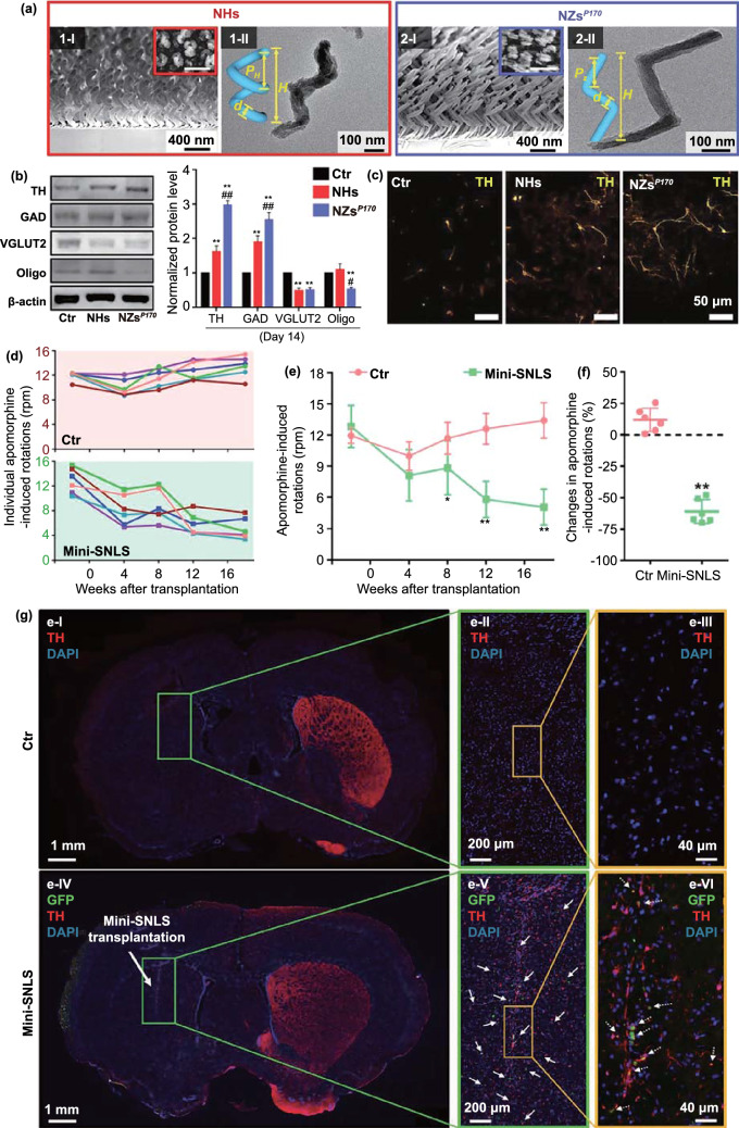
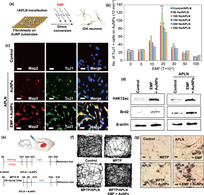
Parkinson's disease (PD), a neurodegenerative disease that shows a high incidence in older individuals, is becoming increasingly prevalent. Unfortunately, there is no clinical cure for PD, and novel anti-PD drugs are therefore urgently required. However, the selective permeability of the blood-brain barrier (BBB) poses a huge challenge in the development of such drugs. Fortunately, through strategies based on the physiological characteristics of the BBB and other modifications, including enhancement of BBB permeability, nanotechnology can offer a solution to this problem and facilitate drug delivery across the BBB. Although nanomaterials are often used as carriers for PD treatment, their biological activity is ignored. Several studies in recent years have shown that nanomaterials can improve PD symptoms via their own nano-bio effects. In this review, we first summarize the physiological features of the BBB and then discuss the design of appropriate brain-targeted delivery nanoplatforms for PD treatment. Subsequently, we highlight the emerging strategies for crossing the BBB and the development of novel nanomaterials with anti-PD nano-biological effects. Finally, we discuss the current challenges in nanomaterial-based PD treatment and the future trends in this field. Our review emphasizes the clinical value of nanotechnology in PD treatment based on recent patents and could guide researchers working in this area in the future.

Keywords: Biomimetic drug delivery; Blood–brain barrier; Nano-biological effects; Nasal delivery; Parkinson’s disease.


Figures

Fig. 1
Fig. 1
Pathology of PD and nano-bio effects useful for its treatment. a The gut microbiota–brain axis that promotes the transport of α-syn from the brain to the gut. The brain-gut communication pathways are of four types: (1) microbial products and metabolites that directly enter the brain; (2) the microbiome, which regulates the immune systems and indirectly influences the CNS; (3) signals sent to the CNS through vagal nerve terminals; (4) gut–blood/lymphatic–brain pathways, through which the microbiome or its metabolites can directly enter the brain. Adapted with permission from Ref. [40]. Copyright 2021 Springe Nature. b The physiological structure of the BBB, which stop drug enter the CNS. Adapted with permission from Ref. [45]. Copyright 2019 Elsevier B.V. c Schematic diagram depicting interactions between major molecular pathways that are implicated in the pathogenesis of Parkinson disease. Adapted with permission from Ref. [4]. Copyright 2017 Springe Nature. d Nano-bio effects of clearing excessive ROS, inhibiting α-syn aggregation, and promoting neuron regeneration for PD treatment
Fig. 2
Fig. 2
Design strategies for nanoplatforms that can cross the BBB (a), and transport pathways used in PD treatment (b)
Fig. 3
Fig. 3
CMT strategy for BBB crossing. a Schematic illustration of the design of the bioinspired delivery system for effective delivery of therapeutic proteins to the CNS. b Pharmacokinetic profiles of mice after intravenous injection of 1 mg mL-1 native BSA or n(BSA) labeled with TAMRA. c Ex vivo images and d normalized fluorescence intensity of dissected tissues including brain, heart, liver, spleen, lung, kidney, and lymph node tissues, from mice treated with TAMRA-labeled BSA and n(BSA). The mice were perfused with PBS and organs were harvested 24 h post-intravenous injection. e RTX plasma and f CSF concentration in mice intravenously injected with 5 mg kg−1 of native RTX or n(RTX), measured by ELISA. g TEM images of CSF obtained from rhesus macaques 24 h after the intravenous administration of 10 mg kg-1 n(HRP). h Plasma and i CSF concentrations of n(HRP) and the j the ratio of CSF versus plasma concentration of n(HRP) in rhesus macaques after the intravenous administration of 2.5, 5.0, and 10 mg kg−1 of n(HRP). Adapted with permission from Ref. [132]. Copyright 2019 WILEY–VCH Verlag GmbH & Co. KgaA, Weinheim
Fig. 4
Fig. 4
Example of RMT. a Preparation and proposed mechanism of RVG-nDMC for PD intervention. b Real-time photoacoustic imaging and c corresponding photoacoustic signal analysis of mice after intravenous injection of nDMC or RVG-Ndmc. d Real-time fluorescence imaging and corresponding fluorescence analysis of mice after intravenous injection of Cy5.5-labeled nDMC or RVG-nDMC. f Representative images of TH, Iba1, and GFAP staining in the SN of mice treated with Cy5.5, nDMC-Cy5.5, or RVG-nDMC-Cy5.5 at 6 h post-injection. White arrows in the enlarged parts of the right column show the presence of NPs in DA neurons and microglia. Red arrows show the presence of NPs outside astrocytes and microglia. Reproduced from Ref. [94]
Fig. 5
Fig. 5
Example of nasal delivery. a Design of self-assembled NanoCA NPs for TFEB-regulated cellular clearance of α-syn in experimental models of PD. b Representative IVIS images showing the biodistribution of NanoCA@TPAAQ in mice after intranasal administration. c Representative photomicrographs of striatal TH immunostaining from the same animals. d Quantification of TH immunoreactivity in the striatum. TH immunoreactivity in midbrain sections from the same animals. f Quantification of surviving TH+ dopaminergic neurons in the SN. Adapted with permission from Ref. [109]. Copyright 2020 American Chemical Society
Fig. 6
Fig. 6
Biomimetic-delivery strategy for BBB crossing. a Parkin Q311(X)A mice (4 Mo. of age) were i.v. injected with 6 × 106 DIR-macrophages and imaged using IVIS. b At the end point (72 h), mice were sacrificed, and perfused; the main organs were removed, and images were obtained using IVIS. c Behavioral tests demonstrating the preservation of locomotory function in Parkin Q311(X) mice upon treatment with GDNF-macrophages at an early stage of disease. d GDNF-BMM could protect dopaminergic neurons in Parkin Q311(X)A mice. Adapted from Ref. [156]
Fig. 7
Fig. 7
Example of magnetic force-mediated brain delivery. a Schematic illustration for Tween-SPIONs crossing the BBB in the presence of a magnet. b The distribution of iron in different brain areas in rats 2 h after tail vein injection. The inset image indicates the position of the magnet. c SPIONs enter the brain by crossing the BBB. Asp: astrocyte processes, End: endothelial cell. d Higher magnification image; the inset shows the size of the SPIONs. Adapted with permission from Ref. [162]. Copyright 2016 American Chemical Society
Fig. 8
Fig. 8
Example of photothermal brain delivery. a The proposed mechanism underlying MSNs-AuNRs@QCT penetration across the BBB under NIR-II (1064 nm) laser irradiation. b Overview of the in vitro BBB Transwell system used to gauge the penetrative capabilities of MSNs-AuNRs@QCT, and the final concentration of QCT in the apical and basolateral chambers in the BBB transwell system. c Photothermal effect on BBB permeability after MSNs-AuNRs@QCT injection and 1064 nm laser irradiation, examined using Evans blue as a BBB permeability indicator. d Representative movement of mice in an open-field box (green). Adapted with permission from Ref. [121]. Copyright 2020 American Chemical Society
Fig. 9
Fig. 9
Example of focused ultrasound-mediated brain delivery for PD treatment. a Improvements in the therapeutic efficacy of curcumin in PD mouse models obtained using CPC combined with ultrasound-targeted microbubble destruction. Schematic of the chemical composition of CPC and the non-invasive localized delivery of CPC NPs to the mouse brain via the ultrasound-targeted microbubble destruction technique for PD therapy. b Photograph of the ultrasound-targeted microbubble destruction setup for the local treatment of the corpus striatum in C57BL/6 mice. c Representative ex vivo fluorescence images obtained at 0.1, 6, 12, and 24 h after intravenous administration (n = 6 per group at each time point). d Representative immunohistochemical staining images of TH+ neurons in the SN in mouse brain sections from different groups (n = 6 per group). Group 1: control; Group 2: only curcumin-loaded cerasomes with no PS 80; Group 3: only CPC with 5% PS 80; Group 4: 5% PS 80-modified cerasomes with no curcumin in combination with ultrasound-targeted microbubble destruction; Group 5: CPC with 5% PS 80 in combination with ultrasound-targeted microbubble destruction. Adapted with permission from Ref. [125]
Fig. 10
Fig. 10
Example of nano-bio effects resulting in the clearance of excessive ROS. a Schematic showing how the PtCu nanozyme scavenges ROS and prevents α-synuclein-induced pathology, neurotoxicity, and cell-to-cell transmission in vitro and in vivo. b Schematic showing how PtCu Nas mimic three redox enzymes (POD: peroxidase, SOD: superoxide dismutase, CAT: catalase). c UV–Vis spectra of TMB in the presence of H2O2 catalyzed by POD-like PtCu Nas. d CAT-like activity of PtCu NPs in reducing H2O2, demonstrated by electron spin resonance (ESR) oximetry; the evolution of the ESR spectra of PDT over time in the presence of 2 Mm H2O2 before and after the addition of PtCu NPs in a closed chamber can be observed. e The SOD-like activity of PtCu Nas in reducing superoxide levels, demonstrated using ESR spectroscopy. f PtCu Nas reduce PFF-induced ROS and quantifies ROS levels. g Timeline of PFF animal experiments with PtCu Nas treatment (top) and the stereotaxic injection sites for PFF and PtCu/Vehicle (bottom). Two-month-old mice were stereotaxically injected with PFF and PtCu/Vehicle and were sacrificed after two months. h Ps129 immunostaining in the substantia nigra (SN) and striatum (ST). Brain sections were stained with anti-Ps129 and anti-TH (tyrosine hydroxylase) antibodies. i Quantification of Ps129 immunostaining. Adapted with permission from Ref. [187]. Copyright 2020 Elsevier Ltd. All rights reserved
Fig. 11
Fig. 11
Example of nano-bio effects resulting in the inhibition of α-syn. a Kinetics of α-syn fibrillization monitored using a ThT fluorescence assay. b TEM images of preformed α-syn fibrils at various time points (6 and 12 h and 1, 3, and 7 days) in the absence (top) and presence (bottom) of GQDs. c Schematic illustration of stereotaxic intrastriatal injection coordinates for α-syn PFFs (5 μg) in C57BL/6 mice. As a treatment, 50 μg of GQDs or PBS were i.p. injected biweekly for 6 months. AP, anteroposterior; ML, mediolateral; DV, dorsoventral; Ctx, cortex; STR, striatum; IHC, immunohistochemistry. d Representative TH immunohistochemistry images of the SN from the α-syn PFF-injected hemisphere in the absence (top) and presence (bottom) of GQDs. e Representative TH immunohistochemistry images of the striatum from the α-syn PFF-injected hemisphere. f Assessments of behavioral deficits based on forepaw activity in the cylinder test (left) and the ability to grasp and descend from a pole (right). g Distribution of LB/LN-like pathology in the CNS of α-syn PFF-injected mice (p-α-syn positive neurons, red dots; p-α-syn positive neurites, red lines). Adapted with permission from Ref. [190]. Copyright 2018 Nature Publishing Group
Fig. 12
Fig. 12
Example of dopaminergic neuron regeneration via stem cells. a Glancing angle deposition (GLAD) of the silica iSECnMs sculptured into (1) NHs and (2) NZsP170: I) scanning electron microscopy (SEM) cross-sectional images (insets: SEM top-down images); II) transmission electron microscopy images of individual nanostructures (insets: diverse structural schemes of NHs and NZs). b Specific differentiation of NSCs on different substrates. Western blotting was used to evaluate the expression of various protein markers of differentiation (TH, GAD, VGLUT2, and Oligo) on day 14. c Immunocytochemical analysis of dopaminergic (DA) neurons induced on different substrates, with representative images of TH staining (yellow). d Individual apomorphine-induced rotations in rats without (control rats, Ctr; pink background) and with transplanted mini-SNLSs (mini-SNLS rats; green background) as a function of time. e Statistical analysis of apomorphine-induced rotations in the Ctr (pink) and mini-SNLS (green) rats. f Changes in the apomorphine-induced rotations in the Ctr (pink) and mini-SNLS (green) rats in the 18th week post-transplantation; g Immunohistochemical analysis of brain coronal sections in I–III) Ctr rats and IV–VI) mini-SNLS rats in the 18th week post-transplantation: TH (red), GFP (green), and DAPI (blue). The boxed area in each image is magnified on the right. Grafted cells (solid arrows in panel [e-V]) and double-labeled cells (dotted arrows in panel [e-VI]) were widely distributed around the primary transplantation site. Reproduced from Ref. [196]
Fig. 13
Fig. 13
Example of dopaminergic neuron regeneration via fibroblasts. a Schematic showing the process of direct lineage reprogramming of fibroblasts into Ida neurons using EMF-induced AuNP magnetization. b Number of TuJ1+ cells generated on magnetized AuNPs under different intensities and frequencies of EMF. c Immunostaining for the mature neuron markers MAP2 and TuJ1 in cells grown on a control AuNP substrate and magnetized AuNP substrate after exposure to 2 × 10–3 T/100 Hz EMF. d Western blotting for H4K12ac and Brd2 in control fibroblasts and EMF-exposed fibroblasts with and without the reprogramming factor APLN. e Schematic of in vivo direct lineage reprogramming using EMF-induced magnetized AuNPs in an MPTP- or 6-OHDA-induced PD mouse model. f Track sheets show the alteration in locomotory function in the MPTP mouse model (control, MPTP, MPTP + APLN and MPTP + APLN + EMF + AuNPs). g Representative image of DAB-TH staining in EMF-induced Ida neurons in the striatum from the control, EMF only, AuNPs only, and EMF + AuNPs groups. Adapted with permission from Ref. [195]. Copyright 2017 Nature Publishing Group

References

    1. Hou Y, Dan X, Babbar M, Wei Y, Hasselbalch SG, et al. Ageing as a risk factor for neurodegenerative disease. Nat. Rev. Neurol. 2019;15:565–581. doi: 10.1038/s41582-019-0244-7. - DOI - PubMed
    1. Kalia LV, Lang AE. Parkinson's disease. The Lancet. 2015;386(9996):896–912. doi: 10.1016/s0140-6736(14)61393-3. - DOI - PubMed
    1. Goedert M, Compston A. Parkinson's disease - the story of an eponym. Nat. Rev. Neurol. 2018;14:57–62. doi: 10.1038/nrneurol.2017.165. - DOI - PubMed
    1. Poewe W, Seppi K, Tanner CM, Halliday GM, Brundin P, et al. Parkinson disease. Nat. Rev. Dis. Primers. 2017;3:17013. doi: 10.1038/nrdp.2017.13. - DOI - PubMed
    1. Przedborski S. The two-century journey of Parkinson disease research. Nat. Rev. Neurosci. 2017;18:251–259. doi: 10.1038/nrn.2017.25. - DOI - PubMed

LinkOut - more resources