. 2022 Jul 8;8(7):1253-1264.
doi: 10.1021/acsinfecdis.2c00006. Epub 2022 Apr 15.

A Highly Potent SARS-CoV-2 Blocking Lectin Protein

Affiliations

A Highly Potent SARS-CoV-2 Blocking Lectin Protein

Recep E Ahan et al. ACS Infect Dis. .

Abstract

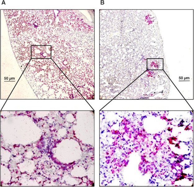
The COVID-19 (coronavirus disease-19) pandemic affected more than 180 million people around the globe, causing more than five million deaths as of January 2022. SARS-CoV-2 (severe acute respiratory syndrome coronavirus 2), the new coronavirus, has been identified as the primary cause of the infection. The number of vaccinated people is increasing; however, prophylactic drugs are highly demanded to ensure secure social contact. A number of drug molecules have been repurposed to fight against SARS-CoV-2, and some of them have been proven to be effective in preventing hospitalization or ICU admissions. Here, we demonstrated griffithsin (GRFT), a lectin protein, to block the entry of SARS-CoV-2 and its variants, Delta and Omicron, into the Vero E6 cell lines and IFNAR-/- mouse models by attaching to the spike protein of SARS-CoV-2. Given the current mutation frequency of SARS-CoV-2, we believe that GRFT protein-based drugs will have a high impact in preventing the transmission of both the Wuhan strain as well as any other emerging variants, including Delta and Omicron variants, causing the high-speed spread of COVID-19.

Keywords: Please add keywords.


Conflict of interest statement

The authors declare no competing financial interest.

Figures

Figure 1
Figure 1
Binding kinetics of rGRFT to S protein of SARS-CoV-2 and the virus itself. (a) Structure of HEK293 produced recombinant SARS-CoV-2 spike protein, and the RBD with attached glycan structures colored blue (side view). (b) Structure and amino acid sequence of rGRFT. (c) Purification validation of rGRFT protein on SDS-PAGE and western blotting gels, single bands represent the expected 14.5 kDA molecular mass of GRFT. (d) Secondary structure changes of rGRFT upon titration with mannose. (e) Molecular binding interaction of SARS-CoV-2 spike protein with rGRFT protein analyzed with ITC. (f) Qualitative ELISA for the interaction of rGRFT protein with SARS-CoV-2 virus. (g) QCM-D-based quantitative analysis of the binding of rGRFT on purified spike (left) and inactivated SARS-CoV-2 (right) immobilized on a carboxylated gold surface.
Figure 2
Figure 2
In vitro antiviral activity of rGRFT against SARS-CoV-2 infection. (a) Schematic representation of rGRFT inhibiting SARS-CoV-2 infection in Vero E6 cells, created with BioRender.com. (b) Determination of the rGRFT IC50 value for SARS-CoV-2 in Vero E6 cells. (c) Effects of rGRFT (5.76 nM) on SARS-CoV-2 replication in Vero E6 cells. (d) Cytotoxicity assessment of rGRFT on Vero E6 cells by MTT (3-[4,5-dimethyl-2-thiazolyl]-2,5-diphenyl-2H-tetrazolium bromide) assay.
Figure 3
Figure 3
Toxicity and immunogenicity of rGRFT in vivo. (a) Body temperature and (b) weight recorded for 14 days in C57BL/6 mice during innocuity testing. (c) Immunogenicity of rGRFT administered to C57BL/6 mice during innocuity testing. Antibodies against rGRFT were significantly lower than those induced by the His-tag in the construct (p < 0.05). (d) Representative tissue sections of the liver (upper left), lung (upper right), kidney (lower left), and spleen (lower right) in C57BL/6 mice on the 14th day following intraperitoneal rGRFT administration (hematoxylin and eosin staining, scale bar: 50 μm).
Figure 4
Figure 4
In vivo antiviral activity of rGRFT against SARS-CoV-2 infection shown for direct or contact-based infection models. (a) IFNAR–/– mice assigned to various study groups. The control group is mice treated with only rGRFT (i.n.) in a cage (n = 3), the prophylaxis group is rGRFT (i.n.)-treated and SARS-CoV-2-inoculated mice in a cage (white mice; n = 3), the protected contact group is consisted of rGRFT-treated mice and SARS-CoV-2-infected mice (white mice and red mice, respectively; n = 4 + 4), and the unprotected contact group is mock (i.n.)-treated mice and SARS-CoV-2-infected mice (brown mice and red mice, respectively; n = 4 + 4). (b) SARS-CoV-2 loads and infective virus titers in lung tissues of individual IFNAR–/– mice assigned to various study groups. rGRFT-treated mice from the protected contact group and mock-treated mice from the unprotected contact group are compared to SARS-CoV-2 loads and infective virus titers; the prophylaxis group (rGRFT-treated and SARS-CoV-2-infected mice) and all SARS-CoV-2-infected mouse are compared to SARS-CoV-2 loads and infective virus titers. The protected contact group values are represented with red color; the unprotected contact group values are represented with blue color; the prophylaxis group values are represented with green color; and the virus inoculation group is represented in orange color.
Figure 5
Figure 5
In situ hybridization of viral RNA in unprotected (A) and protected (B) animals following SARS-CoV-2 infection.
Figure 6
Figure 6
rGRFT inhibitory activity against SARS-CoV-2 variants in vitro with the Vero E6 infection model. The calculated IC50 value of rGRFT for Delta (a) and Omicron (b) variants.

References

    1. Gorbalenya A. E.; Baker S. C.; Baric R. S.; de Groot R. J.; Drosten C.; Gulyaeva A. A.; Haagmans B. L.; Lauber C.; Leontovich A. M.; Neuman B. W.; et al. The species Severe acute respiratory syndrome-related coronavirus: classifying 2019-nCoV and naming it SARS-CoV-2. Nat. Microbiol. 2020, 5, 536–544. 10.1038/s41564-020-0695-z. - DOI - PMC - PubMed
    2. Wu F.; Zhao S.; Yu B.; Chen Y. M.; Wang W.; Song Z. G.; Hu Y.; Tao Z. W.; Tian J. H.; Pei Y. Y.; Yuan M. L.; Zhang Y. L.; Dai F. H.; Liu Y.; Wang Q. M.; Zheng J. J.; Xu L.; Holmes E. C.; Zhang Y. Z. A new coronavirus associated with human respiratory disease in China. Nature 2020, 579, 265–269. 10.1038/s41586-020-2008-3. - DOI - PMC - PubMed
    3. Zhou P.; Yang X. L.; Wang X. G.; Hu B.; Zhang L.; Zhang W.; Si H. R.; Zhu Y.; Li B.; Huang C. L.; Chen H. D.; Chen J.; Luo Y.; Guo H.; Jiang R. D.; Liu M. Q.; Chen Y.; Shen X. R.; Wang X.; Zheng X. S.; Zhao K.; Chen Q. J.; Deng F.; Liu L. L.; Yan B.; Zhan F. X.; Wang Y. Y.; Xiao G. F.; Shi Z. L. A pneumonia outbreak associated with a new coronavirus of probable bat origin. Nature 2020, 579, 270–273. 10.1038/s41586-020-2012-7. - DOI - PMC - PubMed
    4. Ghebreyesus T.WHO Director-General’s opening remarks at the media briefing on COVID-19-2020. 2020.
    1. Mercatelli D.; Giorgi F. M. Geographic and Genomic Distribution of SARS-CoV-2 Mutations. Front. Microbiol. 2020, 11, 1800.10.3389/fmicb.2020.01800. - DOI - PMC - PubMed
    1. Rasmussen A. L.; Popescu S. V. SARS-CoV-2 transmission without symptoms. Science 2021, 371, 1206–1207. 10.1126/science.abf9569. - DOI - PubMed
    1. Chan J. F. W.; Kok K. H.; Zhu Z.; Chu H.; To K. K. W.; Yuan S. F.; Yuen K. Y. Genomic characterization of the 2019 novel human-pathogenic coronavirus isolated from a patient with atypical pneumonia after visiting Wuhan. Emerg. Microbes Infect. 2020, 9, 221–236. 10.1080/22221751.2020.1719902. - DOI - PMC - PubMed
    1. Lan J.; Ge J. W.; Yu J. F.; Shan S. S.; Zhou H.; Fan S. L.; Zhang Q.; Shi X. L.; Wang Q. S.; Zhang L. Q.; Wang X. Structure of the SARS-CoV-2 spike receptor-binding domain bound to the ACE2 receptor. Nature 2020, 581, 215–220. 10.1038/s41586-020-2180-5. - DOI - PubMed
    2. Yan R. H.; Zhang Y. Y.; Li Y. N.; Xia L.; Guo Y. Y.; Zhou Q. Structural basis for the recognition of SARS-CoV-2 by full-length human ACE2. Science 2020, 367, 1444–1448. 10.1126/science.abb2762. - DOI - PMC - PubMed

Publication types

Supplementary concepts