

Accelerated longitudinal cortical atrophy in OEF/OIF/OND veterans with severe PTSD and the impact of comorbid TBI
- PMID: 35426972
- PMCID: PMC9294300
- DOI: 10.1002/hbm.25877
Accelerated longitudinal cortical atrophy in OEF/OIF/OND veterans with severe PTSD and the impact of comorbid TBI
Abstract
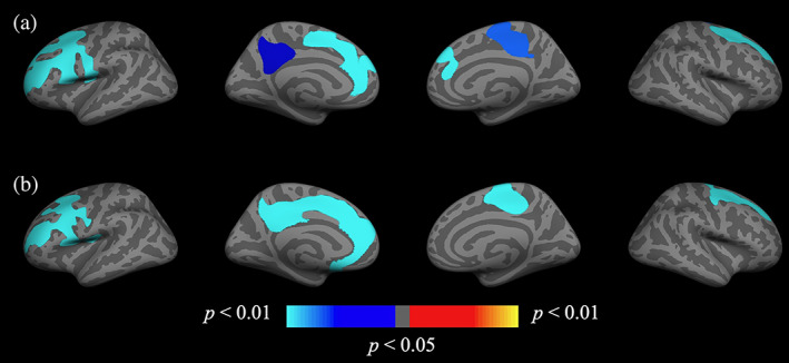
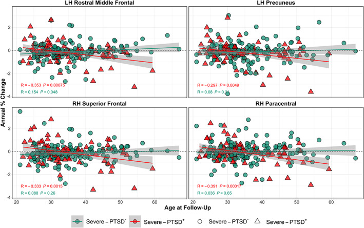
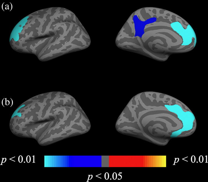
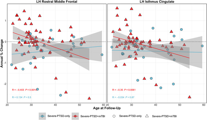
Veterans who deployed in support of Operation Enduring Freedom (OEF), Iraqi Freedom (OIF), and New Dawn (OND) commonly experience severe psychological trauma, often accompanied by physical brain trauma resulting in mild traumatic brain injury (mTBI). Prior studies of individuals with posttraumatic stress disorder (PTSD) have revealed alterations in brain structure, accelerated cellular aging, and impacts on cognition following exposure to severe psychological trauma and potential interactive effects of military-related mTBI. To date, however, little is known how such deployment-related trauma changes with time and age of injury of the affected veteran. In this study, we explored changes in cortical thickness, volume, and surface area after an average interval of approximately 2 years in a cohort of 254 OEF/OIF/OND Veterans ranging in age from 19 to 67 years. Whole-brain vertex-wise analyses revealed that veterans who met criteria for severe PTSD (Clinician-Administered PTSD Scale ≥60) at baseline showed greater negative longitudinal changes in cortical thickness, volume, and area over time. Analyses also revealed a significant severe-PTSD by age interaction on cortical measures with severe-PTSD individuals exhibiting accelerated cortical degeneration with increasing age. Interaction effects of comorbid military-related mTBI within the severe-PTSD group were also observed in several cortical regions. These results suggest that those exhibiting severe PTSD symptomatology have accelerated atrophy that is exacerbated with increasing age and history of mTBI.
Keywords: FreeSurfer; aging; cortical thickness; longitudinal; mild traumatic brain injury; posttraumatic stress disorder.
Published 2022. This article is a U.S. Government work and is in the public domain in the USA. Human Brain Mapping published by Wiley Periodicals LLC.
Conflict of interest statement
The authors declare no conflicts of interest.
Figures
Similar articles
-
Influence of Mild Traumatic Brain Injury (TBI) and Posttraumatic Stress Disorder (PTSD) on Pain Intensity Levels in OEF/OIF/OND Veterans.Pain Med. 2016 Nov;17(11):2017-2025. doi: 10.1093/pm/pnw042. Epub 2016 Apr 3. Pain Med. 2016. PMID: 27040665
-
Retrospective and Prospective Memory Among OEF/OIF/OND Veterans With a Self-Reported History of Blast-Related mTBI.J Int Neuropsychol Soc. 2018 Apr;24(4):324-334. doi: 10.1017/S1355617717001217. Epub 2017 Dec 29. J Int Neuropsychol Soc. 2018. PMID: 29284552
-
Reduced lateral prefrontal cortical volume is associated with performance on the modified Iowa Gambling Task: A surface based morphometric analysis of previously deployed veterans.Psychiatry Res Neuroimaging. 2017 Sep 30;267:1-8. doi: 10.1016/j.pscychresns.2017.06.014. Epub 2017 Jun 28. Psychiatry Res Neuroimaging. 2017. PMID: 28672256
-
Complicating factors associated with mild traumatic brain injury: impact on pain and posttraumatic stress disorder treatment.J Clin Psychol Med Settings. 2011 Jun;18(2):145-54. doi: 10.1007/s10880-011-9239-2. J Clin Psychol Med Settings. 2011. PMID: 21626354 Review.
-
Self-report measures to identify post traumatic stress disorder and/or mild traumatic brain injury and associated symptoms in military veterans of Operation Enduring Freedom (OEF)/Operation Iraqi Freedom (OIF).Neuropsychol Rev. 2012 Mar;22(1):35-53. doi: 10.1007/s11065-012-9191-4. Epub 2012 Feb 19. Neuropsychol Rev. 2012. PMID: 22350740 Review.
Cited by
-
Early attentional processing and cortical remapping strategies of tactile stimuli in adults with an early and late-onset visual impairment: A cross-sectional study.PLoS One. 2024 Jul 9;19(7):e0306478. doi: 10.1371/journal.pone.0306478. eCollection 2024. PLoS One. 2024. PMID: 38980866 Free PMC article.
-
Posttraumatic Stress and Traumatic Brain Injury: Cognition, Behavior, and Neuroimaging Markers in Vietnam Veterans.J Alzheimers Dis. 2023;95(4):1427-1448. doi: 10.3233/JAD-221304. J Alzheimers Dis. 2023. PMID: 37694363 Free PMC article.
-
Post-traumatic stress disorder: evolving conceptualization and evidence, and future research directions.World Psychiatry. 2025 Feb;24(1):52-80. doi: 10.1002/wps.21269. World Psychiatry. 2025. PMID: 39810662 Free PMC article.
-
Genetic Risk for Alzheimer Disease and Plasma Tau Are Associated With Accelerated Parietal Cortex Thickness Change in Middle-Aged Adults.Neurol Genet. 2023 Feb 1;9(1):e200053. doi: 10.1212/NXG.0000000000200053. eCollection 2023 Feb. Neurol Genet. 2023. PMID: 36742995 Free PMC article.
-
Comorbid neurotrauma increases neurodegenerative-relevant cognitive, motor, and autonomic dysfunction in patients with rapid eye movement sleep behavior disorder: a substudy of the North American Prodromal Synucleinopathy Consortium.Sleep. 2024 Jun 13;47(6):zsae007. doi: 10.1093/sleep/zsae007. Sleep. 2024. PMID: 38181205 Free PMC article.
References
-
- Akiki, T. J. , Averill, C. L. , Wrocklage, K. M. , Schweinsburg, B. , Cobb Scott, J. , Martini, B. , Averill, L. A. , Southwick, S. M. , Krystal, J. H. , & Abdallah, C. G. (2017). The association of PTSD symptom severity with localized hippocampus and amygdala abnormalities. Chronic Stress, 1, 2470547017724069. 10.1177/2470547017724069 - DOI - PMC - PubMed
-
- American Psychiatric Association . (2013). Diagnostic and statistical manual of mental disorders: DSM‐5. American Psychiatric Association.
-
- Averill, L. A. , Abdallah, C. G. , Pietrzak, R. H. , Averill, C. L. , Southwick, S. M. , Krystal, J. H. , & Harpaz‐Rotem, I. (2017). Combat exposure severity is associated with reduced cortical thickness in combat veterans: A preliminary report. Chronic Stress, 1, 247054701772471. 10.1177/2470547017724714 - DOI - PMC - PubMed
-
- Clausen, A. N. , Clarke, E. , Phillips, R. D. , Haswell, C. , VA Mid‐Atlantic MIRECC Workgroup , & Morey, R. A. (2020). Combat exposure, posttraumatic stress disorder, and head injuries differentially relate to alterations in cortical thickness in military veterans. Neuropsychopharmacology, 45(3), 491–498. 10.1038/s41386-019-0539-9 - DOI - PMC - PubMed
Publication types
MeSH terms
LinkOut - more resources
Full Text Sources
Medical
Miscellaneous
